Th17 cell-derived IL-17A promoted tumor progression via STAT3/NF-κB/Notch1 signaling in non-small cell lung cancer
- PMID: 30377557
- PMCID: PMC6205058
- DOI: 10.1080/2162402X.2018.1461303
Th17 cell-derived IL-17A promoted tumor progression via STAT3/NF-κB/Notch1 signaling in non-small cell lung cancer
Erratum in
-
Correction.Oncoimmunology. 2021 Jan 20;10(1):1857947. doi: 10.1080/2162402X.2020.1857947. Oncoimmunology. 2021. PMID: 33537168 Free PMC article.
Abstract
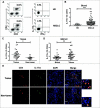
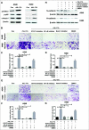
Non-small cell lung cancer (NSCLC) accounts for the majority of all lung cancer cases, which is the leading cause of cancer deaths worldwide. IL-17░A, the major effector cytokine derived from Th17 cells, is a key cytokine in tumor pathogenesis and modulates tumor progression. We aimed to identify whether IL-17░A derived from Th17 cells promotes the progression of NSCLC. Here we found that the level of Th17 cells was increased in NSCLC and IL-17░A was mainly produced by CD4+ cells (Th17 cells) in NSCLC. IL-17░A enhanced the migration, invasion and stemness of NSCLC via STAT3/NF-κB/Notch1 signaling. Blockade of this signaling inhibited the migration, invasion and stemness of NSCLC mediated by IL-17░A. Th17 cells in NSCLC were closely associated with poor prognosis of NSCLC patients. Our results indicated that Th17 cell-derived IL-17░A plays an important role in tumor progression of NSCLC via STAT3/NF-κB/Notch1 signaling. Therefore, therapeutic strategies against this pathway would be valuable to be developed for NSCLC treatment.
Keywords: Th17 cells; Tumor microenvironment; interleukin-17A (IL-17A); non-small cell lung cancer (NSCLC); tumor progression.
Figures


Similar articles
-
IL-10 derived from M2 macrophage promotes cancer stemness via JAK1/STAT1/NF-κB/Notch1 pathway in non-small cell lung cancer.Int J Cancer. 2019 Aug 15;145(4):1099-1110. doi: 10.1002/ijc.32151. Epub 2019 Feb 7. Int J Cancer. 2019. PMID: 30671927
-
IL-17A/IL-17RA promotes invasion and activates MMP-2 and MMP-9 expression via p38 MAPK signaling pathway in non-small cell lung cancer.Mol Cell Biochem. 2019 May;455(1-2):195-206. doi: 10.1007/s11010-018-3483-9. Epub 2018 Dec 18. Mol Cell Biochem. 2019. PMID: 30564960 Clinical Trial.
-
IL-6 promotes metastasis of non-small-cell lung cancer by up-regulating TIM-4 via NF-κB.Cell Prolif. 2020 Mar;53(3):e12776. doi: 10.1111/cpr.12776. Epub 2020 Feb 5. Cell Prolif. 2020. PMID: 32020709 Free PMC article.
-
The IL-17-Th1/Th17 pathway: an attractive target for lung cancer therapy?Expert Opin Ther Targets. 2016 Nov;20(11):1339-1356. doi: 10.1080/14728222.2016.1206891. Epub 2016 Jul 11. Expert Opin Ther Targets. 2016. PMID: 27353429 Review.
-
The Molecular Role of IL-35 in Non-Small Cell Lung Cancer.Front Oncol. 2022 May 26;12:874823. doi: 10.3389/fonc.2022.874823. eCollection 2022. Front Oncol. 2022. PMID: 35719927 Free PMC article. Review.
Cited by
-
Interleukin-17A mediates tobacco smoke-induced lung cancer epithelial-mesenchymal transition through transcriptional regulation of ΔNp63α on miR-19.Cell Biol Toxicol. 2022 Apr;38(2):273-289. doi: 10.1007/s10565-021-09594-0. Epub 2021 Apr 3. Cell Biol Toxicol. 2022. PMID: 33811578
-
IL-17A regulates autophagy and promotes osteoclast differentiation through the ERK/mTOR/Beclin1 pathway.PLoS One. 2023 Feb 16;18(2):e0281845. doi: 10.1371/journal.pone.0281845. eCollection 2023. PLoS One. 2023. PMID: 36795736 Free PMC article.
-
Mechanistic exploration of Traditional Chinese Medicine regulation on tumor immune microenvironment in the treatment of triple-negative breast cancer: based on CiteSpace and bioinformatics analysis.Front Immunol. 2025 Jan 10;15:1443648. doi: 10.3389/fimmu.2024.1443648. eCollection 2024. Front Immunol. 2025. PMID: 39867914 Free PMC article.
-
Cancer stem cell-immune cell crosstalk in tumour progression.Nat Rev Cancer. 2021 Aug;21(8):526-536. doi: 10.1038/s41568-021-00366-w. Epub 2021 Jun 8. Nat Rev Cancer. 2021. PMID: 34103704 Free PMC article. Review.
-
Respiratory Tract Oncobiome in Lung Carcinogenesis: Where Are We Now?Cancers (Basel). 2023 Oct 11;15(20):4935. doi: 10.3390/cancers15204935. Cancers (Basel). 2023. PMID: 37894302 Free PMC article. Review.
References
-
- Pierce BL, Ballard-Barbash R, Bernstein L, Baumgartner RN, Neuhouser ML, Wener MH, Baumgartner KB, Gilliland FD, Sorensen BE, McTiernan A, et al. . Elevated biomarkers of inflammation are associated with reduced survival among breast cancer patients. J Clin Oncol. 2009;27:3437–44. doi: 10.1200/JCO.2008.18.9068. PMID:19470939 - DOI - PMC - PubMed
Publication types
LinkOut - more resources
Full Text Sources
Research Materials
Miscellaneous
