

The evolution of image reconstruction for CT-from filtered back projection to artificial intelligence
- PMID: 30377791
- PMCID: PMC6443602
- DOI: 10.1007/s00330-018-5810-7
The evolution of image reconstruction for CT-from filtered back projection to artificial intelligence
Abstract
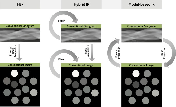
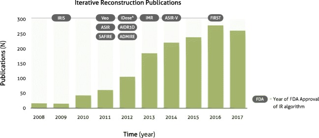
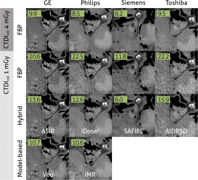
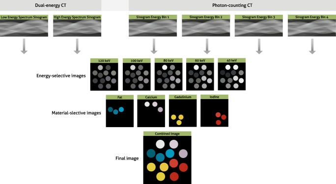
The first CT scanners in the early 1970s already used iterative reconstruction algorithms; however, lack of computational power prevented their clinical use. In fact, it took until 2009 for the first iterative reconstruction algorithms to come commercially available and replace conventional filtered back projection. Since then, this technique has caused a true hype in the field of radiology. Within a few years, all major CT vendors introduced iterative reconstruction algorithms for clinical routine, which evolved rapidly into increasingly advanced reconstruction algorithms. The complexity of algorithms ranges from hybrid-, model-based to fully iterative algorithms. As a result, the number of scientific publications on this topic has skyrocketed over the last decade. But what exactly has this technology brought us so far? And what can we expect from future hardware as well as software developments, such as photon-counting CT and artificial intelligence? This paper will try answer those questions by taking a concise look at the overall evolution of CT image reconstruction and its clinical implementations. Subsequently, we will give a prospect towards future developments in this domain. KEY POINTS: • Advanced CT reconstruction methods are indispensable in the current clinical setting. • IR is essential for photon-counting CT, phase-contrast CT, and dark-field CT. • Artificial intelligence will potentially further increase the performance of reconstruction methods.
Keywords: Artificial intelligence; Image reconstruction; Tomography, x-ray.
Conflict of interest statement
Guarantor
The scientific guarantor of this publication is Peter B. Noël, PhD.
Conflict of interest
The authors of this manuscript declare no relationships with any companies, whose products or services may be related to the subject matter of the article.
Statistics and biometry
One of the authors has significant statistical expertise; however, no complex statistical methods were necessary for this paper.
Informed consent
Written informed consent was not required for this study because this concerns a review paper.
Ethical approval
Institutional Review Board approval was not required because this concerns a review paper.
Methodology
• Review paper
Figures
References
-
- Ambrose J, Hounsfield G. Computerized transverse axial tomography. Br J Radiol. 1973;46:148–149. - PubMed
-
- Hounsfield GN. Computerized transverse axial scanning (tomography). 1. Description of system. Br J Radiol. 1973;46:1016–1022. - PubMed
-
- OECD . Health at a glance 2017: OECD indicators. Paris: OECD Publishing; 2017.
-
- de Graaf FR, Schuijf JD, van Velzen JE, et al. Diagnostic accuracy of 320-row multidetector computed tomography coronary angiography in the non-invasive evaluation of significant coronary artery disease. Eur Heart J. 2010;31:1908–1915. - PubMed
-
- Hata A, Yanagawa M, Honda O et al (2018) Effect of matrix size on the image quality of ultra-high-resolution CT of the lung: comparison of 512 x 512, 1024 x 1024, and 2048 x 2048. Acad Radiol. 10.1016/j.acra.2017.11.017 - PubMed
Publication types
MeSH terms
Grants and funding
LinkOut - more resources
Full Text Sources
Other Literature Sources
Medical
Research Materials
