

Tenocyte cell density, migration, and extracellular matrix deposition with amniotic suspension allograft
- PMID: 30378182
- PMCID: PMC6587843
- DOI: 10.1002/jor.24173
Tenocyte cell density, migration, and extracellular matrix deposition with amniotic suspension allograft
Abstract
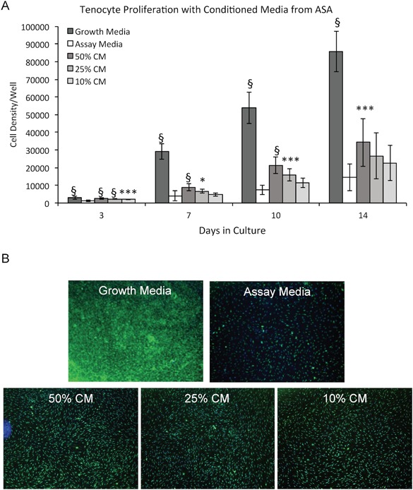
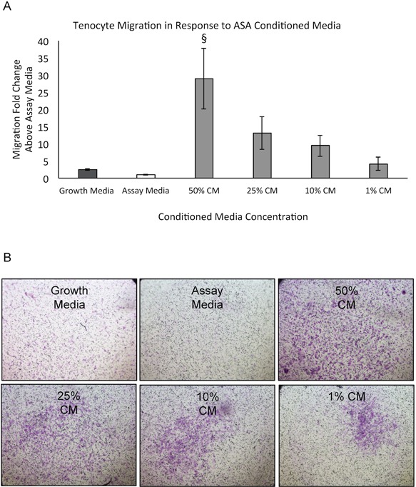
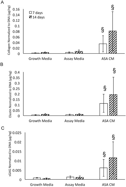
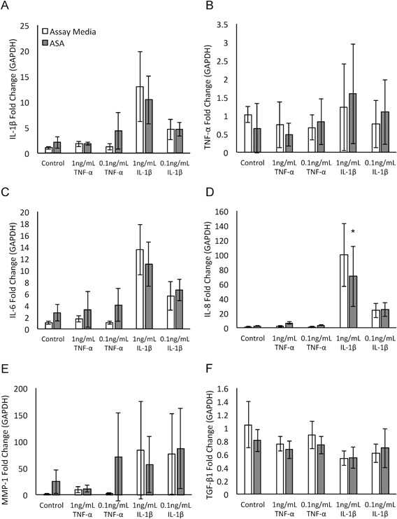
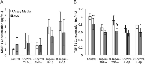
Amniotic suspension allografts (ASA), derived from placental tissues, contain particulated amniotic membrane and amniotic fluid cells. Recently, ASA and other placental-derived allografts have been used in orthopaedic applications, including tendinopathies and tendon injuries. The purpose of this study was to determine the potential effects of ASA on tenocyte cell density, migration, and responses to inflammatory stimuli. Tenocyte cell density was measured using AlamarBlue over multiple time points, while migration was determined using a Boyden chamber assay. Deposition of ECM markers were measured using BioColor kits. Gene expression and protein production of cytokines and growth factors following stimulus with pro-inflammatory IL-1β and TNF-α was measured using qPCR and ELISAs. Conditioned media (CM) was made from ASA and used for all assays in this study. In vitro, ASA CM treatment significantly promoted tenocyte increases in cell density and migration compared to assay media controls. ASA CM also increased the deposition of extracellular matrix (ECM) proteins, including collagen, elastin, and sGAG. Following inflammatory stimulation and treatment with ASA CM, tenocytes downregulated IL-8 gene expression, a pro-inflammatory cytokine normally elevated during the inflammatory phase of tendon healing. Additionally, tenocytes treated with ASA CM had significantly lower protein levels of TGF-β1 compared to controls. This study evaluated ASA and its effect on tenocytes; specifically, treatment with ASA resulted in increased cell density, more robust migration and matrix deposition, and some alteration of inflammatory targets. © 2018 The Authors. Journal of Orthopaedic Research® Published by Wiley Periodicals, Inc. on behalf of Orthopaedic Research Society. J Orthop Res 37:412-420, 2019.
Keywords: amniotic suspension allograft; regenerative; repair; tendon; tenocytes.
© 2018 The Authors. Journal of Orthopaedic Research® Published by Wiley Periodicals, Inc. on behalf of Orthopaedic Research Society.
Figures
References
-
- Sabella N. 1913. Use of fetal membranes in skin grafting. Med Rec 83:478–480.
-
- Riboh JC, Saltzman BM, Yanke AB, et al. 2016. Human amniotic membrane‐derived products in sports medicine: basic science, early results, and potential clinical applications. Am J Sports Med 44:2425–2434. - PubMed
-
- Bryant‐Greenwood GD. 1998. The extracellular matrix of the human fetal membranes: structure and function. Placenta 19:1–11. - PubMed
-
- Hao Y, Ma DH‐K, Hwang DG, et al. 2000. Identification of antiangiogenic and antiinflammatory proteins in human amniotic membrane. Cornea 19:348–352. - PubMed
Publication types
MeSH terms
Substances
Grants and funding
LinkOut - more resources
Full Text Sources
