Review
. 2019 Mar;8(2):e337.
doi: 10.1002/wdev.337. Epub 2018 Oct 31.

Evolution and development of the fish jaw skeleton

Affiliations
Review

Evolution and development of the fish jaw skeleton

April DeLaurier. Wiley Interdiscip Rev Dev Biol. 2019 Mar.

Abstract

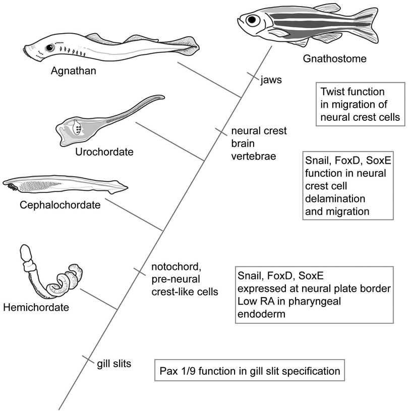
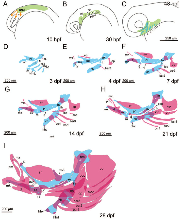
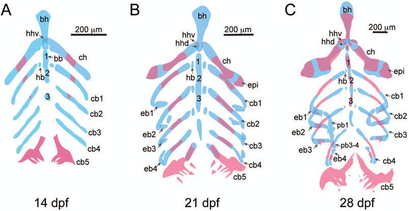
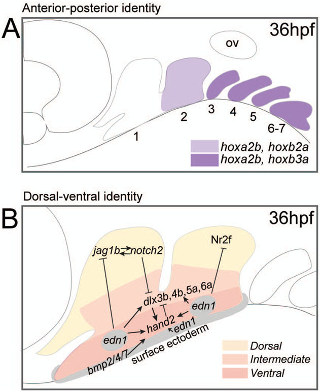
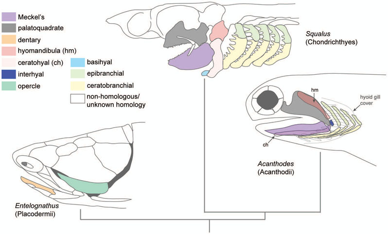
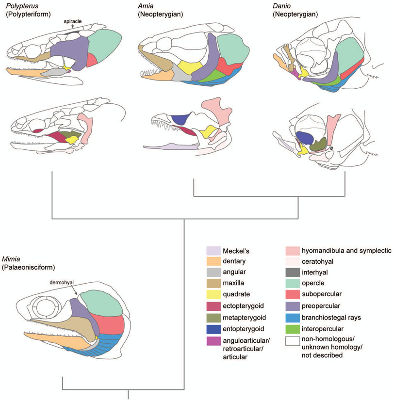
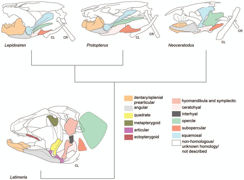
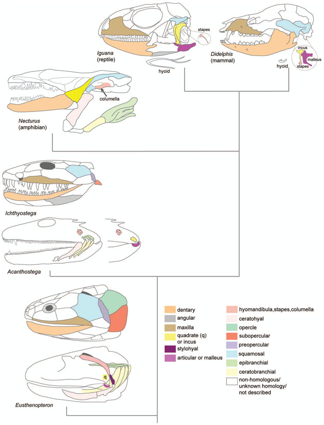
The evolution of the jaw represents a key innovation in driving the diversification of vertebrate body plans and behavior. The pharyngeal apparatus originated as gill bars separated by slits in chordate ancestors to vertebrates. Later, with the acquisition of neural crest, pharyngeal arches gave rise to branchial basket cartilages in jawless vertebrates (agnathans), and later bone and cartilage of the jaw, jaw support, and gills of jawed vertebrates (gnathostomes). Major events in the evolution of jaw structure from agnathans to gnathostomes include axial regionalization of pharyngeal elements and formation of a jaw joint. Hox genes specify the anterior-posterior identity of arches, and edn1, dlx, hand2, Jag1b-Notch2 signaling, and Nr2f factors specify dorsal-ventral identity. The formation of a jaw joint, an important step in the transition from an un-jointed pharynx in agnathans to a hinged jaw in gnathostomes involves interaction between nkx3.2, hand2, and barx1 factors. Major events in jaw patterning between fishes and reptiles include changes to elements of the second pharyngeal arch, including a loss of opercular and branchiostegal ray bones and transformation of the hyomandibula into the stapes. Further changes occurred between reptiles and mammals, including the transformation of the articular and quadrate elements of the jaw joint into the malleus and incus of the middle ear. Fossils of transitional jaw phenotypes can be analyzed from a developmental perspective, and there exists potential to use genetic manipulation techniques in extant taxa to test hypotheses about the evolution of jaw patterning in ancient vertebrates. This article is categorized under: Comparative Development and Evolution > Evolutionary Novelties Early Embryonic Development > Development to the Basic Body Plan Comparative Development and Evolution > Body Plan Evolution.

Keywords: jaw; morphogenesis; neural crest; skeleton; vertebrates.


Conflict of interest statement

No conflicts of interest

Figures

Figure 1:
Figure 1:
Evolution of gills slits and neural crest in hemichordates and chordates. Gill slits first appear in hemichordates, associated with the expression of Pax1/9 homologs. Snail, FoxD, and SoxE factors are expressed at the neural plate border in pre-vertebrate chordates, at locations where neural crest later evolves in vertebrates. In vertebrates, these factors function in neural crest cell delamination and migration. Low retinoic acid (RA) in pharyngeal endoderm specifies the anterior pharynx in pre-vertebrate chordates. Twist is associated with neural crest cell migration in gnathostomes, but does not function in this process in agnathans. Hemichordate redrawn from an image by C.B. Cameron, used with permission.
Figure 2:
Figure 2:
Stages of neural crest migration, pharyngeal arch patterning, and formation of the pharyngeal skeleton in zebrafish. A) Cranial neural crest cell migration begins at approximately 10hpf. B) By 30hpf the first through seventh pharyngeal arches (1-7) and first four pouches (p1-p4) are formed. Primordia for the posterior arches and pouches are present but arches and pouches are indistinct. C) By 48 hpf the precursors for skeletal elements (blue) can be detected within the arch mesenchyme (green). D) At 3dpf, cartilaginous elements of the first through fifth pharyngeal arches are present. Ventral midline elements are present, as is the opercle. E and F) Cartilaginous elements enlarge and more dermal bones form. The opercle becomes a fan-shape and by 6 or 7 dpf ossification of the hyosymplectic and ceratohyal begins. G-I) 14dpf-28dpf cartilaginous elements increase in size and cartilaginous elements undergo ossification. Dermal bones form and expand to encase cartilaginous elements. Cartilage is shown in blue, bone is shown in red. Anguloarticular (aa), branchiostegal ray (bsr), ceratobranchial (cb1-3), ceratohyal (ch), coronomeckelian (cm), cranial neural crest cells (CNC), dentary (d), dorsal hypohyal (hhd), ventral hypohyal (hhv), ectopterygoid (ec), entopterygoid (en), epihyal (epi), hyomandibula (hm), hyosymplectic (hs), interhyal (ih), interopercle (iop), maxilla (mx), Meckel’s cartilage (mk), metapterygoid (mpt), opercle (op), palatine (p), palatoquadrate (pq), premaxilla (pm), preopercle (pop), quadrate (q), retroarticular (ra), subopercle (sop), symplectic (sy).
Figure 3:
Figure 3:
Structures of the branchial skeleton. A) 14 dpf, ventral view of basibranchials (bb, 1-3), basihyal (bh), ceratobranchials (cb1-5), ceratohyal (ch), dorsal hypohyal (hhd), hypobranchials (hb), and ventral hypohyal (hhv). B) 21 dpf, ventral view, epibranchials (ep1-4) are present. C) 28 dpf, epibranchials have increased in size and are mineralizing, and pharyngobranchials (pb1-4) are present.
Figure 4:
Figure 4:
Genetic mechanisms regulating anterior-posterior and dorsal-ventral patterning of the pharyngeal arches in zebrafish. A) Anterior-posterior patterning: expression of Hox paralogs in pharyngeal arches at approximately 36hpf. The first pharyngeal arch is hox-negative. The second pharyngeal arch expresses hoxa2b and hoxb2a, and third through seventh arches express hoxa2b and hoxb3a. Hoxa4a, hoxb4a, are hoxd4a are expressed in posterior gill arches, although the boundaries of expression in specific arches are not clear (not shown). B) Dorsal-ventral patterning: Jag1b-Notch2 signalling and Nr2f genes function in the dorsal domain, dlx3b, dlx4a, dlx5a, and dlx6a are expressed in the intermediate domain. Hand2 is expressed in the ventral domain. Bmp factors are expressed in the ventral epithelia. Edn1 is expressed in ventral pharyngeal arches and mesenchymal cores of arches. Factors function to activate or inhibit one another to specify boundaries of dorsal-ventral identity. Notch2 is expressed throughout the pharyngeal arches at 36 hpf, although Jag1b activates Notch2 in the dorsal domain. Adapted with copyright permission (Alexander et al., 2011; Barske et al., 2018; Hunter & Prince, 2002; Laue et al., 2008; Craig T. Miller et al., 2003; Punnamoottil et al., 2008; Talbot et al., 2010; Zuniga et al., 2011, 2010).
Figure 5:
Figure 5:
Jaw patterning in cartilaginous fish classes Placodermii, Chondrichthyes, and Acanthodii. The fossil placoderm Entelognathus has a bony dermal head skeleton with evidence of an opercle and dentary. Other elements are also present such as maxillary bones, but not shown. Modern Squalus (dogfish shark) features cartilaginous elements only. Acanthodes has a cartilaginous endoskeleton with overlying dermal bone elements, including a hyoid gill covered in bony branchiostegal rays (grey outlines). The hyomandibula is beneath the palatoquadrate and the ceratohyal is beneath the Meckel’s cartilage. Note the presence of an interhyal and symmetry between epibranchial and ceratobranchial cartilages. Adapted with copyright permission (Janvier, 1996; Kardong, 2012; Zhu et al., 2013).
Figure 6:
Figure 6:
Jaw patterning in actinopterygian fishes. For Polypterus, Amia, and Danio, dermatocranial bones are illustrated at the top of figure and viscerocranial elements below. For Mimia, only dermatocranial elements are shown. The fossil palaeonisciform Mimia has both cartilage ossification and dermal bone element representing basic features of actinopterygians. Polypterus, an extant form of polypteriform actinopterygian has transformed the first gill slit into a spiracle for respiration. The ceratohyal is found in two parts, and anterior and posterior element. Amia, representative of an early neopterygian actinopterygian has a sturdy dermal skeleton overlying a hyomandibula with a separate symplectic element and interopercle bone. Danio, a representative neopterygian teleost has a substantially reduced maxilla and dentary compared to earlier forms of actinopterygians, adapted for protrusion of the maxilla during suction feeding. Adapted with copyright permission (Gardiner, 1984; Jarvik, 1980; Jollie, 1984a).
Figure 7:
Figure 7:
Jaw patterning in sarcopterygian fishes. In Latimeria, the hyomandibula and symplectic are separate elements. In Lepidosiren, Protopterus, Neoceratodus (Rhipdistians), the hyomandibula and symplectic are absent. The opercle is a plate-like structure in Neoceratodus, but a blade-like structure in Lepidosiren and Protopterus. Rhipdistians have a novel element, the squamosal forming the adult jaw joint. This arises near the larval quadrate from a separate primordium. In Rhipdistians cranial ribs (CR) articulate with the neurocranium and assist with respiration. CL = clavicle. Adapted with copyright permission (Criswell, 2015; Dutel, Herrel, et al., 2015).
Figure 8:
Figure 8:
Jaw patterning in rhipidistian fishapods and labyrinthodonts. In Eusthenopteron, a prehistoric sarcopterygian fish, the hyomandibula is a blade-like element adjacent to the spiracle. There is no symplectic. The ceratohyal is in two parts. In Ichthyostega and Acanthostega, the squamosal is a cranial vault bone, and the opercular series is much reduced, where the subopercle and preopercle bones are very small elements. Fossil evidence of the deeper elements of the jaw in Acanthostega reveals a small hyomandibula and evidence of gill structures. In the amphibian Necturus, the ceratohyal is present, as are ceratobranchials and epibranchials, associated with gill structures. A columella (hyomandibula, stapes) is present. In the modern Iguana, the hyomandibula has become a hearing organ, the stapes that connects the quadrate to the inner ear. The second arch-derived hyoid is a separate throat structure. In Eusthenopteron, Acanthostega, and reptiles, the jaw joint is between the articular and quadrate. In mammals such as Didelphis (possum) the lower jaw is exclusively formed from the dentary forming a joint with the squamosal. Besides the hyomandibula-derived stapes, the quadrate has become transformed into the incus bone, and the articular has become the malleus of the middle ear (Valdezate et al., 2015). The styloid process is a projection from the temporal bone and is derived from the second pharyngeal arch (Kent and Carr, 2001) (not shown). Adapted with copyright permission (Clack, 2002; Jarvik, 1980; Kardong, 2012; Porter & Witmer, 2015).

Similar articles

Cited by

References

    1. Abitua PB, Wagner E, Navarrete IA, & Levine M (2012). Identification of a rudimentary neural crest in a non-vertebrate chordate. Nature, 492(7427), 104. 10.1038/nature11589 - DOI - PMC - PubMed
    1. Alexander C, Zuniga E, Blitz IL, Wada N, Le Pabic P, Javidan Y, … Schilling TF (2011). Combinatorial roles for BMPs and Endothelin 1 in patterning the dorsal-ventral axis of the craniofacial skeleton. Development (Cambridge, England), 138(23), 5135–5146. 10.1242/dev.067801 - DOI - PMC - PubMed
    1. Amores A, Force A, Yan YL, Joly L, Amemiya C, Fritz A, … Postlethwait JH (1998). Zebrafish hox clusters and vertebrate genome evolution. Science (New York, N.Y.), 282(5394), 1711–1714. - PubMed
    1. Anderson PSL, Friedman M, Brazeau MD, & Rayfield EJ (2011). Initial radiation of jaws demonstrated stability despite faunal and environmental change. Nature, 476(7359), 206–209. 10.1038/nature10207 - DOI - PubMed
    1. Anthwal N, Urban DJ, Luo ZX, Sears KE, & Tucker AS (2017). Meckel’s cartilage breakdown offers clues to mammalian middle ear evolution. Nature Ecology & Evolution, 1(4), 93. 10.1038/s41559-017-0093 - DOI - PMC - PubMed

Further Reading

    1. Clack JA (2002). Gaining Ground: The Origin and Evolution of Tetrapods. Indiana University Press.
    1. Eames BF, DeLaurier A, Ullmann B, Huycke TR, Nichols JT, Dowd J, … Kimmel CB (2013). FishFace: interactive atlas of zebrafish craniofacial development at cellular resolution. BMC Developmental Biology, 13, 23. 10.1186/1471-213X-13-23 - DOI - PMC - PubMed
    1. Frisdal A, & Trainor PA (2014). Development and evolution of the pharyngeal apparatus. Wiley Interdisciplinary Reviews. Developmental Biology, 3(6), 403–418. 10.1002/wdev.147 - DOI - PMC - PubMed
    1. Schubert FR, Singh AJ, Afoyalan O, Kioussi C, & Dietrich S (2018). To roll the eyes and snap a bite - function, development and evolution of craniofacial muscles. Seminars in Cell & Developmental Biology. 10.1016/j.semcdb.2017.12.013 - DOI - PubMed

Publication types