. 2018 Dec;7(23):e1800672.
doi: 10.1002/adhm.201800672. Epub 2018 Oct 31.

A Bioprinted Cardiac Patch Composed of Cardiac-Specific Extracellular Matrix and Progenitor Cells for Heart Repair

Affiliations

A Bioprinted Cardiac Patch Composed of Cardiac-Specific Extracellular Matrix and Progenitor Cells for Heart Repair

Donald Bejleri et al. Adv Healthc Mater. 2018 Dec.

Abstract

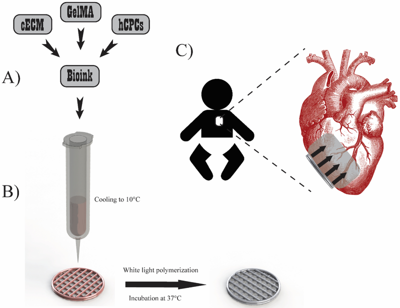
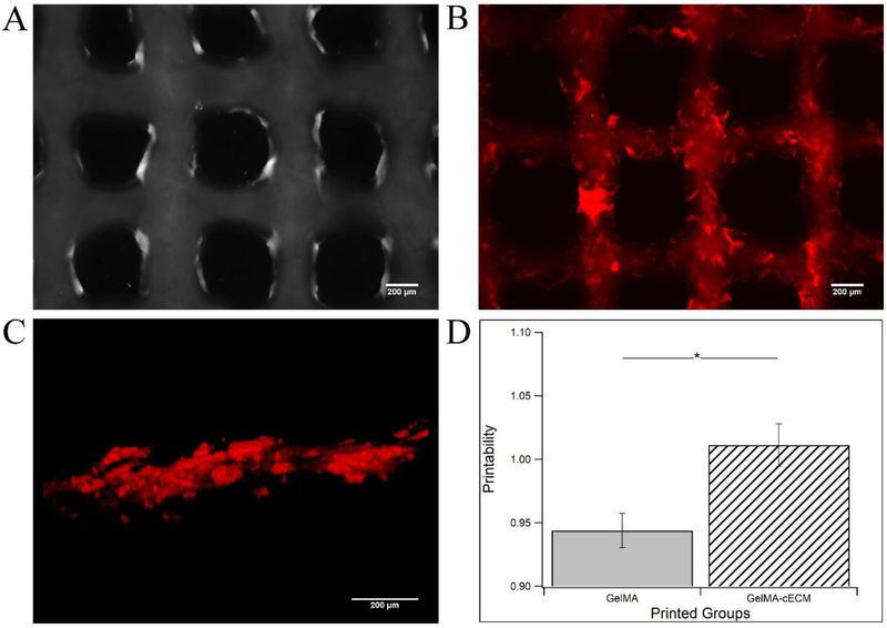
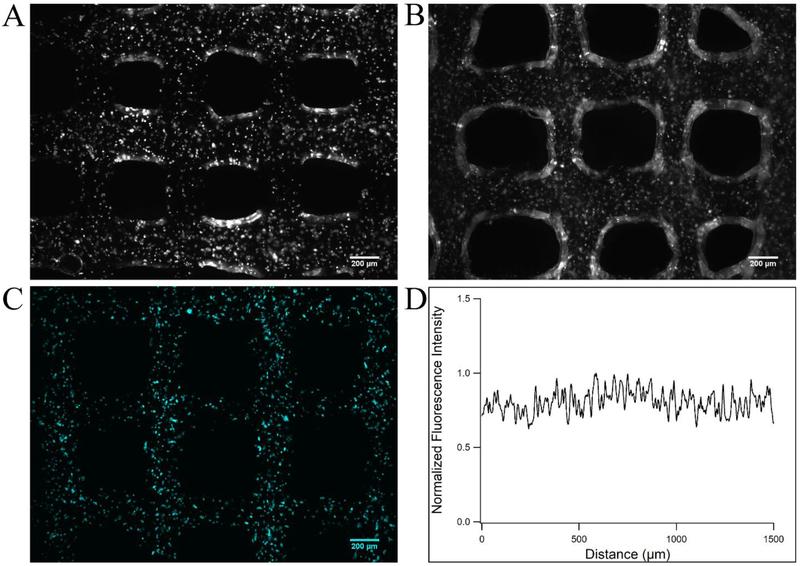
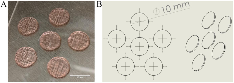
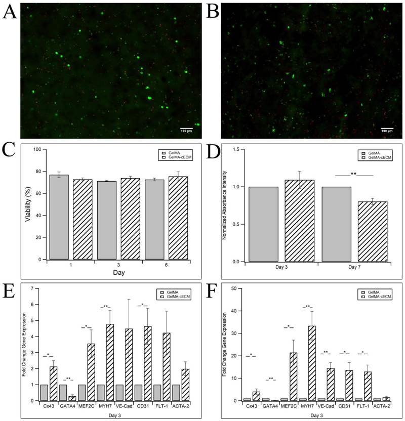
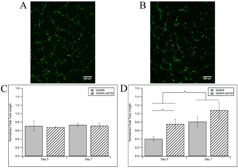
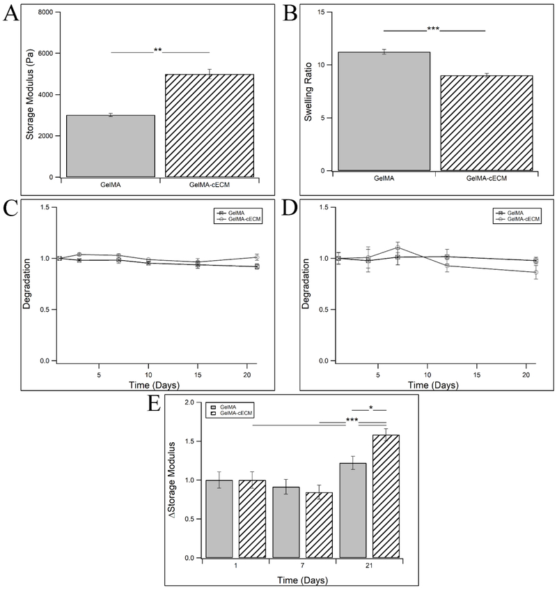
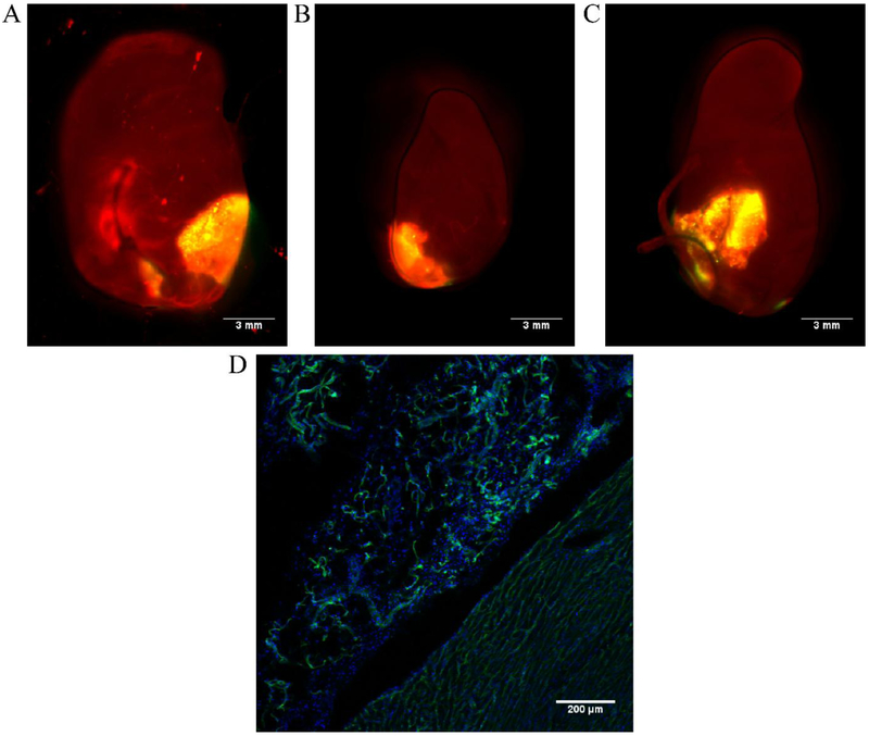
Congenital heart defects are present in 8 of 1000 newborns and palliative surgical therapy has increased survival. Despite improved outcomes, many children develop reduced cardiac function and heart failure requiring transplantation. Human cardiac progenitor cell (hCPC) therapy has potential to repair the pediatric myocardium through release of reparative factors, but therapy suffers from limited hCPC retention and functionality. Decellularized cardiac extracellular matrix hydrogel (cECM) improves heart function in animals, and human trials are ongoing. In the present study, a 3D-bioprinted patch containing cECM for delivery of pediatric hCPCs is developed. Cardiac patches are printed with bioinks composed of cECM, hCPCs, and gelatin methacrylate (GelMA). GelMA-cECM bioinks print uniformly with a homogeneous distribution of cECM and hCPCs. hCPCs maintain >75% viability and incorporation of cECM within patches results in a 30-fold increase in cardiogenic gene expression of hCPCs compared to hCPCs grown in pure GelMA patches. Conditioned media from GelMA-cECM patches show increased angiogenic potential (>2-fold) over GelMA alone, as seen by improved endothelial cell tube formation. Finally, patches are retained on rat hearts and show vascularization over 14 d in vivo. This work shows the successful bioprinting and implementation of cECM-hCPC patches for potential use in repairing damaged myocardium.

Keywords: bioprinting; cardiac extracellular matrix; cardiac patches; cardiac progenitor cells; pediatric heart failure.


Figures

Figure 1.
Figure 1.
Printing overview. A) Bioink preparation involved combining cECM, hCPCs, and GelMA to form naturally derived and cell laden materials for printing. B) Printing methodology involved cooling the bioink to 10°C in the 3D bioprinter barrels to allow GelMA polymerization for improved printability. Patches were printed with infill patterns of 90° intersecting filaments and contour. Patches were polymerized via white light to induce radical polymerization of GelMA, followed by incubation at 37°C for at least 1 hour to induce cECM polymerization. C) Patch implementation will involve pericardially inserting the patch to the RV of pediatric patients, where the patch will release key pro-regenerative paracrine factors.
Figure 2.
Figure 2.
Printability analysis of GelMA-cECM bioinks. A) Bright-field image of printed test grids of GelMA. B) Fluorescence image of printed test grids of GelMA-cECM with staining for cECM by AF568. C) 3D fluorescence close-up view of printed filament of GelMA-cECM, with staining for cECM by AF568. D) Printability comparison between GelMA and GelMA-cECM bioinks. * = p-value < 0.03, given by paired t-test, n = 5.
Figure 3.
Figure 3.
Printing hCPC-containing bioinks. A) Bright-field image of printed test grids of GelMA bioinks containing hCPCs, taken 1 hour after printing. B) Bright-field image of printed test grids of GelMA-cECM bioinks containing hCPCs. C) Fluorescence image of printed test grids of GelMA-cECM with hCPCs stained with DiD. D) Normalized fluorescence intensity of line scans performed on stained hCPC test grids. Line scans were performed across several filaments.
Figure 4.
Figure 4.
Printed patches. A) Printed patches of 10 mm diameter and 0.6 mm height. Patches are printed uniformly from patch to patch, and the grid infill pattern can be seen. Patches are pink post-printing due to inclusion of photoinitiator Eosin Y, and become clear post-polymerization. B) CAD model sketch used for patch printing.
Figure 5.
Figure 5.
hCPC functionality within printed patches. A) Characteristic live/dead fluorescence image of hCPCs in GelMA patches, with live cells marked green (Calcien AM) and dead cells marked red (EtD) at 1 day after formation. B) Characteristic live/dead fluorescence image of hCPCs in GelMA-cECM patches at 1 day after formation. C) Viability of hCPCs in printed patches at 1, 3, and 6 days. D) Proliferation of hCPCs in printed patches at 1 and 7 days, where absorbance intensity is normalized to the measured absorbance of hCPCs in GelMA patches in all experiments. E) Fold change gene expression over hCPCs in GelMA patches for Cx43, GATA4, MEF2C, MYH7, VE-Cad, CD31, FLT-1, and ACTA-2 at day 3. F) Fold change gene expression over hCPCs in GelMA patches for Cx43, GATA4, MEF2C, MYH7, VE-Cad, CD31, FLT-1, and ACTA-2 at day 7. * = p-value < 0.05, ** = p-value < 0.005, given by ANOVA with Tukey’s post-test, n = 3–6 for all samples at all timepoints.
Figure 6.
Figure 6.
Angiogenic potential of cardiac patches. Characteristic HUVEC tube formation after 6 hours when grown with conditioned media collected at day 7 from cell-laden A) GelMA and B) GelMA-cECM patches. Total HUVEC tube length normalized to positive controls for C) cell-free and D) cell-laden patches. * = p-value < 0.05, given by ANOVA with Tukey’s post-test, n = 3-6 for all samples at all timepoints.
Figure 7.
Figure 7.
Material analysis of printed patches. A) Viscoelastic storage moduli of GelMA and GelMA-cECM. B) Swelling ratio of GelMA and GelMA-cECM patches. C) Degradation of patches, measured as the sample weight compared to initial weight of the patches post-swelling. D) Degradation of cell-free materials in cell culture media, measured as the sample storage modulus compared to the initial modulus of the material post-swelling. E) Remodeling of hCPC-laden materials grown in cFB conditioned media, measured as the sample storage modulus compared to the initial modulus of the material post-swelling. * = p-value < 0.05, ** = p-value < 0.01, *** = p-value < 0.005, given by ANOVA with Tukey’s post-test, n = 3 for all samples in all subfigures.
Figure 8.
Figure 8.
In vivo patch retention. hCPC-laden GelMA-cECM patches (yellow) are retained after 7 (A, suture method) and 14 (B, pericardial tucking method and C, suture method) days following implantation. D) Immunohistological analysis of vasculature formation (green) and cells (blue) after 14 days in vivo.

References

    1. Rossano JW, Kim JJ, Decker JA, Price JF, Zafar F, Graves DE, Morales DL, Heinle JS, Bozkurt B, Denfield SW, Circulation. 2010, 122, A1374.
    1. Robbins JM, Bird TM, Tilford JM, Cleves MA, Hobbs CA, Grosse SD, Correa A, MMWR. 2007, 56, 25. - PubMed
    1. Bove EL, Pediatr. Cardiol. 1998, 19, 308. - PubMed
    1. Segers VF, Lee RT, Nature 2008, 451, 937. - PubMed
    1. Bolli R, Chugh AR, D’Amario D, Loughran JH, Stoddard MF, Ikram S, Beache GM, Wagner SG, Leri A, Hosoda T, The Lancet 2011, 378, 1847. - PMC - PubMed

Publication types

MeSH terms