

Cardioprotection during ischemia by coronary collateral growth
- PMID: 30379567
- PMCID: PMC6383359
- DOI: 10.1152/ajpheart.00145.2018
Cardioprotection during ischemia by coronary collateral growth
Abstract
Ischemic heart diseases (IHD) cause millions of deaths around the world annually. While surgical and pharmacological interventions are commonly used to treat patients with IHD, their efficacy varies from patient to patient and is limited by the severity of the disease. One promising, at least theoretically, approach for treating IHD is induction of coronary collateral growth (CCG). Coronary collaterals are arteriole-to-arteriole anastomoses that can undergo expansion and remodeling in the setting of coronary disease when the disease elicits myocardial ischemia and creates a pressure difference across the collateral vessel that creates unidirectional flow. Well-developed collaterals can restore blood flow in the ischemic area of the myocardium and protect the myocardium at risk. Moreover, such collaterals are correlated to reduced mortality and infarct size and better cardiac function during occlusion of coronary arteries. Therefore, understanding the process of CCG is highly important as a potentially viable treatment of IHD. While there are several excellent review articles on this topic, this review will provide a unified overview of the various aspects related to CCG as well as an update of the advancements in the field. We also call for more detailed studies with an interdisciplinary approach to advance our knowledge of CCG. In this review, we will describe growth of coronary collaterals, the various factors that contribute to CCG, animal models used to study CCG, and the cardioprotective effects of coronary collaterals during ischemia. We will also discuss the impairment of CCG in metabolic syndrome and the therapeutic potentials of CCG in IHD.
Keywords: cardioprotection; coronary circulation; coronary collateral growth; ischemia.
Conflict of interest statement
No conflicts of interest, financial or otherwise, are declared by the authors.
Figures
References
-
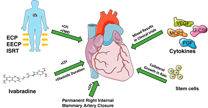
- Arora RR, Chou TM, Jain D, Fleishman B, Crawford L, McKiernan T, Nesto RW. The multicenter study of enhanced external counterpulsation (MUST-EECP): effect of EECP on exercise-induced myocardial ischemia and anginal episodes. J Am Coll Cardiol 33: 1833–1840, 1999. doi:10.1016/S0735-1097(99)00140-0. - DOI - PubMed
-
- Benjamin EJ, Blaha MJ, Chiuve SE, Cushman M, Das SR, Deo R, de Ferranti SD, Floyd J, Fornage M, Gillespie C, Isasi CR, Jiménez MC, Jordan LC, Judd SE, Lackland D, Lichtman JH, Lisabeth L, Liu S, Longenecker CT, Mackey RH, Matsushita K, Mozaffarian D, Mussolino ME, Nasir K, Neumar RW, Palaniappan L, Pandey DK, Thiagarajan RR, Reeves MJ, Ritchey M, Rodriguez CJ, Roth GA, Rosamond WD, Sasson C, Towfighi A, Tsao CW, Turner MB, Virani SS, Voeks JH, Willey JZ, Wilkins JT, Wu JH, Alger HM, Wong SS, Muntner P; American Heart Association Statistics Committee and Stroke Statistics Subcommittee . Heart Disease and Stroke Statistics−2017 update: a report from the American Heart Association. Circulation 135: e146–e603, 2017. [Corrections in Circulation 135: e646 and 136: e196, 2017.] doi:10.1161/CIR.0000000000000485. - DOI - PMC - PubMed
-
- Buschmann EE, Brix M, Li L, Doreen J, Zietzer A, Li M, Buschmann I, Hillmeister P. Adaptation of external counterpulsation based on individual shear rate therapy improves endothelial function and claudication distance in peripheral artery disease. Vasa 45: 317–324, 2016. doi:10.1024/0301-1526/a000544. - DOI - PubMed
Publication types
MeSH terms
Grants and funding
LinkOut - more resources
Full Text Sources
Other Literature Sources
Miscellaneous