. 2019 Jan 1;316(1):H212-H223.
doi: 10.1152/ajpheart.00652.2017. Epub 2018 Oct 31.

Bromine inhalation mimics ischemia-reperfusion cardiomyocyte injury and calpain activation in rats

Affiliations

Bromine inhalation mimics ischemia-reperfusion cardiomyocyte injury and calpain activation in rats

Shama Ahmad et al. Am J Physiol Heart Circ Physiol. .

Abstract

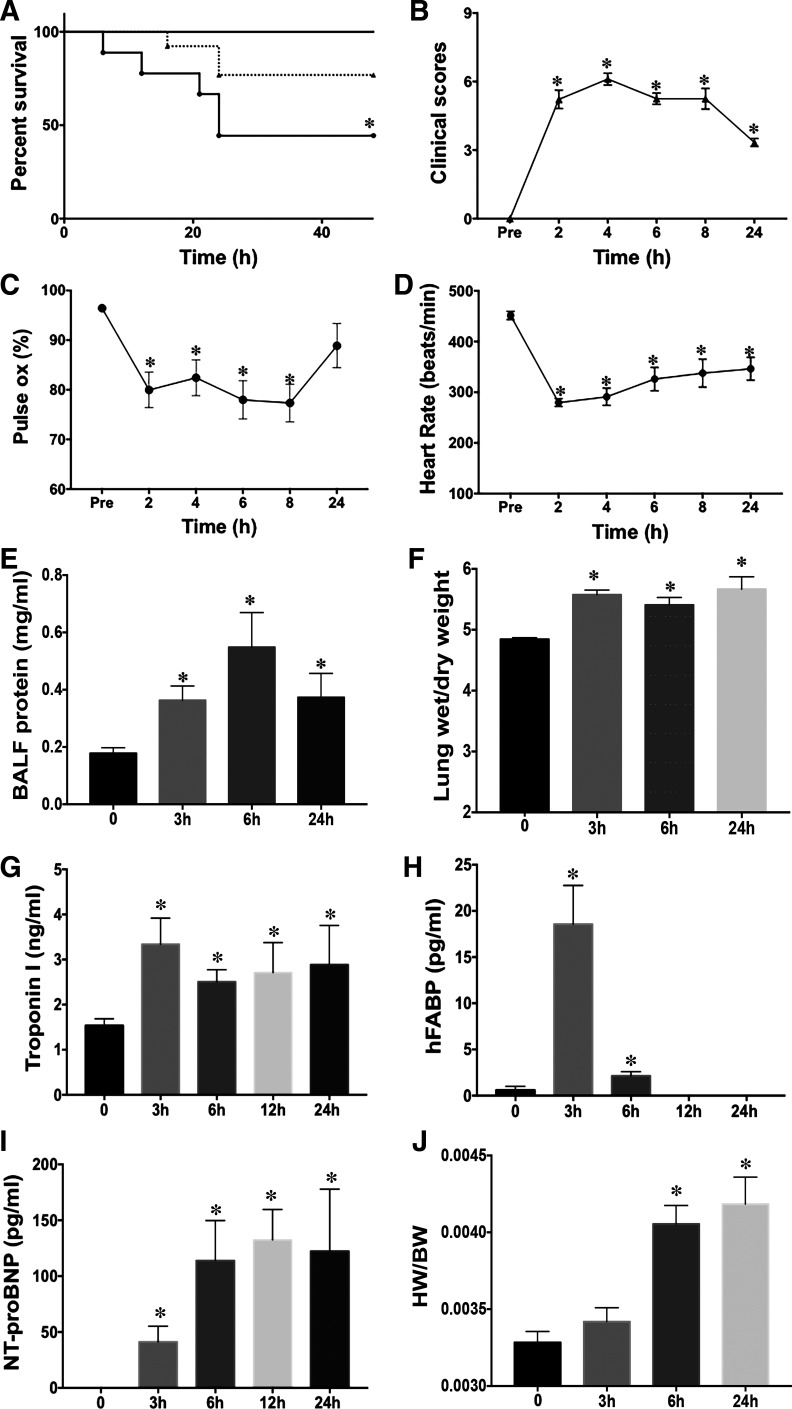
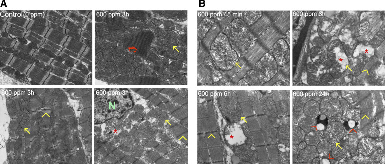
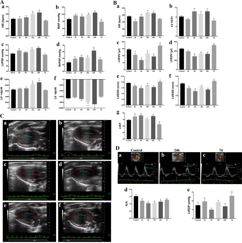
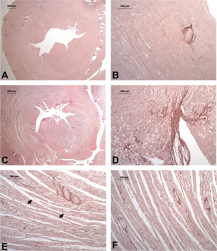
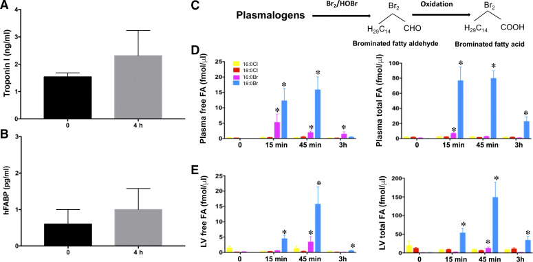
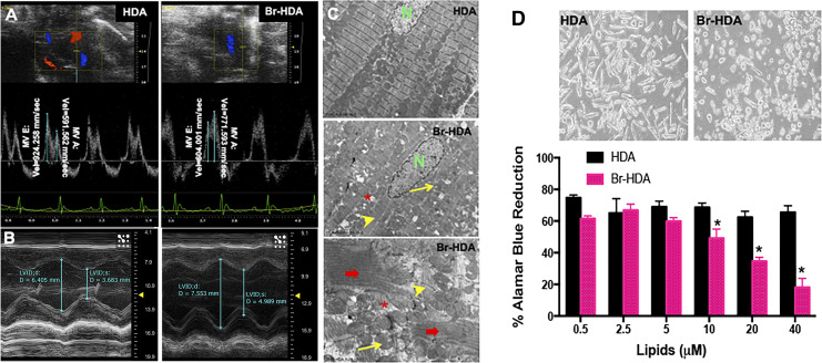
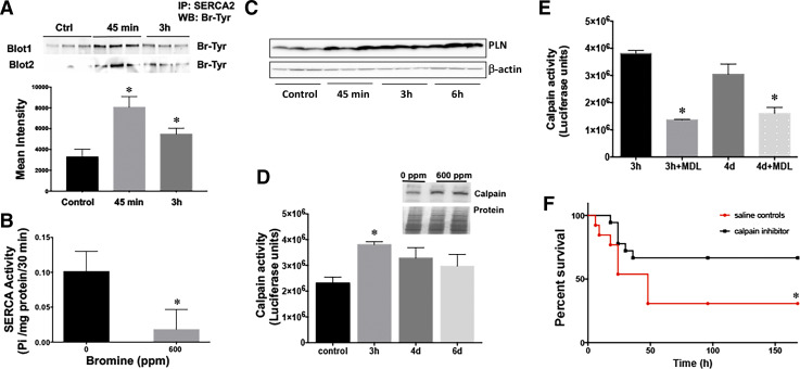
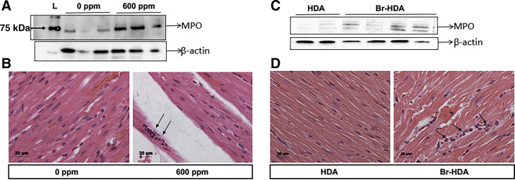
Halogens are widely used, highly toxic chemicals that pose a potential threat to humans because of their abundance. Halogens such as bromine (Br2) cause severe pulmonary and systemic injuries; however, the mechanisms of their toxicity are largely unknown. Here, we demonstrated that Br2 and reactive brominated species produced in the lung and released in blood reach the heart and cause acute cardiac ultrastructural damage and dysfunction in rats. Br2-induced cardiac damage was demonstrated by acute (3-24 h) increases in circulating troponin I, heart-type fatty acid-binding protein, and NH2-terminal pro-brain natriuretic peptide. Transmission electron microscopy demonstrated acute (3-24 h) cardiac contraction band necrosis, disruption of z-disks, and mitochondrial swelling and disorganization. Echocardiography and hemodynamic analysis revealed left ventricular (LV) systolic and diastolic dysfunction at 7 days. Plasma and LV tissue had increased levels of brominated fatty acids. 2-Bromohexadecanal (Br-HDA) injected into the LV cavity of a normal rat caused acute LV enlargement with extensive disruption of the sarcomeric architecture and mitochondrial damage. There was extensive infiltration of neutrophils and increased myeloperoxidase levels in the hearts of Br2- or Br2 reactant-exposed rats. Increased bromination of sarco(endo)plasmic reticulum Ca2+-ATPase (SERCA) and increased phosphalamban after Br2 inhalation decreased cardiac SERCA activity by 70%. SERCA inactivation was accompanied by increased Ca2+-sensitive LV calpain activity. The calpain-specific inhibitor MDL28170 administered within 1 h after exposure significantly decreased calpain activity and acute mortality. Bromine inhalation and formation of reactive brominated species caused acute cardiac injury and myocardial damage that can lead to heart failure. NEW & NOTEWORTHY The present study defines left ventricular systolic and diastolic dysfunction due to cardiac injury after bromine (Br2) inhalation. A calpain-dependent mechanism was identified as a potential mediator of cardiac ultrastructure damage. This study not only highlights the importance of monitoring acute cardiac symptoms in victims of Br2 exposure but also defines calpains as a potential target to treat Br2-induced toxicity.

Keywords: calcium; calpains; cardiac; halogen; neutrophil; sarco(endo)plasmic reticulum Ca-ATPase.


Conflict of interest statement

No conflicts of interest, financial or otherwise, are declared by the authors.

Figures

Fig. 1.
Fig. 1.
Bromine (Br2) inhalation increases mortality and causes respiratory distress and acute cardiopulmonary injury in survivors. Rats were exposed to Br2 (600 ppm, 30 or 45 min) and returned to room air. Rats were observed for clinical symptoms for 48 h, and Kaplan-Meier curves were generated for survival data (dotted line, 600 ppm for 30 min; solid line, 600 ppm for 45 min dose) (A). B−D: deterioration in clinical scores (B), decrease in O2 saturation (C) and heart rate (D). Lung injury was determined by increased bronchoalveolar lavage fluid (BALF) protein (E) and lung wet-to-dry weight ratios (F). Cardiac injury was indicated by increased troponin (G) and heart-type fatty acid-binding protein (hFABP) (H). Increased NH2-terminal pro-brain natriuretic peptide (NT-proBNP; I) in plasma and increases in heart-to-body weight ratios (HW/BW; J) also indicated heart damage. Data are means ± SE; n = 8 for each group. *P < 0.05 vs. 0 ppm.
Fig. 2.
Fig. 2.
Bromine (Br2) inhalation causes disruption of the cardiac cytoskeleton and loss of the normal highly organized linear mitochondrial sarcomere integrity. A: representative transmission electron microscopy (TEM; ×3,200) images demonstrating a normal control heart (top left). The remaining three images demonstrate myofibrillar loss, contraction band necrosis (red arrow), loss of I bands, and disruption of z-disks (yellow arrowheads) in the left ventricle 3 h after Br2 exposure in addition to mitochondrial swelling (yellow arrows) and cristae lysis (red asterisk) [nucleus (N)]. B: representative TEM image at higher magnification (×17,000) demonstrating the time course of inhaled Br2-induced mitochondrial swelling and cristae lysis. At longer time intervals there were lipid granules (small black dots) and extensive mitochondrial vacuolization (red arrowhead).
Fig. 3.
Fig. 3.
Acute (3–24 h) and delayed (7-day) effects of Bromine (Br2) inhalation (600 ppm, 45 min) on hemodynamics and cardiac function (n = 8 per group). A: heart rate (HR) during hemodynamic measurements (a), mean arterial pressure (MAP; b), left ventricular (LV) peak systolic pressure (LVPSP; c), right ventricular peak systolic pressure (RVPSP; d), maximal rate of LV pressure rise (+dP/dt; e), and maximal rate of LV pressure fall (−dP/dt; f). Data are means ± SE. *P < 0.05 vs. 0 ppm. B: HR during echo/Doppler measurements (a), LV velocity of circumferential shortening (VCFr; b), LV end-systolic volume (LVESV; c), LV end-diastolic volume (LVEDV; d), LV end-systolic diameter (LVESD; e), LV end-diastolic diameter (LVEDD; f), and LV ejection fraction (LVEF; e). Data are means ± SE; n = 8 for each group. *P < 0.05 vs. 0 ppm. C: representative parasternal long-axis images of the LV of a control rat (a) and rats 3 h (b), 6 h (c), 12 h (d), 24 h (e), and 7 days (f) after Br2 exposure. D: representative image of LV diastolic indexes measured by transmitral pulse Doppler of a control rat (a) and rats 24 h (b) and 7 days (c) after Br2 exposure. MV, mitral valve; E, early filling flow velocity; A, late filling flow velocity, IVRT, isovolumic relaxation time. The ratio of peak velocities of early to late transmitral waves (E/A; d) and LV end-diastolic pressure (LVEDP; e) are shown. Data are means ± SE; n = 8 for each group. *P < 0.05 vs. 0 ppm.
Fig. 4.
Fig. 4.
Picrosirius red (PSR) staining for collagen in rats 7 days after bromine (Br2) exposure. PSR-stained images of ×1 (A) and ×10 (B) unexposed control demonstrate minimal interstitial and endocardial collagen with homogeneous staining and compact myocardium. In contrast, the Br2-exposed heart at 7 days demonstrates a patchy endocardial fibrosis at ×1 (C) and ×10 (D). E and F: ×10 longitudinal images of the midmyocardium demonstrate the diffuse fracturing and increased interstitial space. Arrows demonstrate the areas of decreased staining in Br2-exposed rats and represent myofibrillar loss compared with the homogeneous staining of normal rats (A and B).
Fig. 5.
Fig. 5.
Role of hypoxia alone in cardiac injury and production of reactive brominated lipids on bromine (Br2) inhalation. Plasma troponin (A) and heart-type fatty acid binding protein (hFABP) (B) did not increase significantly in rats exposed to normoxia (21% oxygen) or hypoxia (10% oxygen) for 4 h. Data are means ± SE; n = 4 for each group. Plasma and heart tissues of Br2-exposed animals were also analyzed for the production of brominated fatty acids (FAs) by mass spectrometry, demonstrating the formation of 2-bromopalmitic acid (pink bars) and 2-bromostearic acid (blue bars). The yellow and red bars are the internal standards. Data are means ± SE; n = 6 for each group. *P < 0.05 vs. 0 ppm (CE).
Fig. 6.
Fig. 6.
Cardiac dysfunction and cardiomyocyte death with 2-bromohexadecanal (Br-HDA). A: left ventricular (LV) diastolic dysfunction was reflected by the decrease in mitral valve (MV) early and late wave velocities 4 h after injection of Br-HDA into the LV cavity. B: Br-HDA caused an increase in LV end-diastolic dimension and end-systolic dimension, resulting in a decrease in fractional shortening. Image demonstrates M-mode echocardiography of the LV before and 4 h after intracardiac injection of Br-HDA. C: Br-HDA caused extensive disruption of the cardiac cytoskeleton and loss of the normal highly organized linear mitochondrial sarcomere integrity. Transmission electron microscopy (×3,200 in the top images and ×8,000 in the bottom image) demonstrated contraction band necrosis (red arrows), loss of I bands, and disruption of z-disk (yellow arrowheads) in the LV of rats administered Br-HDA as well as mitochondrial swelling (yellow arrows) and cristae lysis (red asterisk). D: Br-HDA-induced cardiac cardiomyocyte death in primary adult rat cardiomyocytes with viability by alamar blue dye reduction. n = 8. *P < 0.05 vs. HDA. Top: representative phase-contrast microscope image of cardiomyocytes treated with 40 μM HDA or Br-HDA.
Fig. 7.
Fig. 7.
Bromine (Br2) exposure causes bromination and inactivation of cardiac sarco(endo)plasmic reticulum Ca2+-ATPase 2 (SERCA2) and activation of cardiac calpain. A: Br2 inhalation induced chemical modification of SERCA protein via bromination of its tyrosine residues. Bottom, quantitation of the immunoblots for bromotyrosine. Data are means ± SE values; n = 6 for each group. *P < 0.05 vs. 0 ppm. B: left ventricular (LV) SERCA protein and activity were decreased in rats exposed to Br2 (600 ppm for 45 min). *P < 0.05 vs. 0 ppm. C: SERCA regulator phosphalamban (PLN) was also increased in the hearts of rats inhaling Br2. D: Br2-induced LV calpain activity. Inset, Western blot image of calpain 2 protein and the Ponceu-stained gel to show protein loading. Data are means ± SE values; n = 6 for each group. *P < 0.05 vs. 0 ppm. E: inhibition of Br2-induced LV calpain activity by the specific calpain inhibitor MDL28170 (1 mg/kg). Data are means ± SE values; n = 6 for each group. *P < 0.05 vs. saline control. F: Kaplan-Meier curves demonstrating that calpain inhibition improves survival after Br2 inhalation 7 days posttreatment. *P < 0.05 vs. saline control.
Fig. 8.
Fig. 8.
Bromine (Br2)- and 2-bromohexadecaneal (Br-HDA)-induced neutrophil accumulation in the heart. Left ventricles (LVs) were collected 20 h after Br2 inhalation (A and B). A: Western blot for myeloperoxidase (MPO). B: hematoxylin and eosin (H&E) staining in a representative images (×20) of Br2 (0 or 600 ppm)-exposed LV sections, where arrows show neutrophils. C and D: LV Western blots and H&E staining 4 h after Br-HDA injection. C: Western blots for MPO demonstrating a similar increase in MPO and neutrophils (H&E staining; D) in Br-HDA compared with HDA.

Similar articles

Cited by

References

    1. Aggarwal S, Lam A, Bolisetty S, Carlisle MA, Traylor A, Agarwal A, Matalon S. Heme attenuation ameliorates irritant gas inhalation-induced acute lung injury. Antioxid Redox Signal 24: 99–112, 2016. doi:10.1089/ars.2015.6347. - DOI - PMC - PubMed
    1. Ahmad S, Ahmad A, Hendry-Hofer TB, Loader JE, Claycomb WC, Mozziconacci O, Schöneich C, Reisdorph N, Powell RL, Chandler JD, Day BJ, Veress LA, White CW. Sarcoendoplasmic reticulum Ca2+ ATPase. A critical target in chlorine inhalation-induced cardiotoxicity. Am J Respir Cell Mol Biol 52: 492–502, 2015. doi:10.1165/rcmb.2014-0005OC. - DOI - PMC - PubMed
    1. Ahmed KA, Nichols AL, Honavar J, Dransfield MT, Matalon S, Patel RP. Measuring nitrate reductase activity from human and rodent tongues. Nitric Oxide 66: 62–70, 2017. doi:10.1016/j.niox.2017.04.001. - DOI - PMC - PubMed
    1. Ahmed MI, Guichard JL, Soorappan RN, Ahmad S, Mariappan N, Litovsky S, Gupta H, Lloyd SG, Denney TS, Powell PC, Aban I, Collawn J, Davies JE, McGiffin DC, Dell’Italia LJ. Disruption of desmin-mitochondrial architecture in patients with regurgitant mitral valves and preserved ventricular function. J Thorac Cardiovasc Surg 152: 1059–1070.e2, 2016. doi:10.1016/j.jtcvs.2016.06.017. - DOI - PMC - PubMed
    1. Albert CJ, Crowley JR, Hsu FF, Thukkani AK, Ford DA. Reactive brominating species produced by myeloperoxidase target the vinyl ether bond of plasmalogens: disparate utilization of sodium halides in the production of alpha-halo fatty aldehydes. J Biol Chem 277: 4694–4703, 2002. doi:10.1074/jbc.M110875200. - DOI - PubMed

Publication types

MeSH terms