Review
. 2019 Jan 27:70:239-255.
doi: 10.1146/annurev-med-081117-010451. Epub 2018 Oct 31.

CRISPR Correction of Duchenne Muscular Dystrophy

Affiliations
Review

CRISPR Correction of Duchenne Muscular Dystrophy

Yi-Li Min et al. Annu Rev Med. .

Abstract

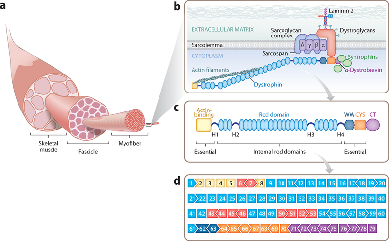
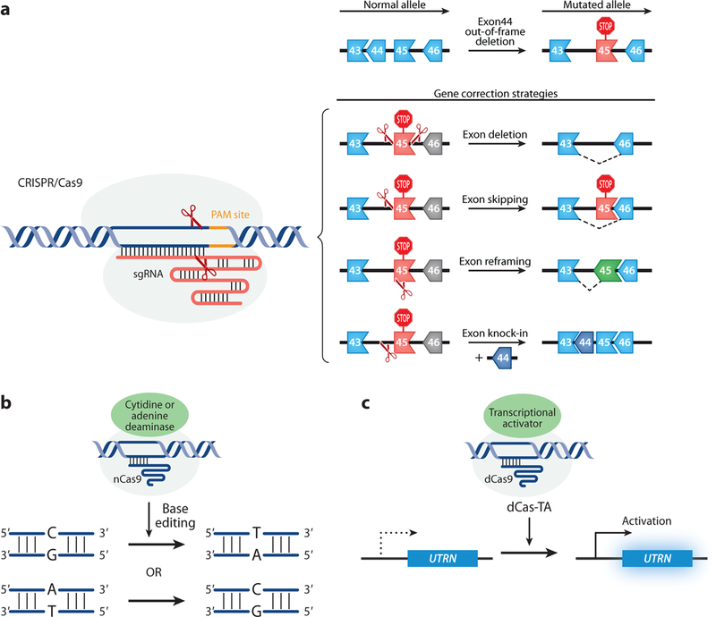
The ability to efficiently modify the genome using CRISPR technology has rapidly revolutionized biology and genetics and will soon transform medicine. Duchenne muscular dystrophy (DMD) represents one of the first monogenic disorders that has been investigated with respect to CRISPR-mediated correction of causal genetic mutations. DMD results from mutations in the gene encoding dystrophin, a scaffolding protein that maintains the integrity of striated muscles. Thousands of different dystrophin mutations have been identified in DMD patients, who suffer from a loss of ambulation followed by respiratory insufficiency, heart failure, and death by the third decade of life. Using CRISPR to bypass DMD mutations, dystrophin expression has been efficiently restored in human cells and mouse models of DMD. Here, we review recent progress toward the development of possible CRISPR therapies for DMD and highlight opportunities and potential obstacles in attaining this goal.

Keywords: CRISPR; dystrophin; muscular dystrophy; skeletal muscle.


Figures

Figure 1
Figure 1
(a) Skeletal muscle is composed of thousands of multinucleated myofibers. Myofibers are held together in groups called fascicles. (b) The dystrophin-glycoprotein complex (DGC) resides on the sarcolemma of the myofiber and acts as an anchor. The N terminus of dystrophin connects with actin filaments and the C terminus interacts with the DGC, providing stability and integrity to the muscle cell. (c) Dystrophin protein structure. The N terminus of dystrophin contains the primary actin-binding domain, whereas the C terminus contains the dystroglycan-, dystrobrevin-, and syntrophin-binding sites. The N and C termini are essential for dystrophin function. The central rod domain acts like a spring between the two ends. The 24 spectrin-like repeats in the rod domain can be shortened to create a functional but less flexible dystrophin. (d ) The exon structure of the dystrophin gene, showing the 79 exons. The open reading frame (ORF) compatibility is shown by the shape of the adjacent exons. The exons are color coded to match the major functional dystrophin protein domains in panel c. The exons within the mutational hotspot regions are indicated in red. Adapted from Reference 21.
Figure 2
Figure 2
CRISPR-mediated editing strategies to correct DMD. (a) CRISPR/Cas9-mediated strategies that require double-strand breaks, including exon deletion, exon skipping, exon reframing, and exon knock-in. (b) CRISPR/nCas9 attached with a cytidine or adenine deaminase carry out a C:G to T:A or A:T to C:G pair base substitution. (c) CRISPR/dCas9-mediated gene regulation. UTRN, a gene that codes for utrophin, a compensatory protein for dystrophin, can be upregulated by CRISPR/dCas9 fused with a transcriptional activator. Abbreviations: CRISPR, clustered regularly interspaced short palindromic repeats; dCas9, catalytically deficient Cas9; DMD, Duchenne muscular dystrophy; nCas9, Cas9 nickase.

Similar articles

Cited by

References

    1. Bonne G, Rivier F, Hamroun D. 2017. The 2018 version of the gene table of monogenic neuromuscular disorders (nuclear genome). Neuromuscul. Disord 27:1152–83 - PubMed
    1. Hoffman EP, Brown RH Jr., Kunkel LM. 1987. Dystrophin: the protein product of the Duchenne muscular dystrophy locus. Cell 51:919–28 - PubMed
    1. Lee T, Takeshima Y, Kusunoki N, et al. 2014. Differences in carrier frequency between mothers of Duchenne and Becker muscular dystrophy patients. J. Hum. Genet 59:46–50 - PMC - PubMed
    1. Darras BT, Urion DK, Ghosh PS. 1993–2018. Dystrophinopathies Seattle: Univ. Washington Press - PubMed
    1. Andrews JG, Wahl RA. 2018. Duchenne and Becker muscular dystrophy in adolescents: current perspectives. Adolesc. Health Med. Ther 9:53–63 - PMC - PubMed

Publication types

MeSH terms