. 2018 Oct 31;16(10):e3000027.
doi: 10.1371/journal.pbio.3000027. eCollection 2018 Oct.

EGFR signaling coordinates patterning with cell survival during Drosophila epidermal development

Affiliations

EGFR signaling coordinates patterning with cell survival during Drosophila epidermal development

Samuel H Crossman et al. PLoS Biol. .

Abstract

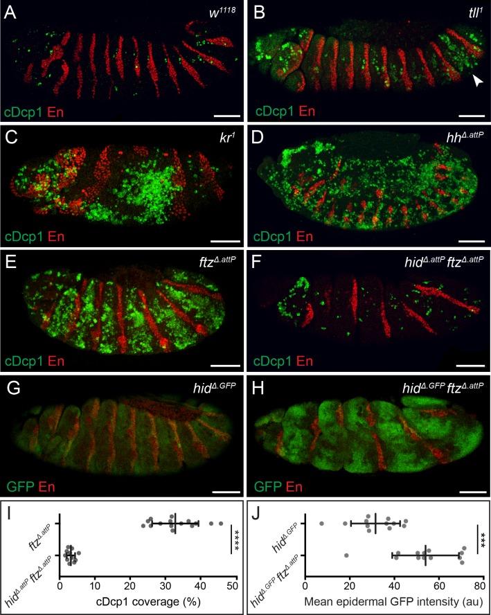
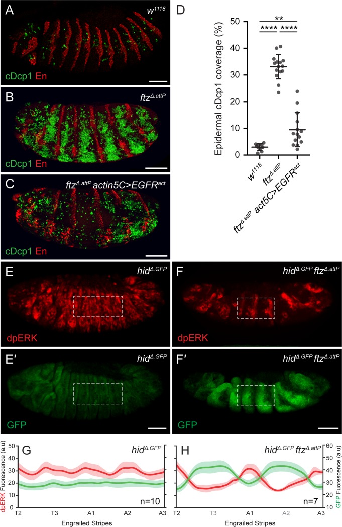
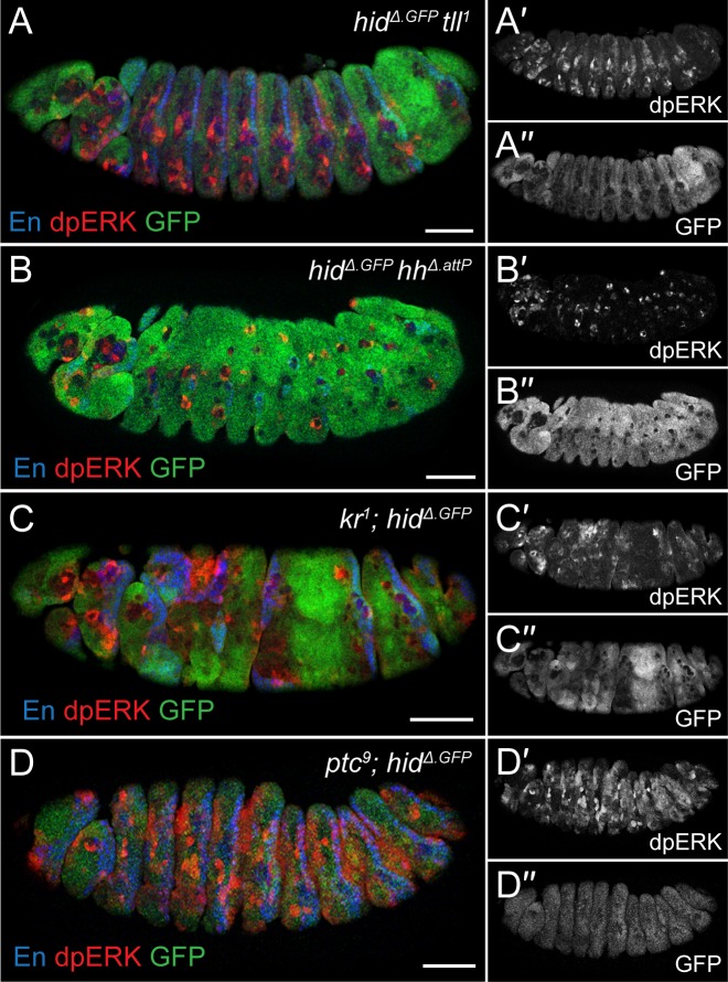
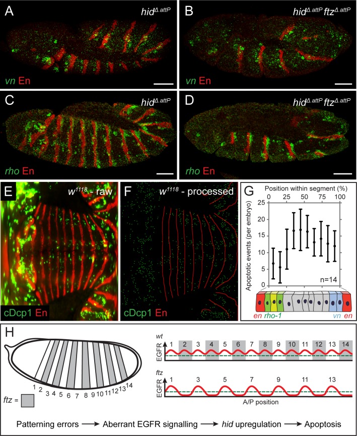
Extensive apoptosis is often seen in patterning mutants, suggesting that tissues can detect and eliminate potentially harmful mis-specified cells. Here, we show that the pattern of apoptosis in the embryonic epidermis of Drosophila is not a response to fate mis-specification but can instead be explained by the limiting availability of prosurvival signaling molecules released from locations determined by patterning information. In wild-type embryos, the segmentation cascade elicits the segmental production of several epidermal growth factor receptor (EGFR) ligands, including the transforming growth factor Spitz (TGFα), and the neuregulin, Vein. This leads to an undulating pattern of signaling activity, which prevents expression of the proapoptotic gene head involution defective (hid) throughout the epidermis. In segmentation mutants, where specific peaks of EGFR ligands fail to form, gaps in signaling activity appear, leading to coincident hid up-regulation and subsequent cell death. These data provide a mechanistic understanding of how cell survival, and thus appropriate tissue size, is made contingent on correct patterning.


Conflict of interest statement

The authors declare that they have no competing interests

Figures

Fig 1
Fig 1. Widespread apoptosis in segmentation mutants is mediated by hid.
(A–E) cDcp1 immunoreactivity (green), in control (A), tll (B), kr (C), hh (D), and ftz (E) stage 13/14 embryos. Segmental enrichment of cDcp1 is detected around the regions where the mutated gene is known to act during normal development. Arrowhead in B indicates the zone of elevated Dcp1 cleavage in the posterior epidermis of tll1 embryos. Anti-Engrailed (En, red) provides a positional reference along the A/P axis throughout. (F) Epidermal cDcp1 immunoreactivity in stage 13 hidΔ.attP ftzΔ.attP double homozygotes is strongly reduced. (G, H) Transcription of hid, as assayed with the hidΔ.GFP reporter, is detected at uniform low levels in a wild-type background (G) but is up-regulated in a banded pattern in hidΔ.GFP ftzΔ.attP double homozygotes. Embryos late stage 12. Scale bars 50 μm. (I) Quantification of cDcp1 levels in ftzΔ.attP single mutant and hidΔ.attP ftzΔ.attP double mutant embryos. (J) Quantification of mean epidermal GFP intensity values throughout the epidermis of hidΔ.GFP single mutant and hidΔ.GFP ftzΔ.attP double mutant embryos. In graphs, means are shown, and error bars display standard deviation (****p < 0.0001, ***p < 0.001, unpaired Student t test). The underlying data for (I) and (J) can be found in S1 Data. A/P, anterior/posterior; cDcp1, cleaved Death caspase-1; ftz, fushi-tarazu; GFP, green fluorescent protein; hh, hedgehog; hid, head involution defective; kr, krüppel; tll, tailless.
Fig 2
Fig 2. Loss of EGFR signaling and hid up-regulation in ftz mutants.
(A–D) cDcp1 immunoreactivity in control w1118 (A), ftzΔ.attP (B), and ftzΔ.attP actin5C-Gal4, UAS–EGFRact (C) stage 13/14 embryos. (D) Quantification of mean cDcp1 levels for genotypes shown in A–C. Error bars display standard deviation (***p < 0.001, **p < 0.01, unpaired Student t test). (E, F) dpERK/GFP immunoreactivity in stage 13 hidΔ.GFP single mutant (E, E′) and hidΔ.GFP ftzΔ.attP double mutant embryos (F, F′). Dashed line indicates representative region of interest for quantification in panels G and H. Scale bars 50 μm. (G, H) Mean dpERK (red) and GFP (green) fluorescence intensity profiles in control hidΔ.GFP (G) and hidΔ.GFP ftzΔ.attP (H) samples. Engrailed expression stripes were used as a spatial marker along the A/P axis (see material and methods). Shaded areas depict standard error of the mean. The underlying data for (D), (G) and (H) can be found in S1 Data. cDcp1, cleaved Death caspase-1; dpERK, phosphorylated Extracellular signal–regulated kinase; EGFR, epidermal growth factor receptor; ftz, fushi-tarazu; GFP, green fluorescent protein; hid, head involution defective; UAS, upstream activation sequence; w1118, white1118.
Fig 3
Fig 3. hid and dpERK are anticorrelated in various patterning mutants.
(A–D) GFP and dpERK immunoreactivity in hidΔ.GFP tll1 (A), hidΔ.GFP hhΔ.attP (B), kr1; hidΔ.GFP (C), and ptc9; hidΔ.GFP (D) stage 13 double mutants. In each instance, dpERK intensity and hidΔ.GFP expression are anticorrelated. Scale bars 50 μm. dpERK, phosphorylated extracellular signal–regulated kinase; GFP, green fluorescent protein; hh, hedgehog; hid, head involution defective; kr, krüppel; ptc, patched; tll, tailless.
Fig 4
Fig 4. Patterned expression of vn and rho-1 ensure epidermal cell survival.
(A, B) Expression of vn (green) in control hidΔ.attP (A) and hidΔ.attP ftzΔ.attP (B) stage 12 homozygotes, as detected by fluorescent in situ hybridisation. hidΔ.attP is included in the background to avoid any confounding effects of apoptosis. (C, D) Expression of rho-1 (green) in hidΔ.attP (C) and hidΔ.attP ftzΔ.attP (D) stage 12 homozygotes, as detected by fluorescent in situ hybridisation. Scale bars 50 μm. (E) Representative raw mSPIM image of a stage 12 w1118 embryo (control) stained with anti-cDcp1 and anti-En (red). Anterior is to the left and posterior to the right, with the ventral midline running horizontally through the middle. (F) Processed version of the image shown in E. cDcp1-positive cells were segmented and reduced to individual pixels (green). Similarly, stripes of en expression were reduced to a skeleton (red) to allow the position of each apoptotic cell to be mapped along the A/P axis of the segment and thus to derive a relative position applicable to all segments. (G) Histogram displaying the frequency of apoptosis along the A/P axis of the segment. Illustration of the en, rho-1, and vn expression domains is included to indicate the approximate sites of ligand production. Error bars show standard deviation. (H) Model explaining the pattern of apoptosis in ftz and other segmentation mutants. In normal development, the segmental expression of EGFR ligands maintains signaling activity above the threshold level required for cell survival (green dashed line). When patterning errors disrupt the landscape of EGFR activity, signaling falls below the survival threshold, triggering hid up-regulation and patterned apoptosis. The underlying data for (G) can be found in S1 Data. A/P, anterior/posterior; cDcp1, cleaved Death caspase-1; EGFR, epidermal growth factor receptor; en, engrailed; ftz, fushi-tarazu; hid, head involution defective; mSPIM, multiview single plane illumination microscopy; rho-1, rhomboid; vn, vein; w1118, white1118.

References

    1. Merino Marisa M, Rhiner C, Lopez-Gay Jesus M, Buechel D, Hauert B, et al. (2015) Elimination of Unfit Cells Maintains Tissue Health and Prolongs Lifespan. Cell 160: 461–476. 10.1016/j.cell.2014.12.017 - DOI - PMC - PubMed
    1. Koto A, Kuranaga E, Miura M (2011) Apoptosis ensures spacing pattern formation of Drosophila sensory organs. Curr Biol 21: 278–287. 10.1016/j.cub.2011.01.015 - DOI - PubMed
    1. Fuchs Y, Steller H (2011) Programmed Cell Death in Animal Development and Disease. Cell 147: 742–758. 10.1016/j.cell.2011.10.033 - DOI - PMC - PubMed
    1. Derry WB, Putzke AP, Rothman JH (2001) Caenorhabditis elegans p53: role in apoptosis, meiosis, and stress resistance. Science 294: 591–595. 10.1126/science.1065486 - DOI - PubMed
    1. Merino MM, Levayer R, Moreno E (2016) Survival of the Fittest: Essential Roles of Cell Competition in Development, Aging, and Cancer. Trends Cell Biol 26: 776–788. 10.1016/j.tcb.2016.05.009 - DOI - PubMed

Publication types

MeSH terms