. 2018 Oct 31;13(10):e0205496.
doi: 10.1371/journal.pone.0205496. eCollection 2018.

Harmonization of exosome isolation from culture supernatants for optimized proteomics analysis

Affiliations

Harmonization of exosome isolation from culture supernatants for optimized proteomics analysis

Agata Abramowicz et al. PLoS One. .

Abstract

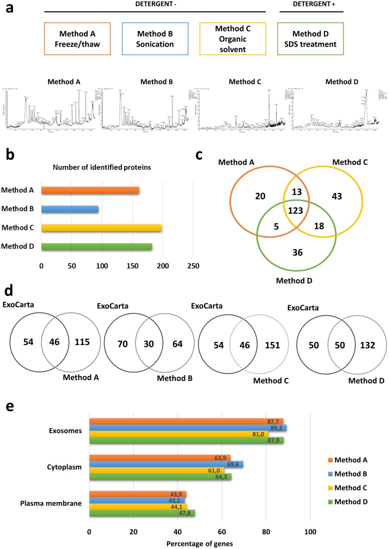
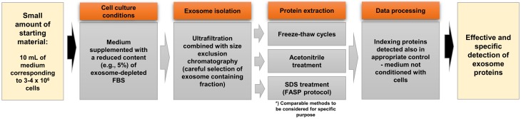
Exosomes, the smallest subset of extracellular vesicles (EVs), have recently attracted much attention in the scientific community. Their involvement in intercellular communication and molecular reprogramming of different cell types created a demand for a stringent characterization of the proteome which exosomes carry and deliver to recipient cells. Mass spectrometry (MS) has been extensively used for exosome protein profiling. Unfortunately, no standards have been established for exosome isolation and their preparation for MS, leading to accumulation of artefactual data. These include the presence of high-abundance exosome-contaminating serum proteins in culture media which mask low-abundance exosome-specific components, isolation methods that fail to yield "pure" vesicles or variability in protein solubilization protocols. There is an unmet need for the development of standards for exosome generation, harvesting, and isolation from cellular supernatants and for optimization of protein extraction methods before proteomics analysis by MS. In this communication, we illustrate the existing problems in this field and provide a set of recommendations that are expected to harmonize exosome processing for MS and provide the faithful picture of the proteomes carried by exosomes. The recommended workflow for effective and specific identification of proteins in exosomes released by the low number of cells involves culturing cells in medium with a reduced concentration of exosome-depleted serum, purification of exosomes by size-exclusion chromatography, a combination of different protein extraction method and removal of serum-derived proteins from the final dataset using an appropriate sample of cell-unexposed medium as a control. Application of this method allowed detection of >250 vesicle-specific proteins in exosomes from 10 mL of culture medium.


Conflict of interest statement

The authors have declared that no competing interests exist.

Figures

Fig 1
Fig 1. Effects of fetal bovine serum (FBS) on cell viability.
The cell viability assessed for the panel of head and neck cancer cell lines cultured for 24 h in a medium with 10% FBS or without FBS supplementation.
Fig 2
Fig 2. Characteristics of exosome-depleted FBS.
In (a) Quantification of exosomes present in the cell culture medium supplemented with STD FBS or ED FBS at the input and in the post-UC fractions (supernatant and pellet) based on ACHE activity. In (b) Western blot of CD63 in the cell culture medium either before (input) or after UC (supernatant); Ponceau S staining demonstrates the serum albumin content. In (c) Coomassie blue staining of electrophoretically-separated proteins from standard (STD) and exosome-depleted (ED) FBS. In (d) Effects of supplementation with FBS on cell viability; FaDu cells were cultured for 24 h in a medium supplemented with STD FBS, ultracentrifuged STD FBS (STD-UC) or ED FBS.
Fig 3
Fig 3. Exosome isolation by the size exclusion chromatography.
In (a) representative immunoblot showing the distribution of exosome markers (CD63, CD9, CD81) and high-abundance serum proteins (illustrated by Ponceau S staining) in the successive SEC fractions of a FaDU culture medium. In (b) total protein concentrations (μg/uL) in the subsequent SEC fractions. In (c) culture medium supplemented with 5% ED FBS and NOT co-cultured with cells was analyzed as in Panel A; “E+” denotes exosome-containing positive control.
Fig 4
Fig 4. Mass spectrometry analysis of the selected SEC fractions # 5–8.
In (a) the number of proteins identified by MS in each SEC fraction; the ratios of putative exosome specific (red) and FBS serum-derived proteins (gray) in each fraction are shown. In (b) the overlap between the top hundred exosomal proteins reported in the ExoCarta database and proteins detected in the “fresh” culture medium supplemented with ED FBS (SEC fractions # 5–8).
Fig 5
Fig 5. A comparison of four different methods for exosome protein extraction.
In (a) the LC profiles of exosome samples processed by each of the four methods. In (b) numbers of exosome proteins identified by MS in each sample. In (c) overlapping and distinct proteins identified using Methods A, C and D. In (d) numbers of exosome proteins overlapping with or distinct from the ExoCarta database. In (e) functional enrichment analysis showing the percentages of the detected proteins that were exosome-related in each method.
Fig 6
Fig 6. Practical hints for effective proteomics analysis of exosomes.

References

    1. Raposo G, Stoorvogel W. Extracellular vesicles: exosomes, microvesicles, and friends. J Cell Biol. 2013;200: 373–83. 10.1083/jcb.201211138 - DOI - PMC - PubMed
    1. Colombo M, Raposo G, Théry C. Biogenesis, secretion, and intercellular interactions of exosomes and other extracellular vesicles. Annu Rev Cell Dev Biol. 2014;30: 255–289. 10.1146/annurev-cellbio-101512-122326 - DOI - PubMed
    1. Whiteside TL. The potential of tumor-derived exosomes for noninvasive cancer monitoring. Expert Rev Mol Diagn. 2015;15: 1293–310. 10.1586/14737159.2015.1071666 - DOI - PMC - PubMed
    1. Alvarez ML, Khosroheidari M, Kanchi Ravi R, DiStefano JK. Comparison of protein, microRNA, and mRNA yields using different methods of urinary exosome isolation for the discovery of kidney disease biomarkers. Kidney Int. 2012;82: 1024–1032. 10.1038/ki.2012.256 - DOI - PubMed
    1. Kalra H, Adda CG, Liem M, Ang CS, Mechler A, Simpson RJ, et al. Comparative proteomics evaluation of plasma exosome isolation techniques and assessment of the stability of exosomes in normal human blood plasma. Proteomics. 2013;13: 3354–3364. 10.1002/pmic.201300282 - DOI - PubMed

Publication types