Review
. 2018 Oct 29;19(11):3385.
doi: 10.3390/ijms19113385.

Anisotropic Gold Nanoparticles in Biomedical Applications

Affiliations
Review

Anisotropic Gold Nanoparticles in Biomedical Applications

Claudia Kohout et al. Int J Mol Sci. .

Abstract

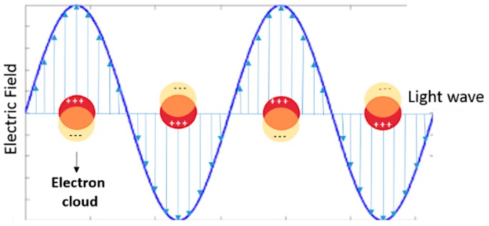
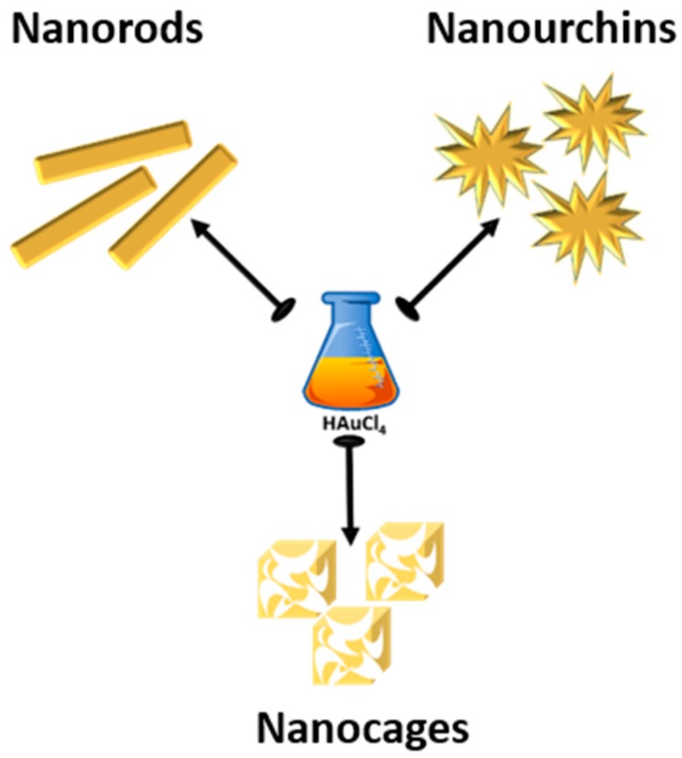
Gold nanoparticles (AuNPs) play a crucial role in the development of nanomedicine, principally due to their unique photophysical properties and high biocompatibility. The possibility to tune and customize the localized surface plasmon resonance (LSPR) toward near-infrared region by modulating the AuNP shape is one of the reasons for the huge widespread use of AuNPs. The controlled synthesis of no-symmetrical nanoparticles, named anisotropic, is an exciting goal achieved by the scientific community which explains the exponential increase of the number of publications related to the synthesis and use of such type of AuNPs. Even with such steps forward and the AuNP translation in clinic being done, some key issues are still remain and they are related to a reliable and scalable production, a full characterization, and to the development of nanotoxicology studies on the long run. In this review we highlight the very recent advances on the synthesis of the main classes of anisotropic AuNPs (nanorods, nanourchins and nanocages) and their use in the biomedical fields, in terms of diagnosis and therapeutics.

Keywords: anisotropic AuNPs; biomedical applications; gold nanoparticles; synthesis.


Conflict of interest statement

The authors declare no conflict of interest.

Figures

Figure 1
Figure 1
Graphical illustration of localized surface plasmon resonance (LSPR) band formation (in yellow circle the negative electron cloud, in red circle the positive electron cloud).
Figure 2
Figure 2
Anisotropic gold nanoparticles considered in the present review: nanorods, nanourchins and nanocages.
Figure 3
Figure 3
Left: Comparing the conventional seed-mediated protocol and the two steps approach. Right: TEM images, scale bars 100 nm. (ac) Different AuNR volume in function of seed concentration. (d) Correlation between seed concentration scaling factor (m) and the final AuNR volume (V). (eg) Effect of the change of surfactant in the second growth solution on the AuNR aspect ratio. (h) UV-vis spectra of the AuNRs e–g. Reproduced from [30] with permission from the American Chemical Society.
Figure 4
Figure 4
Schematic illustration of the production of the red blood cell membrane camouflaged gold nanorods (RBCM-AuNRs). Reproduced from [37] with permission from the Royal Chemical Society.
Figure 5
Figure 5
Scheme of seedless synthesis technique based on the use of 5-bromosalicylic acid and hydroquinone. Reproduced from [38] with permission from the John Wiley and Sons.
Figure 6
Figure 6
(a,b) TEM images of the coral-shaped nanoparticles (scale bar; (a) 50 nm; (b) 10 nm). (c) HR-TEM (scale bar, 5.0 nm). (dg) HR-TEM images of distorted gold nanorods and clusters in early stages of coral-shaped particle formation (scale bar, 5.0 nm). (hm) Sequence of in situ liquid cell TEM time (scale bar, 20 nm) showing the early stages of coral-shaped particle formation (insets of h, i are the magnification of TEM images, scale bar at 10 nm). (n) Schematic representation for the multibranched formation of gold nanoparticles induced by peptoids. Reproduced from [51] with permission from the Nature Publisher.
Figure 7
Figure 7
Left: Scheme of the fluidic production of multibranched AuNUs. The precursors loaded in the syringes can be mixed in the T-mizer (mixing zone), in the reactor (1 m) the AuNUs form and drop directly in an excess of ligand for proper stabilization. Right: (A) TEM image of a single multibranched gold nanoparticle immobilized on titania; (B) HR-TEM image of a single branch interacting with a titania. Reproduced from [56] with permission from John Wiley and Sons.
Figure 8
Figure 8
(a) Procedure for the synthesis of AuNCs by starting form polysterene beads. (bd) SEM images of each intermediate step (scale bar 200 nm). Reproduced from [65] with permission from Elsevier.
Figure 9
Figure 9
Schematic representation of the production of multibranched-hollow gold nanoparticles, based on self-assembled polymer template. Reproduced from [67] with permission from the American Chemical Society.
Figure 10
Figure 10
Two mechanisms at the base of the Raman signal enhancement: (a) electromagnetic and (b) chemical.
Figure 11
Figure 11
Schematic process for Au-AgAu core-shell structures synthesis. Reproduced from [80] with permission from the American Chemical Society.
Figure 12
Figure 12
Up: scheme of the formation of poly(ADP-ribose)(PAR), catalyzed by poly(ADP-ribose) polymerase (PARP-1). Down: Scheme of the colorimetric strategy based on AuNRs without and with PARP-1 detector inhibitor. Reproduced from [87] with permission from Elsevier.
Figure 13
Figure 13
Scheme of construction and release mechanism of pH-trigger release system. Reproduced from [102] with permission from Ivyspring International Publisher.
Figure 14
Figure 14
In vivo studies in A375 tumor-bearing mice of GNR@SiO2-PFP nanorattles. (a) Thermographic images and (b) Temperature change after laser irradiation at 24 h post-injection (* P < 0.05). (c) Tumor growth after PTT treatment (*** P < 0.001) and (d) tumor weight of mice on day 18 after treatments (*** P < 0.001). (e) A375 tumor-bearing mice (red arrow) after treatments. Reproduced from [122] with permission from John Wiley and Sons.

References

    1. Yang X., Yang M., Pang B., Vara M., Xia Y. Gold Nanomaterials at Work in Biomedicine. Chem. Rev. 2015;115:10410–10488. doi: 10.1021/acs.chemrev.5b00193. - DOI - PubMed
    1. Compostella F., Pitirollo O., Silvestri A., Polito L. Glyco-gold nanoparticles: Synthesis and applications. Beilstein J. Org. Chem. 2017;13 doi: 10.3762/bjoc.13.100. - DOI - PMC - PubMed
    1. Louis C., Pluchery O. In: Gold Nanoparticles for Physics, Chemistry and Biology. Louis C., Pluchery O., editors. Imperial College Press; London, UK: 2017. 2nd ed.
    1. Attia Y.A., Buceta D., Requejo F.G., Giovanetti L.J., López-Quintela M.A. Photostability of gold nanoparticles with different shapes: The role of Ag clusters. Nanoscale. 2015;7:11273–11279. doi: 10.1039/C5NR01887K. - DOI - PubMed
    1. Mie G. Beiträge zur Optik trüber Medien, speziell kolloidaler Metallösungen. Ann. Phys. 1908;330:377–405. doi: 10.1002/andp.19083300302. - DOI

LinkOut - more resources