Review
. 2018 Oct 30;10(11):408.
doi: 10.3390/cancers10110408.

The Emerging Role of the Microenvironment in Endometrial Cancer

Affiliations
Review

The Emerging Role of the Microenvironment in Endometrial Cancer

Subhransu S Sahoo et al. Cancers (Basel). .

Abstract

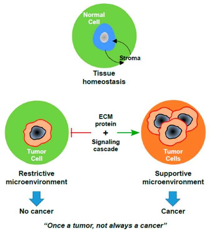
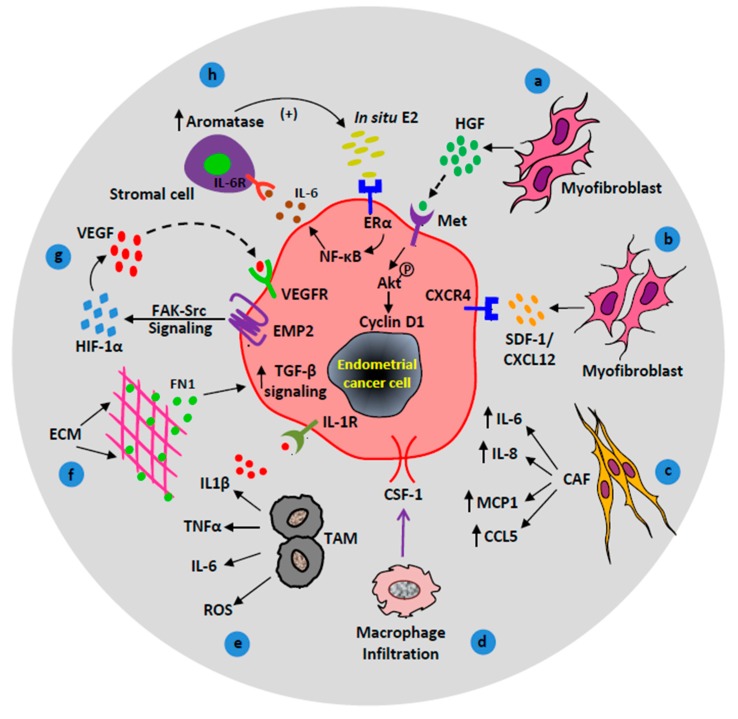
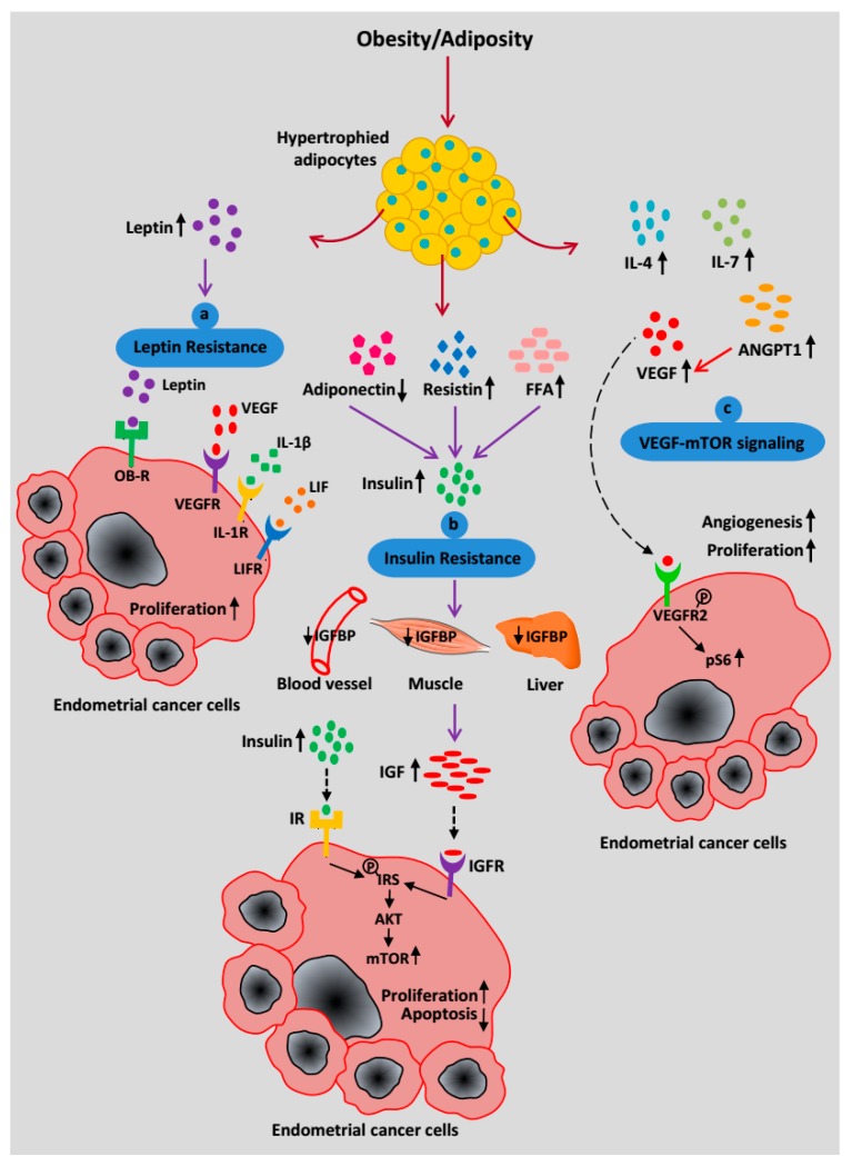
Endometrial cancer (EC) is one of the most frequently diagnosed cancers in women, and despite recent therapeutic advances, in many cases, treatment failure results in cancer recurrence, metastasis, and death. Current research demonstrates that the interactive crosstalk between two discrete cell types (tumor and stroma) promotes tumor growth and investigations have uncovered the dual role of the stromal cells in the normal and cancerous state. In contrast to tumor cells, stromal cells within the tumor microenvironment (TME) are genetically stable. However, tumor cells modify adjacent stromal cells in the TME. The alteration in signaling cascades of TME from anti-tumorigenic to pro-tumorigenic enhances metastatic potential and/or confers therapeutic resistance. Therefore, the TME is a fertile ground for the development of novel therapies. Furthermore, disrupting cancer-promoting signals from the TME or re-educating stromal cells may be an effective strategy to impair metastatic progression. Here, we review the paradoxical role of different non-neoplastic stromal cells during specific stages of EC progression. We also suggest that the inhibition of microenvironment-derived signals may suppress metastatic EC progression and offer novel potential therapeutic interventions.

Keywords: TME; chemoprevention; endometrial cancer; metastasis; stromal cells.


Conflict of interest statement

The authors have nothing to disclose.

Figures

Figure 1
Figure 1
The paradox of cancer development. Upon loss of tissue homeostasis, the progression of an occult tumor to frank carcinoma requires significant changes in the microenvironment. ECM: Extracellular Matrix.
Figure 2
Figure 2
Contributions of activated/recruited stromal cells in endometrial cancer (EC) progression. (a) Hepatocyte growth factor (HGF) stimulates proliferation of EC cells via the HGF/c-MET/AKT signaling pathway. (b) Myofibroblasts promote tumor growth via CXCR4/CXCL12 signaling axis. (c) Cancer-associated fibroblasts (CAF) secrete cytokines (IL-6, IL-8, and MCP-1) and chemokines (CCL5/RANTES) to promote cancer progression. (d) Macrophage response, colony-stimulating factor 1 (CSF1) signals macrophage infiltration to the endometrial reactive stroma. (e) Tumor-associated macrophages contribute to endometrial carcinogenesis via the production of cytokines (IL1β, TNFα, IL-6) and reactive oxygen species (ROS). (f) Extracellular matrix (ECM) protein, fibronectin upregulates transforming growth factor-beta (TGF-β) signaling in EC cells which facilitates EC metastasis. (g) Under hypoxic conditions, epithelial membrane protein-2 (EMP2) enhances angiogenesis through focal adhesion kinase (FAK)-Src and hypoxia-inducible factor 1-alpha (HIF-1α) signaling pathway. (h) A positive feedback loop between in situ estrogen (E2), IL-6, and aromatase upregulate EC cell proliferation.
Figure 3
Figure 3
Systemic effects of increased adiposity on EC progression. (a) A high level of circulating leptin binds to its receptor OB-R to trigger a signaling pathway as well as recruit pro-angiogenic factors such as VEGF, IL-1β, and LIF to induce cancer progression and metastasis. (b) Increased free fatty acids (FFA), resistin, and decreased adiponectin secretion contribute to insulin resistance, which leads to an increase in insulin synthesis. Hyperinsulinemia is associated with decreased bioavailability of IGFBP and the simultaneous increase in IGF-1 production. Insulin and IGF-1 signal through IR and IGFR respectively to promote EC progression via mTOR activation. (c) Hypertrophied adipocytes secrete an increasing amount of pro-inflammatory cytokines (IL-4, IL-7), ANGPT1, and VEGF to infiltrate endothelial cells, which facilitates angiogenesis. VEGF acts as a key mediator of the EC cell-adipocyte interaction and binds to its receptor, VEGFR2, on the EC cell surface. Phosphorylation of VEGFR2 activates downstream targets and upregulates the mTOR pathway through a high pS6 level.

References

    1. Siegel R.L., Miller K.D., Jemal A. Cancer statistics, 2016. CA Cancer J. Clin. 2016;66:7–30. doi: 10.3322/caac.21332. - DOI - PubMed
    1. Bonnans C., Chou J., Werb Z. Remodelling the extracellular matrix in development and disease. Nat. Rev. Mol. Cell Biol. 2014;15:786–801. doi: 10.1038/nrm3904. - DOI - PMC - PubMed
    1. Lu P., Weaver V.M., Werb Z. The extracellular matrix: A dynamic niche in cancer progression. J. Cell Biol. 2012;196:395–406. doi: 10.1083/jcb.201102147. - DOI - PMC - PubMed
    1. Hanahan D., Coussens L.M. Accessories to the crime: Functions of cells recruited to the tumor microenvironment. Cancer Cell. 2012;21:309–322. doi: 10.1016/j.ccr.2012.02.022. - DOI - PubMed
    1. Hanahan D., Weinberg R.A. Hallmarks of cancer: The next generation. Cell. 2011;144:646–674. doi: 10.1016/j.cell.2011.02.013. - DOI - PubMed

LinkOut - more resources