. 2018 Dec 19;140(50):17444-17455.
doi: 10.1021/jacs.8b06354. Epub 2018 Dec 5.

Oxidative Post-translational Modifications Accelerate Proteolytic Degradation of Calprotectin

Affiliations

Oxidative Post-translational Modifications Accelerate Proteolytic Degradation of Calprotectin

Jules R Stephan et al. J Am Chem Soc. .

Abstract

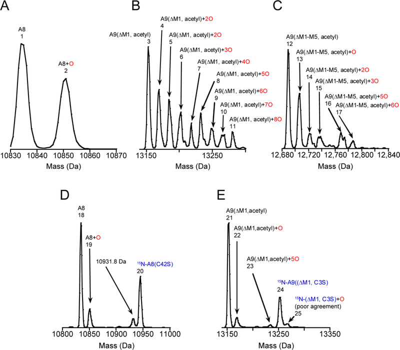
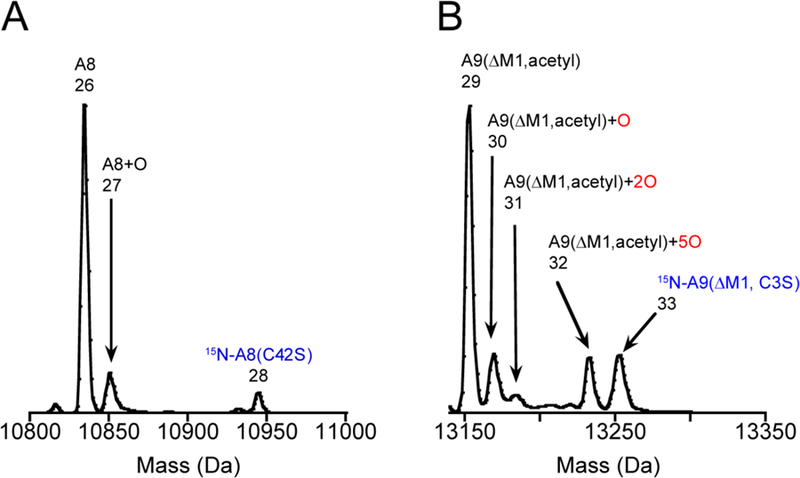
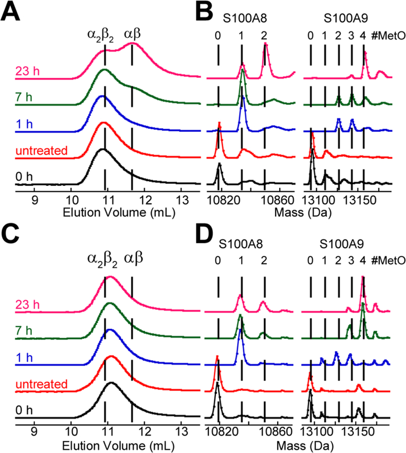
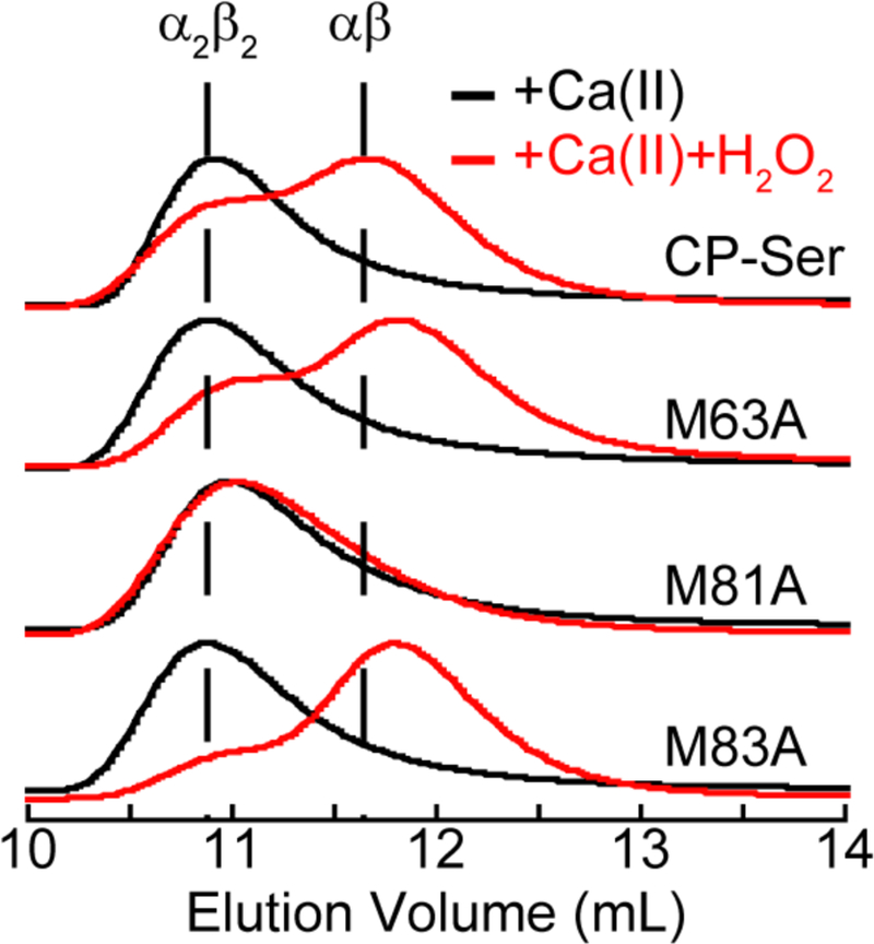
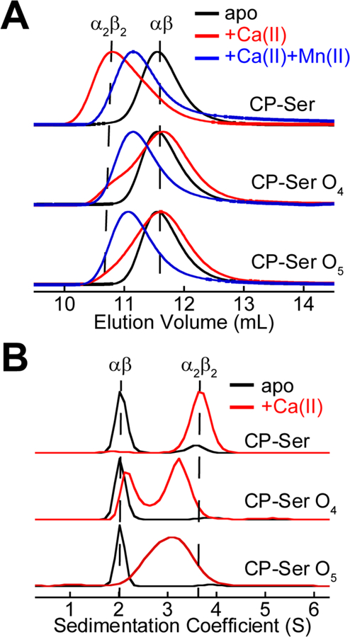
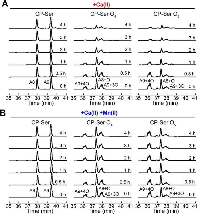
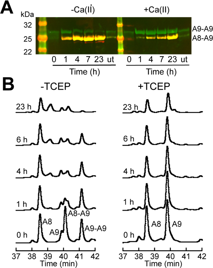
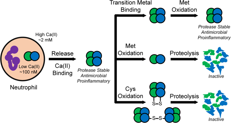
Oxidative post-translational modifications affect the structure and function of many biomolecules. Herein we examine the biophysical and functional consequences of oxidative post-translational modifications to human calprotectin (CP, S100A8/S100A9 oligomer, MRP8/MRP14 oligomer, calgranulins A/B oligomer). This abundant metal-sequestering protein contributes to innate immunity by starving invading microbial pathogens of transition metal nutrients in the extracellular space. It also participates in the inflammatory response. Despite many decades of study, little is known about the fate of CP at sites of infection and inflammation. We present compelling evidence for methionine oxidation of CP in vivo, supported by using 15N-labeled CP-Ser (S100A8(C42S)/S100A9(C3S)) to monitor for adventitious oxidation following human sample collection. To elucidate the biochemical and functional consequences of oxidative post-translational modifications, we examine recombinant CP-Ser with methionine sulfoxide modifications generated by exposing the protein to hydrogen peroxide. These oxidized species coordinate transition metal ions and exert antibacterial activity. Nevertheless, oxidation of M81 in the S100A9 subunit disrupts Ca(II)-induced tetramerization and, in the absence of a transition metal ion bound at the His6 site, accelerates proteolytic degradation of CP. We demonstrate that native CP, which contains one Cys residue in each full-length subunit, forms disulfide bonds within and between S100A8/S100A9 heterodimers when exposed to hydrogen peroxide. Remarkably, disulfide bond formation accelerates proteolytic degradation of CP. We propose a new extension to the working model for extracellular CP where post-translational oxidation by reactive oxygen species generated during the neutrophil oxidative burst modulates its lifetime in the extracellular space.


Figures

Figure 1
Figure 1
A heterodimer unit from the crystal structure (PDB: 1XK4) of Mn(II)-, Ca(II)-, and Na(I)-bound CP-Ser. The green and blue chains are S100A8 and S100A9, respectively. Mn(II), Ca(II), and Na(I) ions are shown in magenta, yellow, and purple, respectively. The transition-metal binding residues are shown with orange sticks.
Figure 2
Figure 2
Deconvoluted mass spectra of human nasal mucus. The S100A8 panels are normalized to the unmodified S100A8 peak and S100A9 panels are normalized to the most abundant S100A9 peak. The observed and theoretical masses for each numbered peak are listed in Table S1. Data from (A) mucus sample 2 expanded around S100A8, (B) mucus sample 2 expanded around S100A9, (C) mucus sample 2 expanded around S100A9(ΔM1–5), (D) mucus sample 22 expanded around S100A8 that shows 15N-A8(C42S), (E) mucus sample 22 expanded around S100A9 that shows 15N-A9(ΔM1, C3S).
Figure 3
Figure 3
Deconvoluted mass spectra of human pus with 15N-CP-Ser added immediately after collection. The observed and theoretical masses for each numbered peak are contained in Table S1. Data from pus sample 2 (A) expanded around S100A8 and (B) expanded around S100A9.
Figure 4
Figure 4
SEC chromatograms and corresponding deconvoluted mass spectra of 30 µM CP-Ser treated with 100 mM H2O2 in the presence of 1.5 mM Ca(II) or both 1.5 mM Ca(II) and 30 µM Mn(II) (75 mM HEPES, 100 mM NaCl, pH 7.5). In the mass spectra, the dashed lines represent the expected masses of the CP subunits with additional oxygen atoms. The chromatograms and mass spectra were normalized to a maximum value of 1. The listed times correspond to the duration of treatment with H2O2 prior to sample analysis. The untreated sample was incubated at 37 °C for 23 h. (A) SEC chromatograms for the Ca(II) only samples. (B) Regions of the deconvoluted mass spectra showing the S100A8 and S100A9 subunits for the Ca(II) only samples. (C) +Ca(II)+Mn(II) SEC. (C) SEC chromatograms for the Ca(II) and Mn(II) samples. (D) Regions of the deconvoluted mass spectra showing the S100A8 and S100A9 subunits for the Ca(II) and Mn(II) samples. In the SEC chromatograms, the Mn(II)- and Ca(II)-bound CP-Ser tetramer elutes slightly later than the Ca(II)-bound CP-Ser tetramer.
Figure 5
Figure 5
SEC chromatograms of 30 µM CP-Ser and the Met-to-Ala variants in the presence of 1.5 mM Ca(II) before (black traces) and after (red traces) treatment with 500 mM H2O2 for 7 h (75 mM HEPES, 100 mM NaCl, pH 7.5). Each chromatogram was normalized to a maximum absorbance of 1.
Figure 6
Figure 6
Biophysical characterization of oxidized CP-Ser. (A) SEC chromatograms of 30 µM CP-Ser, CP-Ser O4, and CP-Ser O5 in the absence and presence of 1.5 mM Ca(II) only or 1.5 mM Ca(II) and 30 µM Mn(II) (75 mM HEPES, 100 mM NaCl, pH 7.5, 20 °C). Each chromatogram was normalized to a maximum absorbance of 1. The dashed lines represent the elution volumes of the tetramer and dimer. (B) Sedimentation velocity distributions of 30 µM CP-Ser, CP-Ser O4, and CP-Ser O5 in the absence and presence of 600 µM Ca(II) analyzed by SEDFIT using the c(s) model (75 mM HEPES, 100 mM NaCl, pH 7.5, 20 °C). The peaks were normalized to a maximum value of 1. The dashed lines represent the S value of the dimer and tetramer. In the experiments with apo protein (black traces), 30 µM EDTA was added to the samples.
Figure 7
Figure 7
Representative HPLC traces from trypsin (0.45 µM) digestions of 30 µM CP-Ser, CP-Ser O4, and CP-Ser O5 (75 mM HEPES, 100 mM NaCl, pH 7.5, 37 °C). (A) In the presence of 1.5 mM Ca(II). (B) In the presence of 1.5 mM Ca(II) and 30 µM Mn(II).
Figure 8
Figure 8
Evaluation of disulfide-linked CP. The abbreviation u.t. stands for untreated. (A) A non-reducing western blot of 30 µM CP treated with 100 µM H2O2 at various points in time in the presence and absence of 1.5 mM Ca(II) (75 mM HEPES, 100 mM NaCl, pH 7.5). S100A8 is red (mouse anti-S100A8), S100A9 is green (goat anti-S100A9), and yellow indicates both subunits. Representative HPLC chromatograms of 30 µM disulfide-linked CP treated with 0.45 µM trypsin in the presence of 1.5 mM Ca(II) (75 mM HEPES, 100 mM NaCl, pH 7.5). Samples were quenched in the absence (left panel) and presence (right panel) of TCEP.
Figure 9
Figure 9
Working model for the function and fate of extracellular CP considering the consequences of post-translational oxidation. Although not depicted, we expect that CP species containing disulfide bonds and MetO modifications without and with bound transition metal ions also exist.

References

    1. Steinbakk M; Naess-Andresen C-F; Lingaas E; Dale I; Brandtzaeg P; Fagerhol MK Lancet 1990, 336, 763–765. - PubMed
    1. Hood MI; Skaar EP Nat. Rev. Microbiol 2012, 10, 525–537. - PMC - PubMed
    1. Corbin BD; Seeley EH; Raab A; Feldmann J; Miller MR; Torres VJ; Anderson KL; Dattilo BM; Dunman PM; Gerads R; Caprioli RM; Nacken W; Chazin WJ; Skaar EP Science 2008, 319, 962–965. - PubMed
    1. Johne B; Fagerhol MK; Lyberg T; Prydz H; Brandtzaeg P; Naess-Andresen CF; Dale I J. Clin. Pathol.: Mol. Pathol 1997, 50, 113–123. - PMC - PubMed
    1. Edgeworth J; Gorman M; Bennett R; Freemont P; Hogg N J. Biol. Chem 1991, 266, 7706–7713. - PubMed

Publication types

MeSH terms