

Safety, Feasibility, and Radiographic Outcomes of the Anterior Meniscal Takedown Technique to Approach Chondral Defects on the Tibia and Posterior Femoral Condyle: A Matched Control Study
- PMID: 30380907
- PMCID: PMC7755970
- DOI: 10.1177/1947603518809409
Safety, Feasibility, and Radiographic Outcomes of the Anterior Meniscal Takedown Technique to Approach Chondral Defects on the Tibia and Posterior Femoral Condyle: A Matched Control Study
Abstract
Objective: Takedown of the anterior meniscus to facilitate exposure of the cartilage defects located on the tibial plateau and/or posterior femoral condyle with subsequent reattachment is being performed clinically; however, clinical evidence is lacking to support the safety of this technique. The aim of this study was therefore to investigate whether meniscal extrusion develops after patients undergo meniscus takedown and transosseous refixation during autologous chondrocyte implantation (ACI).
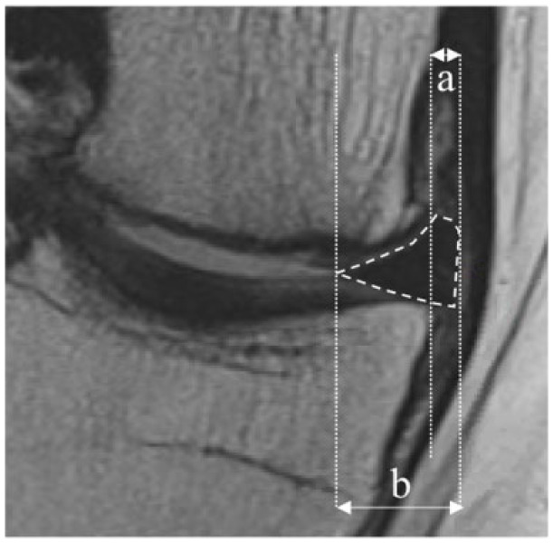
Design: We analyzed data from 124 patients with a mean follow-up of 6.8 ± 2.5 years. Sixty-two patients who underwent (ACI) with anterior meniscus takedown and refixation by the senior surgeon (TM), were compared with a matched control group of patients who underwent ACI without meniscus takedown. Meniscal extrusion was investigated by measuring the absolute value and the relative percentage of extrusion (RPE) on 1.5-T magnetic resonance images (MRI) at final follow-up. The number of menisci with radial displacement greater or lesser than 3 mm was determined. In cases where a preoperative MRI was available, both pre- and postoperative meniscal extrusion was evaluated (n = 30) in those patients undergoing meniscal takedown.
Results: There was no significant difference in either absolute meniscus extrusion, RPE, or extrusion rate in patients with and without meniscus takedown. Among patients with meniscal takedown and both pre- and postoperative MRI scans, absolute meniscus extrusion, RPE, and extrusion rate showed no significant differences.
Conclusion: Meniscal takedown and subsequent transosseous refixation is a safe and effective technique for exposure of the tibial plateau and posterior femoral condyle.
Keywords: cartilage repair; knee surgery; meniscal extrusion; meniscal repair; surgical exposure.
Conflict of interest statement
Figures

References
-
- Ogura T, Mosier BA, Bryant T, Minas T. A 20-year follow-up after first-generation autologous chondrocyte implantation. Am J Sports Med. 2017;45(12):2751-61. - PubMed
-
- Biant LC, Bentley G, Vijayan S, Skinner JA, Carrington RW. Long-term results of autologous chondrocyte implantation in the knee for chronic chondral and osteochondral defects. Am J Sports Med. 2014;42(9):2178-83. - PubMed
-
- Aldrian S, Zak L, Wondrasch B, Albrecht C, Stelzeneder B, Binder H, et al. Clinical and radiological long-term outcomes after matrix-induced autologous chondrocyte transplantation: a prospective follow-up at a minimum of 10 years. Am J Sports Med. 2014;42(11):2680-8. - PubMed
-
- Bentley G, Biant LC, Vijayan S, Macmull S, Skinner JA, Carrington RW. Minimum ten-year results of a prospective randomised study of autologous chondrocyte implantation versus mosaicplasty for symptomatic articular cartilage lesions of the knee. J Bone Joint Surg Br. 2012;94(4):504-9. - PubMed
Publication types
MeSH terms
LinkOut - more resources
Full Text Sources
Medical
