. 2018 Nov 5;115(40):666-673.
doi: 10.3238/arztebl.2018.0666.

Autoantibody-Mediated Encephalitis

Affiliations

Autoantibody-Mediated Encephalitis

Klaus-Peter Wandinger et al. Dtsch Arztebl Int. .

Abstract

Background: Acute and subacute disturbances of wakefulness and cognitive function are common neurological manifestations in the hospital and in outpatient care. An important element of the differential diagnosis was described only a few years ago: autoimmune encephalitis, a condition whose diagnosis and treatment pose an interdisciplinary challenge.

Methods: This review is based on pertinent publications from the years 2005-2017 that were retrieved by a selective search in PubMed, and on the authors' personal experience and case reports.

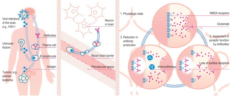
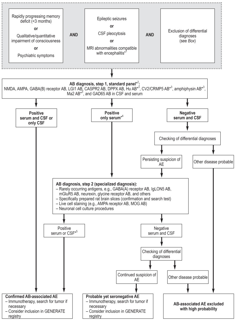
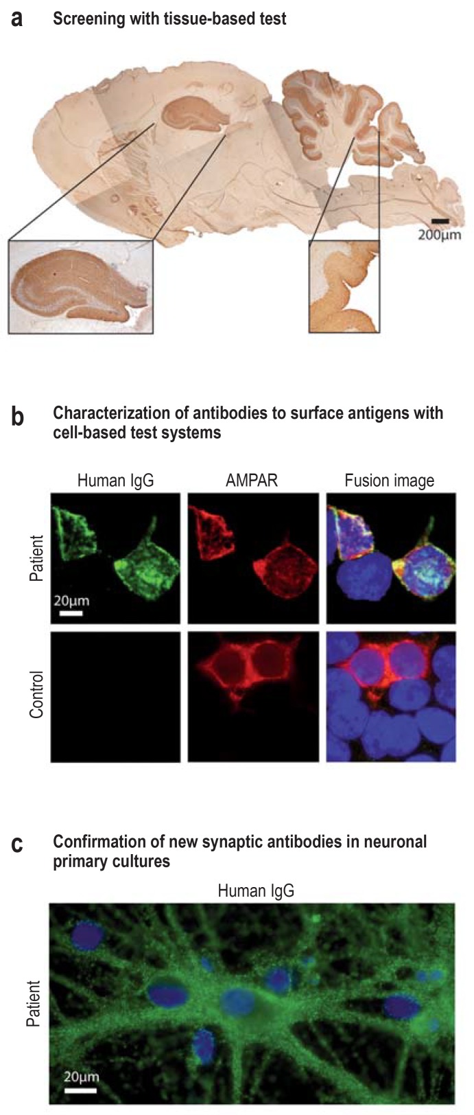
Results: The incidence of autoimmune encephalitis in Germany is estimated at 8-15 cases per million persons per year. In some patients with psychotic manifestations or impaired consciousness of acute or subacute onset, an autoimmune patho - genesis can be demonstrated by the laboratory detection of autoantibodies against neuronal target antigens (e.g., glutamate receptors). Testing of this type should be performed in patients with inflammatory changes in the cerebrospinal fluid or on magnetic resonance imaging (MRI), or those who have had an otherwise unexplained first epileptic seizure or status epilepticus. The cumulative sensitivity of testing for all potentially causative antineuronal antibodies in patients with clinically defined autoimmune encephalitis is estimated at 60-80 %. Figures on cumulative specificity are currently unavailable.

Conclusion: The detection of antineuronal antibodies in patients with the corresponding appropriate symptoms implies the diagnosis of autoimmune encephalitis. Observational studies have shown that rapidly initiated immunosuppressive treatment improves these patients' outcomes. Further studies are needed to determine the positive predictive value of antineuronal antibody detection and to develop further treatment options under randomized and controlled conditions.


Figures

Figure 1
Figure 1
Pathomechanism of synaptic encephalitis: the example of anti-NMDA receptor encephalitis Trigger factors: The misdirected autoimmune reaction may be triggered by the expression of N-methyl-D-aspartate (NMDA) receptors in ovarian teratomas or by the secretion of these receptors in the inflamed brain, e.g., following herpes simplex-I (HSV-I) encephalitis. In many cases, however, the trigger factor is unknown. Establishment of immune reaction: While the systemic immune reaction does not lead to sufficient antibody levels in the brain in the presence of an intact blood–brain barrier, the passage of antibody-producing plasma cells (e.g., triggered by infection) brings about high concentrations of antibodies in the cerebrospinal fluid. Antagonistic effect of antibodies on synapses: The presence of the autoantibodies causes internalization of the receptors and impairment of glutamate transfer. Immunotherapy and/or plasmapheresis lead to reduction of the antibody concentration in the synapse. This enables newly formed receptors to reach the surface of the neuron in sufficient numbers. (Reproduction by kind permission of Katja Duwe-Schrinner, Duwe.Design)
Figure 2
Figure 2
Proposed diagnostic algorithm for investigation of autoimmune encephalitis (AE) (based on [8]) *1 A particularly strong sign of encephalitis on magnetic resonance imaging (MRI) is bilateral edema of the mesial temporal lobe. Unspecific changes are most common, however, and MRI serves especially to exclude differential diagnoses. *2 Standardized, commercially available test kits (immunofluorescence tests with tissue and fixed, transfected cells, ELISA, immunoblotting). *3 Serum testing is sufficient for these antibodies. *4 Immunofluorescence results with commercial cell-based assays with low titers (<1 : 40) but without corresponding CSF findings may be false positive. *5 For rarely occurring antibodies, individual decisions are necessary in the event of isolated serum positivity without corresponding CSF findings. AB, Antibodies; CSF, cerebrospinal fluid; GENERATE, German Network for Research on Autoimmune Encephalitis; for further abbreviations see foot of Table
eFigure:
eFigure:
Experimental analyses In the case of persistent clinical suspicion in the face of a negative result, or to check an implausible positive result, more detailed analysis of antineuronal antibodies can be carried out using experimental methods in specialized laboratories. a) A tissue-based test using specially prepared sagittal slices of rat brain serves for both screening and confirmation. This panel shows the hippocampal and cerebellar staining after incubation of serum (1 : 200) from a patient suspected of having autoimmune encephalitis. The results point to the presence of a synaptic autoantibody with an unknown target antigen. Antihuman secondary antibody and DAB immunohistochemistry. Scale bar: 200 µm. b) In contrast to commercial cell-based test kits, unfixed cell cultures could be used for antigen-specific testing in the specialized laboratory (live cell staining without potential fixation artifacts). This panel shows HEK293T cells (HEK, human embryonal kidney), which express AMPA receptors on their surface after transfection with specific cDNA and were stained with serum (green, 1 : 40) from a patient with anti-AMPA receptor encephalitis or a control patient. Incubation with a monoclonal antibody to AMPA receptors (red) is shown for comparison. The fusion images show that colocalization of both colors (yellow) occurs, which confirms the presence of AMPA receptor specific IgG antibodies in this patient’s serum. Scale bar: 20 µm. c) Neuronal primary cell cultures are used to confirm a new neuronal surface antibody (e.g., from panel a). The example of an anti-IgLON5 antibody (in green) illustrates the binding of the autoantibody to the surface of the neurons and their dendrites. The nuclei of the neurons and the adjacent glial cells are stained blue (neuronal, embryonal, murine hippocampal primary culture DIV21; incubation with serum 1 : 200 in culture with unfixed, unpermeabilized neurons and staining with green-marked secondary antihuman IgG antibodies). Scale bar: 20 µm. AMPAR, a-Amino-3-hydroxy-5-methyl-4-isoxazole propionic acid; DAB, 3,3’-diaminobenzidine; IgG, immunoglobulin G; DIV21, 21 days in vitro IgLON5, Iglon family member 5

References

    1. Royl G, Ploner CJ, Möckel M, Leithner C. [Neurological chief complaints in an emergency room] Nervenarzt. 2010;81:1226–1230. - PubMed
    1. Ryan DJ, O‘Regan NA, Caoimh RÓ, et al. Delirium in an adult acute hospital population: predictors, prevalence and detection. BMJ Open. 2013;3 e001772. - PMC - PubMed
    1. Inouye SK, Westendorp RGJ, Saczynski JS. Delirium in elderly people. Lancet. 2014;383:911–922. - PMC - PubMed
    1. Leypoldt F, Armangue T, Dalmau J. Autoimmune encephalopathies. Ann N Y Acad Sci. 2015;1338:94–114. - PMC - PubMed
    1. Escudero D, Guasp M, Ariño H, et al. Antibody-associated CNS syndromes without signs of inflammation in the elderly. Neurology. 2017;89:1471–1475. - PMC - PubMed