. 2018 Oct 31;10(465):eaah4066.
doi: 10.1126/scitranslmed.aah4066.

Inflammasome inhibition prevents α-synuclein pathology and dopaminergic neurodegeneration in mice

Affiliations

Inflammasome inhibition prevents α-synuclein pathology and dopaminergic neurodegeneration in mice

Richard Gordon et al. Sci Transl Med. .

Abstract

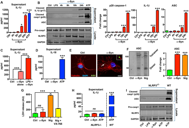
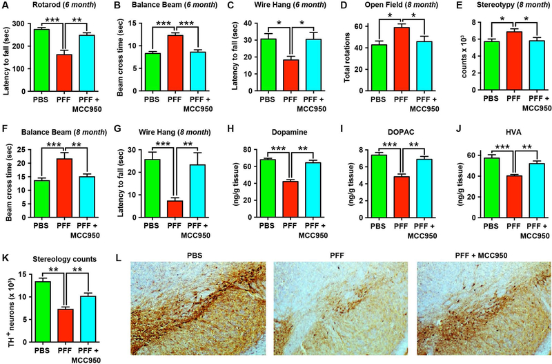
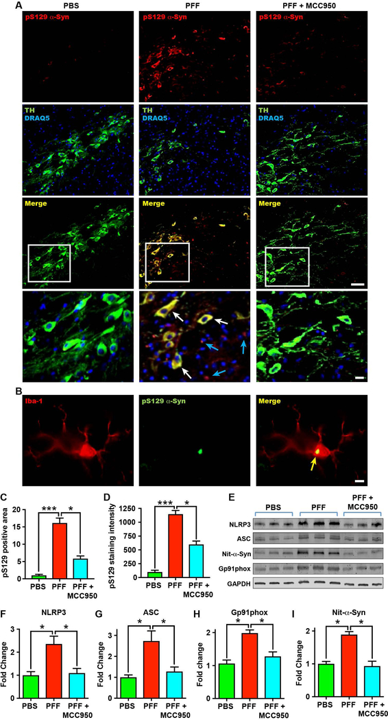
Parkinson's disease (PD) is characterized by a profound loss of dopaminergic neurons in the substantia nigra, accompanied by chronic neuroinflammation, mitochondrial dysfunction, and widespread accumulation of α-synuclein-rich protein aggregates in the form of Lewy bodies. However, the mechanisms linking α-synuclein pathology and dopaminergic neuronal death to chronic microglial neuroinflammation have not been completely elucidated. We show that activation of the microglial NLR family pyrin domain containing 3 (NLRP3) inflammasome is a common pathway triggered by both fibrillar α-synuclein and dopaminergic degeneration in the absence of α-synuclein aggregates. Cleaved caspase-1 and the inflammasome adaptor protein apoptosis-associated speck-like protein containing a C-terminal caspase recruitment domain (ASC) were elevated in the substantia nigra of the brains of patients with PD and in multiple preclinical PD models. NLRP3 activation by fibrillar α-synuclein in mouse microglia resulted in a delayed but robust activation of the NLRP3 inflammasome leading to extracellular interleukin-1β and ASC release in the absence of pyroptosis. Nanomolar doses of a small-molecule NLRP3 inhibitor, MCC950, abolished fibrillar α-synuclein-mediated inflammasome activation in mouse microglial cells and extracellular ASC release. Furthermore, oral administration of MCC950 in multiple rodent PD models inhibited inflammasome activation and effectively mitigated motor deficits, nigrostriatal dopaminergic degeneration, and accumulation of α-synuclein aggregates. These findings suggest that microglial NLRP3 may be a sustained source of neuroinflammation that could drive progressive dopaminergic neuropathology and highlight NLRP3 as a potential target for disease-modifying treatments for PD.


Conflict of interest statement

Competing interests: A.A.B.R., L.A.O., K.S., and M.A.C., are a co-inventors on patent applications for NLRP3 inhibitors which have been licensed to Inflazome Ltd, a biotechnology company that is developing drugs to address clinical unmet needs in inflammatory disease by targeting inflammasomes (WO/2016/131098 and WO/2017/140778, Sulfonylureas and related compounds and use of same). M.A.C. and L.A.O. are co-founders and shareholders of Inflazome, and are acting as CEO and CSO respectively. M.A.C also holds a fractional Professorial Research Fellow appointment at The University of Queensland. K.S. was a member of the Scientific Advisory Board for Inflazome Ltd from 2016 to 2017. The remaining authors declare no competing interests.

Figures

Fig. 1.
Fig. 1.. Extensive inflammasome activation and microglial NLRP3 expression is observed in PD patient brains and animal models.
(A) Western blot and (B) densitometric analysis for caspase-1 and ASC from substantia nigra tissue lysates obtained from PD patients and control subjects (n=5–6/group). (C, D) Immunohistochemistry of key inflammasome components NLRP3 (green in C) and ASC (red in D), and Iba-1-positive microglia in substantia nigra tissue sections of postmortem PD patients and age-matched controls. Magnification x40, scale bar 20μm. (E, F) Immunohistochemistry within the striatum of mice 3 days after 6-OHDA or PBS injection, showing NLRP3 (green in E) and ASC (red in F) localized to hypertrophic activated Iba-1-positive microglia in 6-OHDA mice. Magnification x40, scale bar 20μm. (G) Western blot and (H) densitometric analysis for cleaved caspase-1 and ASC in ipsilateral striatal tissue of 6-OHDA and PBS-injected mice at 3 days after injection (n=4 mice/group). (I) Western blot and (J) densitometric analysis for cleaved caspase-1 and ASC in substantia nigra tissue from MitoPark mice (MP) and littermate controls (Ctrl) at 12 and 24 weeks of age (n=2–4 mice/group). (K) Western blot and (L) densitometric analysis for cleaved caspase-1 and ASC in ipsilateral striatal tissue from PBS- and α-synuclein PFF-injected mice at 30 days after injection (n=9 mice/group). Data shown as means ± SEM. *P < 0.05, **P < 0.01 and ***P < 0.001 by Student’s (panels H and J) or Mann-Whitney (panels B and L) t-tests.
Fig. 2.
Fig. 2.. Fibrillar α-synuclein drives delayed NLRP3 inflammasome activation and extracellular ASC release.
(A) Time course of IL-1β secretion in control (Ctrl), or LPS-primed primary microglia exposed to fibrillar α-synuclein (α-Syn; 10 μM). ATP (5 mM) treatment for 1 h was used as a positive control. (B) Western blot (left) and densitometric analysis (right) for cleaved caspase-1 (p20), cleaved IL-1β, and ASC in the supernatants of primed microglia treated with α-synuclein for 24 h. Expression of pro-caspase-1, NLRP3 and GAPDH were determined in cell lysates and 1 h ATP treatment (5 mM) was used as a positive control. (C) Comparison of α-synuclein-mediated IL-1β secretion in un-primed or LPS-primed primary microglia at 24 h. (D) IL-18 secretion in microglia treated with α-synuclein for 24 h compared with ATP (1 h). (E) Immunofluorescence staining for ASC (green) in control or α-synuclein-activated microglia showing the formation of a characteristic inflammasome ASC speck in green. Scale bar 10μm. Inset (top right) shows magnified view of ASC speck identified by arrow. (F) Western blot (left) and band quantification (right) for detection of oligomeric ASC following chemical crosslinking. Nigericin (Nig; 10 μM, 1 h) was used as a positive control. (G) LDH release assay for quantification of caspase-1 dependent pyroptosis (ns, not significant). Nigericin and VX-765 (20 μM) were used as a positive controls. (H) Supernatant IL-1β and (I) western blots for cleaved caspase-1 and ASC release from NLRP3−/− microglia. Wildtype (WT) microglia activated with ATP was used for comparison. Data represented as mean ± SEM from at least 3 independent experiments. *P <0.05, ***P < 0.001 by one-way ANOVA with Bonferroni’s post-hoc test (panels B, C, F, H), or Kruskal-Wallis test with Dunn’s post-hoc test (panels A, D, G).
Fig. 3.
Fig. 3.. Oral dosing of the potent NLRP3 inhibitor MCC950 is active in the CNS and blocks inflammasome activation in multiple pre-clinical PD models.
(A) Dose-response curve for inhibition of ATP-induced NLRP3 inflammasome activation by MCC950 in LPS-primed microglia (n=3–4). (B) Fibrillar α-synuclein (α-Syn)-mediated microglial IL-1β secretion in the presence or absence of MCC950 (100 nM) (n=8–11). (C) Western blots and (D) densitometric analysis for cleaved caspase-1 (p20), cleaved IL-1β (p17) and ASC in the supernatants of α-synuclein-activated microglia co-treated with MCC950 (n=3). (E) Western blot and (F) densitometric quantification of oligomeric ASC (both dimers (D) and tetramers (T)) in microglia treated as indicated (n=3). (G) Pharmacokinetics of MCC950 over 24 h in plasma (ng/ml) or perfused mouse brain (ng/g) following oral gavage (20 mg/kg) (n=3–4 mice/time point). The equivalent microglial IC50 of MCC950 (~3 ng/ml) is indicated by the dashed line. (H-J) Western blot and densitometric quantification of cleaved caspase-1 in (H) ipsilateral striatal tissue lysates of α-synuclein PFF-injected mice at 30 days (n=6 mice/group); (I) substantia nigra tissue lysates of 12-week old MitoPark (MP) mice (n=5–7 mice/group); and (J) ipsilateral striatal tissue lysates of 6-OHDA (6-OH) lesioned mice at 7 days (n=9 mice/group). Data shown as mean ± SEM. *P < 0.05, **P < 0.01, ***P < 0.001 by one-way ANOVA with Bonferroni’s post-hoc test (panels F, H-J), or Kruskal-Wallis test with Dunn’s post-hoc test (panels B, D).
Fig. 4.
Fig. 4.. NLRP3 inhibition with oral MCC950 treatment protects against nigrostriatal dopaminergic degeneration and behavioral deficits in the 6-OHDA model of PD.
(A) Amphetamine-induced ipsilateral rotations quantified at 21 days in PBS-injected, 6-OHDA (6-OH)-injected, or 6-OHDA-injected mice treated with MCC950 (20mg/kg, daily oral gavage) (n=8–16 mice/group). (B) Balance beam footslips quantified 14 days after PBS or 6-OHDA injection (n=8–12 mice/group). (C-E) Striatal dopamine and its metabolites DOPAC and HVA 28 days after PBS or 6-OHDA injection (n=7–8 mice/group). (F) Striatal denervation in 6-OHDA mice shown with DAB immunohistochemistry for tyrosine hydroxylase (TH). Representative images are shown. (G) Western blot and quantitation for TH in the ipsilateral striatum of PBS or 6-OHDA injected mice at 28 days (n=3 mice/group). (H) Representative images and (I) stereological estimates for TH-positive substantia nigra dopaminergic neurons in MCC950-treated and untreated 6-OHDA mice (n=4 mice/group). (J) Amphetamine-induced ipsilateral rotations at 21-days after PBS or 6-OHDA injection in age-matched wildtype (WT) and NLRP3−/− mice (n=4/mice group). (K-M) Dopamine, DOPAC, and HVA in the ipsilateral striatum of wildtype and NLRP3−/− mice (n=6/group) 28-days after PBS or 6-OHDA injection. Data shown as mean ± SEM. *P < 0.05, **P < 0.01, ***P < 0.001 by Student’s (panel J, L) or Mann-Whitney (panel K, M) t-tests, or by one-way ANOVA with Bonferroni’s post-hoc test (panel A, B, D, G) or Kruskal-Wallis test with Dunn’s post-hoc test (panels C, E), or by two-way ANOVA with Bonferroni’s post-hoc test (panel I). Ns, not significant.
Fig. 5.
Fig. 5.. Chronic NLRP3 inhibition with oral MCC950 protects against motor deficits and dopaminergic degeneration in the α-synuclein PFF model of PD.
(A) Rotarod test in α-synuclein PFF-injected mice at 6 months following treatment with or without MCC950 in the drinking water (0.3 mg/ml) (n=15–17 mice/group). PBS-injected mice were used as controls. (B) Balance beam performance measured as time taken to cross the beam in PFF mice at 6 months (n=15–17 mice/group). (C) Wire-hang test in PFF mice (n=15–17 mice/group) at 6 months. (D, E) Open-Field activity at 8 months post PBS or PFF inoculation measuring rotational counts (D) and stereotypic behaviour (E) (n=10 mice/group). (F, G) Balance beam, and wire-hang tests on a separate cohort of PBS-injected control mice, or MCC950-treated and untreated PFF mice at 8 months (n=6–8 mice/group). (H-J) Dopamine, DOPAC, and HVA in the ipsilateral striatum of PFF mice at 8 months (n=8 mice/group). (K) Stereological estimates and (L) representative images for tyrosine hydroxylase (TH)-positive dopaminergic neurons in the substantia nigra of 8-month old PFF mice (n=5–6 mice/group). Data shown as mean ± SEM. *P < 0.05, **P < 0.01, ***P < 0.001 by one-way ANOVA with Bonferroni’s post-hoc test (panels B-E, G, K), or Kruskal-Wallis test with Dunn’s post-hoc test (panels A, F, H-J).
Fig. 6.
Fig. 6.. NLRP3 therapeutic inhibition with MCC950 ameliorates pathological α-synuclein accumulation in PFF mice.
(A) Representative immunohistochemistry images for phosphorylated serine 129 α-synuclein (pS129 α-Syn) (red), dopaminergic neurons labelled with tyrosine hydroxylase (TH, green), and nuclei stained with DRAQ5 (blue), in the substantia nigra. Images show PBS-injected mice, α-synuclein PFF-injected mice, and α-synuclein PFF-injected mice treated with MCC950 (drinking water, 0.3 mg/ml) for 8 months. Scale bar 30μm. Bottom panels indicate magnified section outlined by white box demonstrating pS129 α-Syn inclusions within dopaminergic neurons (white arrows – yellow merge), as well as not associated with dopaminergic neurons (blue arrows) in untreated PFF mice (scale bar 10μm). (B) Representative immunohistochemistry image for Iba-1 (microglia, red) and pS129 α-Syn (green) in PFF mice showing co-localization with microglia (yellow arrow) (scale bars 5μm). (C, D) Quantification of pS129 α-Syn-positive staining area (C) and staining intensity (D) in the substantia nigra region of stained sections of PFF mice after 8 months (n=7–8 mice/group). (E) Western blot and (F-I) quantitation for NLRP3, ASC, gp91phox and nitrosylated α-synuclein (Nit-α-Syn) in the substantia nigra of PFF mice after 8 months (n=4 mice/group). Data shown as mean ± SEM. *P < 0.05, **P < 0.01, ***P < 0.001 by Kruskal-Wallis test with Dunn’s post-hoc test.

References

    1. Poewe W, Seppi K, Tanner CM, Halliday GM, Brundin P, Volkmann J, Schrag AE, Lang AE, Parkinson disease. Nat Rev Dis Primers 3, 17013 (2017). - PubMed
    1. Brettschneider J, Del Tredici K, Lee VM, Trojanowski JQ, Spreading of pathology in neurodegenerative diseases: a focus on human studies. Nat Rev Neurosci 16, 109–120 (2015). - PMC - PubMed
    1. Luk KC, Kehm V, Carroll J, Zhang B, O’Brien P, Trojanowski JQ, Lee VM, Pathological alpha-synuclein transmission initiates Parkinson-like neurodegeneration in nontransgenic mice. Science 338, 949–953 (2012). - PMC - PubMed
    1. Obeso JA, Rodriguez-Oroz MC, Goetz CG, Marin C, Kordower JH, Rodriguez M, Hirsch EC, Farrer M, Schapira AH, Halliday G, Missing pieces in the Parkinson’s disease puzzle. Nat Med 16, 653–661 (2010). - PubMed
    1. Peelaerts W, Bousset L, Van der Perren A, Moskalyuk A, Pulizzi R, Giugliano M, Van den Haute C, Melki R, Baekelandt V, alpha-Synuclein strains cause distinct synucleinopathies after local and systemic administration. Nature 522, 340–344 (2015). - PubMed

Publication types

MeSH terms

Substances