. 2018 Nov 13;115(46):E10888-E10897.
doi: 10.1073/pnas.1809548115. Epub 2018 Oct 31.

Caspase-8 induces cleavage of gasdermin D to elicit pyroptosis during Yersinia infection

Affiliations

Caspase-8 induces cleavage of gasdermin D to elicit pyroptosis during Yersinia infection

Joseph Sarhan et al. Proc Natl Acad Sci U S A. .

Abstract

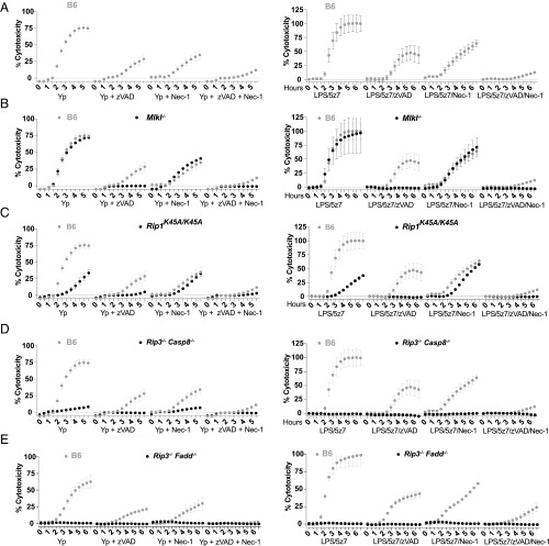
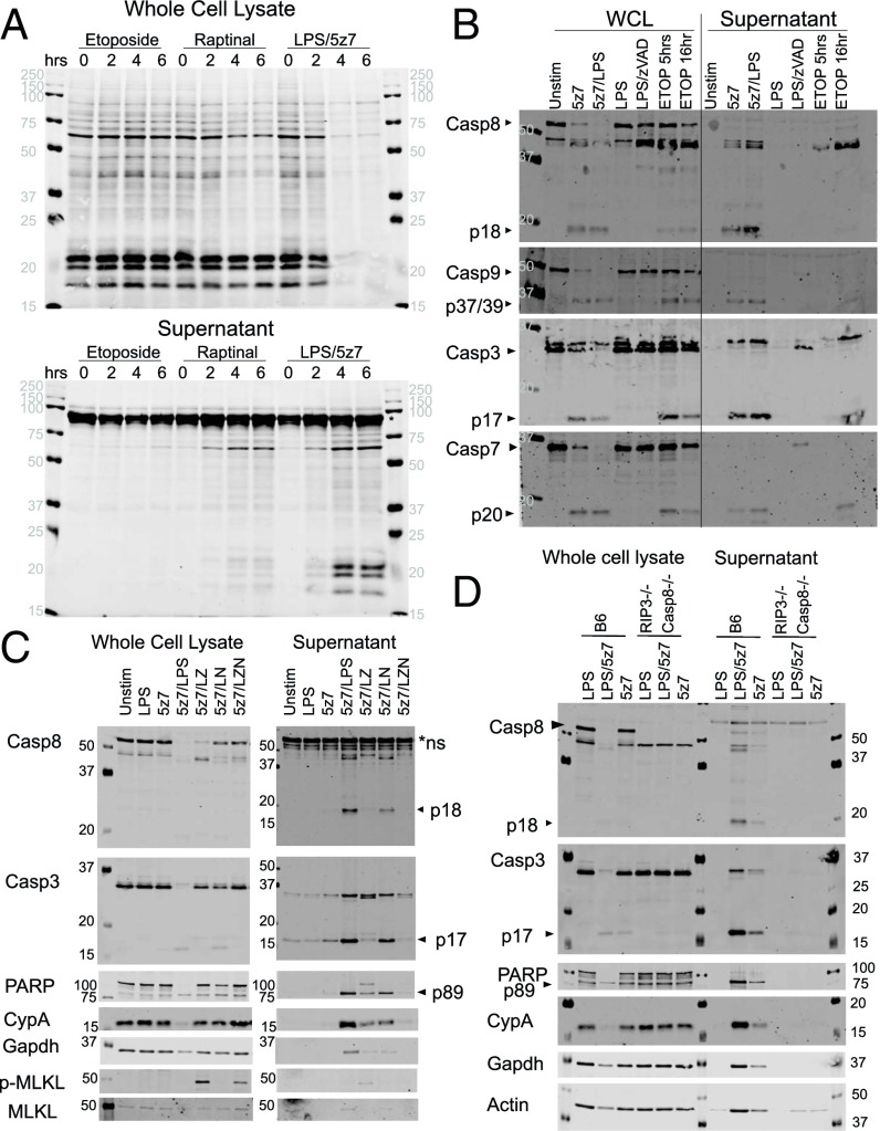
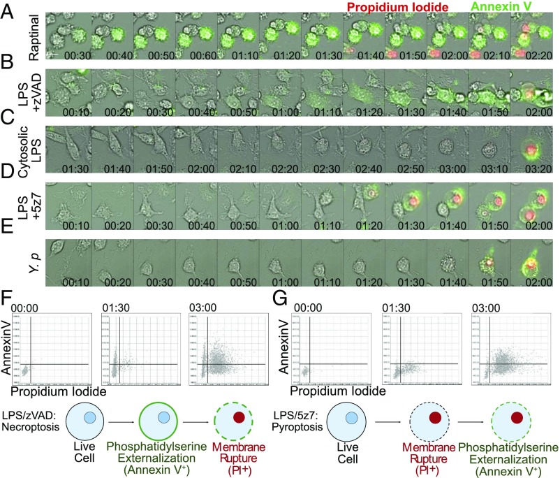
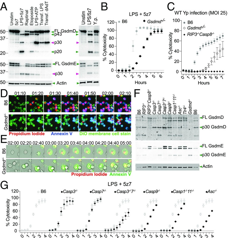
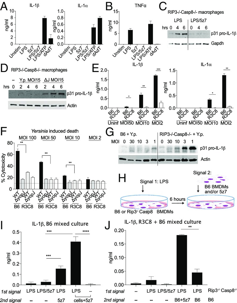
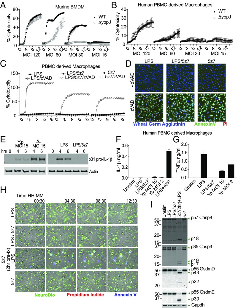
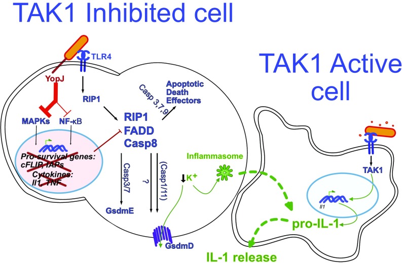
Cell death and inflammation are intimately linked during Yersinia infection. Pathogenic Yersinia inhibits the MAP kinase TGFβ-activated kinase 1 (TAK1) via the effector YopJ, thereby silencing cytokine expression while activating caspase-8-mediated cell death. Here, using Yersinia pseudotuberculosis in corroboration with costimulation of lipopolysaccharide and (5Z)-7-Oxozeaenol, a small-molecule inhibitor of TAK1, we show that caspase-8 activation during TAK1 inhibition results in cleavage of both gasdermin D (GSDMD) and gasdermin E (GSDME) in murine macrophages, resulting in pyroptosis. Loss of GsdmD delays membrane rupture, reverting the cell-death morphology to apoptosis. We found that the Yersinia-driven IL-1 response arises from asynchrony of macrophage death during bulk infections in which two cellular populations are required to provide signal 1 and signal 2 for IL-1α/β release. Furthermore, we found that human macrophages are resistant to YopJ-mediated pyroptosis, with dampened IL-1β production. Our results uncover a form of caspase-8-mediated pyroptosis and suggest a hypothesis for the increased sensitivity of humans to Yersinia infection compared with the rodent reservoir.

Keywords: TAK1; Yersinia; caspase-8; gasdermin; pyroptosis.


Conflict of interest statement

The authors declare no conflict of interest.

Figures

Fig. 1.
Fig. 1.
During TAK1 inhibition, TLR stimulation drives caspase-8–dependent cell death, with necroptosis as a backup mechanism. BMDMs from C57BL/6 (B6) and various genetically modified animals were stimulated with Y. pseudotuberculosis IP2666 (Left) or LPS (10 ng/mL)/5z7 (125 nM) (Right). Y. pseudotuberculosis was infected at an MOI of 30. Percentage cytotoxicity was calculated by 4× microscopy via counting PI+ nuclei per field of view, normalized to 100% lysis by 0.1% Triton X-100. Caspases were inhibited with zVAD treatment (50 μM), and RIP kinase activity was inhibited with Nec1 (10 μM). All inhibitors were added simultaneously at time 0. The cytotoxicity curve of each genotype is superimposed over B6 cytotoxicity curves (gray) for clarity of comparison. Data in AD are from one experiment utilizing the same set of B6 controls; data in E are from an independent experiment. All kinetic cytotoxicity data are representative of at least three independent experiments. See related SI Appendix, Fig. S1.
Fig. 2.
Fig. 2.
TAK1 inhibition results in necrotic cell death, exhibiting pan-caspase activation and the externalization of cytosolic content. (A) BMDMs were stimulated with etoposide (150 μM), raptinal (10 μM), or LPS (10 ng/mL)/5z7 (125 nM) for 0–6 h. Cellular lysates (Upper) and precipitated supernatant (Lower) were run on SDS/PAGE gels, and total protein was stained by LiCOR total protein stain. The asterisk indicates serum proteins from residual FBS in the culture medium. (B) BMDMs were stimulated as indicated for 5 h, except for etoposide, which was stimulated for an additional 16 h. Cellular lysate and precipitated supernatant were run on SDS/PAGE gels and were probed for pro- and cleaved forms of various apoptotic caspases. (C) BMDMs were stimulated with LPS/5z7 for 5 h in the presence of zVAD (50 μM) or Nec1 (10 μM) to block caspase or RIP kinase activity, respectively. Cellular lysate and precipitated supernatant were run on SDS/PAGE gels and probed for caspases, the caspase substrate PARP, cytosolic proteins cyclophilin A (CypA), Gapdh, phosphorylated (S345) MLKL (p-MLKL), and total MLKL. (D) BMDMs from B6 and Rip3−/− Casp8−/− animals were stimulated with LPS, LPS/5z7, or 5z7 for 5 h. Cellular lysate and precipitated supernatant were run on SDS/PAGE gels and probed for caspases, the caspase substrate PARP, cytosolic proteins cyclophilin A (CypA), Gapdh, and actin. All Western blots are representative of three or more experiments.
Fig. 3.
Fig. 3.
Yersinia- and LPS/5z7-driven cell death resembles pyroptosis by morphology. (AE) B6 BMDMs were stimulated as indicated. Y.p., Y. pseudotuberculosis, MOI 5. Shown are 10-min frames from time-lapse microscopy performed at 40× magnification of annexin V and PI dual staining during the progression of cell death. Twelve frames preceding membrane rupture are shown to depict the 2 h of cell-death progression leading to loss of membrane integrity. (Scale bar: 10 μm.) (F and G, Upper) Cells stained with wheat germ agglutinin, annexin V, and PI were imaged at 4× magnification at 2-min intervals, with 4,000 cells per field of view. Annexin V and PI signals were extracted from wheat germ agglutinin-labeled cells, converted into numerical values, and plotted on the y axis for annexin V and on the x axis for PI. Representative time points of 0, 1.5, and 3 h are shown. (Lower) Cartoons depict our simplified model of the progression of phosphatidylserine externalization and membrane rupture during necroptosis or LPS/5z7-driven cell death. All imaging experiments are representative of three or more experiments.
Fig. 4.
Fig. 4.
Caspase-8 induces GsdmD cleavage to drive pyroptosis, curtailing apoptosis. (A) Cleavage products of GsdmD and GsdmE in cell lysate when BMDMs are stimulated with various cell-death triggers or Yersinia infection. The time point shown is 3 h post stimulation or infection with Y. pseudotuberculosis at an MOI of 25. (B) Cell-death kinetics by PI incorporation in B6 and Gsdmd-/ BMDMs stimulated with LPS/5z7. (C) Cell-death kinetics by PI incorporation in B6, Gsdmd-/, and Rip3−/−Casp8−/− BMDMs infected with Yersinia (MOI 25). (D) Time-lapse microscopy of B6 and GsdmD−/− BMDMs triple stained with Neuro-DiO, annexin V, and PI imaged at 20× magnification. Cells were stimulated with LPS/5z7. Image series depict the hour leading up to membrane rupture in GsdmD−/− BMDMs. (Scale bars: 10 μm.) (E) Time-lapse microscopy of B6 and GsdmD−/− BMDMs triple stained with annexin V and PI imaged at 40× magnification. Cells were infected with Yersinia (MOI 5). Imaged series depict the 3 h leading to membrane rupture in GsdmD−/− BMDMs. (Scale bars: 10 μm.) (F) Cleavage products of GsdmD and GsdmE in cell lysate of BMDMs from various knockouts when stimulated with LPS/5z7 for 2 h. (G) LPS/5z7-driven cell-death kinetics by PI incorporation in BMDMs deficient for various caspases or ASC. The cytotoxicity curve of each genotype is superimposed over B6 cytotoxicity curves for clarity of comparison. All kinetic cytotoxicity data, Western blots, and imaging experiments are representative of three or more experiments.
Fig. 5.
Fig. 5.
IL-1 secretion requires two distinct cell populations to generate signal 1 and signal 2. (A) Supernatant IL-1α and IL-1β measured by ELISA 6 h post stimulation by LPS/5z7 compared with canonical inflammasome stimuli. (B) TNF secretion measured by ELISA 6 h post stimulation by LPS/5z7 compared with canonical inflammasome stimuli. (C and D) Pro–IL-1β from cell lysates of Rip3−/−Casp8−/− BMDMs stimulated with agonists (C) or infected with Yersinia (MOI 15) (D) at the indicated time points. (E) Supernatant IL-1α andIL-1β measured by ELISA 16 h post Yersinia infection of B6 and Rip3−/−Casp8−/− BMDMs as a function of decreasing bacterial MOI. (F) Yersinia YopJ-induced cytotoxicity in B6 and Rip3−/−Casp8−/− BMDMs at 6 h post infection as a function of decreasing bacterial MOI. (G) YopJ-sufficient Yersinia infected at decreasing MOIs. Pro–IL-1β synthesis was measured from cell lysate of B6 and Rip3−/−Casp8−/− BMDMs (16 h). (H) Experimental schematics for I and J. (I) IL-1β ELISA from B6 BMDMs asynchronously stimulated with the indicated signals with a 1-h interval between the first and second stimulus. The last two conditions represent the addition of B6 BMDMs and 5z7 for the second signal. (J) IL-1β ELISA from mixed cultures of B6 and Rip3−/−Casp8−/− BMDMs asynchronously stimulated with the indicated signals with 1 h between the first and second stimulus. The first population of cells consists of Rip3−/−Casp8−/− BMDMs plated overnight and experiencing the first signal for 1 h before the addition of the second signal. The second signal is either absent or is present in B6 BMDMs with or without 5z7. Time-point cytotoxicity quantifications and ELISA data are shown as ± SD from three independent experiments compared using Student’s two-tailed t test: ns, nonsignificant (P > 0.05); *P < 0.05; **P < 0.01; ***P < 0.001. All Western blot data are representative of three or more experiments.
Fig. 6.
Fig. 6.
Human macrophages are resistant to TAK1 inhibition-induced cell death with no consequent secretion of IL-1. (A) Yersinia-induced murine macrophage cell death at various bacterial MOIs; MOIs of 60–30 show YopJ-specific cell death. (B) Yersinia-induced death of human monocyte-derived macrophages. Bacteria were infected at multiple MOIs with no YopJ-dependent killing observed. (C) Human PBMC-derived macrophages were treated with LPS, LPS/5z7, or 5z7 in the presence or absence of zVAD (50 μM). Kinetics of cell death was measured by PI+ nuclei. (D) Representative images of triple-stained human PBMC-derived macrophages taken at 4× magnification. PBMC-derived macrophages were stimulated as in C. The image shown is at 7.5 h post stimulation. (Scale bar: 100 m.) (E) Pro–IL-1β from cell lysates of human PBMC-derived macrophages challenged with Yersinia at the indicated MOI and time points (Left) or stimulated with LPS ± 5z7 (Right). (F and G) Supernatant IL-1β (F) and TNF (G) from human PBMC-derived macrophages stimulated with LPS/5z7 or Yersinia at MOIs that elicit robust IL-1α/β secretion from murine macrophages. (H) Human PBMC-derived macrophages were stimulated with the indicated conditions and kinetically imaged with triple staining (Neuro-DiO, PI, and annexin V). The second row from the bottom involved a 2-h 5z7 pretreatment before LPS stimulation. (I) Human PBMC-derived macrophages were stimulated with the indicated conditions for 4 h and were analyzed by Western blotting for caspase-8 and caspase-3 cleavage as well as GSDMD and GSDME. The second-to-last condition involved 2 h of 5z7 pretreatment before LPS stimulation. All kinetic cytotoxicity data, Western blots, ELISAs, and imaging experiments are representative of three or more experiments.
Fig. 7.
Fig. 7.
Model of Yersinia-induced cell death and IL-1 production. Two mouse macrophages are illustrated. The macrophage on the left (a TAK1-inhibited dying cell) is sufficiently intoxicated with Yersinia YopJ to inhibit TAK1. The bacterium is also recognized by TLR4, which signals via RIP1 to form a cell-death complex composed of RIP1, caspase-8, and, likely, FADD. This death complex drives GsdmD cleavage and activation partially via caspase-1/11 and GsdmE cleavage via caspase-3 and caspase-7, both of which result in cell membrane permeabilization and subsequent cell death. There are likely additional effectors of cell death-driven activation, which in conjunction with GsdmD may lead to potassium flux out of the cell and subsequent inflammasome activation. A second macrophage that is not sufficiently intoxicated by YopJ is necessary for the production of pro–IL-1 cytokines. Upon contact with the dying cell or cellular components, IL-1 maturation occurs. See Discussion for details.

References

    1. O’Neill LA, Golenbock D, Bowie AG. The history of toll-like receptors–Redefining innate immunity. Nat Rev Immunol. 2013;13:453–460. - PubMed
    1. Reddick LE, Alto NM. Bacteria fighting back: How pathogens target and subvert the host innate immune system. Mol Cell. 2014;54:321–328. - PMC - PubMed
    1. Park JM, et al. Signaling pathways and genes that inhibit pathogen-induced macrophage apoptosis–CREB and NF-kappaB as key regulators. Immunity. 2005;23:319–329. - PubMed
    1. Chen G, Goeddel DV. TNF-R1 signaling: A beautiful pathway. Science. 2002;296:1634–1635. - PubMed
    1. Kelliher MA, et al. The death domain kinase RIP mediates the TNF-induced NF-kappaB signal. Immunity. 1998;8:297–303. - PubMed

Publication types

MeSH terms