. 2019 Jan 4;93(2):e01639-18.
doi: 10.1128/JVI.01639-18. Print 2019 Jan 15.

Mutations in Influenza A Virus Neuraminidase and Hemagglutinin Confer Resistance against a Broadly Neutralizing Hemagglutinin Stem Antibody

Affiliations

Mutations in Influenza A Virus Neuraminidase and Hemagglutinin Confer Resistance against a Broadly Neutralizing Hemagglutinin Stem Antibody

Kristina L Prachanronarong et al. J Virol. .

Abstract

Influenza A virus (IAV), a major cause of human morbidity and mortality, continuously evolves in response to selective pressures. Stem-directed, broadly neutralizing antibodies (sBnAbs) targeting the influenza virus hemagglutinin (HA) are a promising therapeutic strategy, but neutralization escape mutants can develop. We used an integrated approach combining viral passaging, deep sequencing, and protein structural analyses to define escape mutations and mechanisms of neutralization escape in vitro for the F10 sBnAb. IAV was propagated with escalating concentrations of F10 over serial passages in cultured cells to select for escape mutations. Viral sequence analysis revealed three mutations in HA and one in neuraminidase (NA). Introduction of these specific mutations into IAV through reverse genetics confirmed their roles in resistance to F10. Structural analyses revealed that the selected HA mutations (S123G, N460S, and N203V) are away from the F10 epitope but may indirectly impact influenza virus receptor binding, endosomal fusion, or budding. The NA mutation E329K, which was previously identified to be associated with antibody escape, affects the active site of NA, highlighting the importance of the balance between HA and NA function for viral survival. Thus, whole-genome population sequencing enables the identification of viral resistance mutations responding to antibody-induced selective pressure.IMPORTANCE Influenza A virus is a public health threat for which currently available vaccines are not always effective. Broadly neutralizing antibodies that bind to the highly conserved stem region of the influenza virus hemagglutinin (HA) can neutralize many influenza virus strains. To understand how influenza virus can become resistant or escape such antibodies, we propagated influenza A virus in vitro with escalating concentrations of antibody and analyzed viral populations by whole-genome sequencing. We identified HA mutations near and distal to the antibody binding epitope that conferred resistance to antibody neutralization. Additionally, we identified a neuraminidase (NA) mutation that allowed the virus to grow in the presence of high concentrations of the antibody. Virus carrying dual mutations in HA and NA also grew under high antibody concentrations. We show that NA mutations mediate the escape of neutralization by antibodies against HA, highlighting the importance of a balance between HA and NA for optimal virus function.

Keywords: broadly neutralizing antibody; hemagglutinin; influenza virus; mutants; neuraminidase; resistance.


Figures

FIG 1
FIG 1
Experimental design and viral amplification for passaging with the broadly neutralizing antibody F10. (A) Schematic of experiment 1 and experiment 2 trajectories. Cyan boxes indicate virus that was passaged in the absence of antibody, with the top three passages as P1, P2, and P3 and additional passages as labeled. Red and orange boxes indicate virus that was passaged in the presence of F10 broadly neutralizing antibody (experiments 1 and 2, respectively). Gray boxes indicate virus that was passaged in the presence of 80R control antibody (experiment 2). (B) Ratios of viral titers (output/input) plotted against passage number (experiment 1, upper panel; experiment 2, lower panel).
FIG 2
FIG 2
Mutations inferred to be evolving under positive selection in the presence of the broadly neutralizing antibody F10. (A) Trajectories of select mutations elicited by viral passaging with F10, with 80R control antibody, or without antibody, in terms of allele frequency. Mutations individually marked as A638G and A639T (gray box) are in perfect linkage and yield N203V, as the wild-type sequence is GGT AAC CAA (AAC, positions 638/639/640; protein, GNQ). The mutant sequence is GGT GTC CAA (GTC, positions 638/639/640; protein, GVQ). (B) The posterior probability distribution of selection coefficients (s) for the mutations for experiments 1 and 2. Specific mutations are listed by influenza viral protein, nucleotide change, and amino acid change. Seg, segment; Syn, synonymous. (C) Posterior distributions of effective population size inferred from WFABC. The effective population size was estimated from time-sampled genomic data assuming neutrality. For F10-treated (F10) and control (ctrl) samples, we respectively estimated Ne to be 208 (99% highest posterior density interval, 162, 249) and 440 (99% highest posterior density interval, 350, 512).
FIG 3
FIG 3
Growth of WT and individual mutant viruses in the presence of F10 or oseltamivir. (A) WT and mutant viruses (HA, NA, and HA-NA double mutant) were grown in the indicated concentrations of F10 and quantified by plaque assay. (B) Viral response to the NA inhibitor oseltamivir was measured for the resistance mutation H275YNA compared to responses of the wild type and the mutation E329KNA. Error bars in panel A indicate the standard deviations.
FIG 4
FIG 4
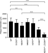
Viral fitness was estimated by plaque size. Plaque diameters of HA and NA mutant viruses in the absence of F10 (n = 20 per virus) are shown. Error bars indicate the standard deviations. A one-way analysis of variance multiple-comparison test was performed (***, P < 0.001; ****, P < 0.0001).
FIG 5
FIG 5
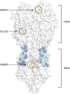
Escape mutations identified in the F10 trajectories mapped onto the structure of HA. The HA trimer is displayed in gray surface representation (PDB accession number 3FKU). The F10 epitope (or footprint) on the HA stem is displayed as sticks and colored according to degree of contacts with the antibody F10. Residues with the greatest contacts are shown in green, intermediate contacts in cyan, and smallest contacts in navy blue. The fusion peptide is shown in orange stick representation between the F10 epitope (footprint), and the locations of escape mutations are labeled.
FIG 6
FIG 6
The N460SHA mutation is located adjacent to the fusion peptide. (A) The structure of HA monomer at neutral pH is shown with respect to the viral envelope and endosomal membrane (PDB accession number 3FKU). The HA1 subunit, which forms the head of HA, is shown in blue, the HA2 subunit, which forms the stem of HA, is in gray, and the fusion peptide is shown in red. (B) The location of mutation N460S is circled on the structure of HA at neutral pH, with the F10 epitope colored as shown in Fig. 5. (C and D) A zoomed-in view of the stem region harboring N460S (C) and of the hydrogen bond between N460 and the fusion peptide indicated with a black dashed line (D). (E) At acidic pH, the fusion peptide dissociates from the stem of HA and inserts into the endosomal membrane (PDB accession number 1HTM). (F) The structure of HA2 at acidic pH is shown, where residue N460 is exposed to the surface and is shown in yellow (PDB accession number 1HTM). (G and H) The N460S residue is shown in more detail.
FIG 7
FIG 7
S123GHA is located in a hinge region of conformational change in an early fusion intermediate of HA1. (A) The surface representation of HA structure at neutral pH where residue S123 is circled and surrounding residues 115 to 129 are displayed in green (PDB accession number 3QQB). (B) The structure of an early fusion intermediate of HA at acidic pH where residue S123 is circled and residues 115 to 129 are shown in yellow (PDB accession number 3QQO). (C) A detailed view of S123 at neutral pH with surrounding residues shown in green to show the early conformational changes that occur in HA1 during fusion. S123 is located in a hinge region of conformational change in HA1, and the direction of the conformational changes that occur at acidic pH is indicated with red arrows (PDB 3QQB). (D) The resulting structure of the early fusion intermediate of HA is shown in yellow (PDB 3QQO).
FIG 8
FIG 8
The N203VHA mutation is located in the receptor binding site. (A) The head region of HA is represented by a gray surface, and the location of mutation N203V is labeled with a circle and shown in yellow. The human receptor analog LSTc is shown as gold sticks (PDB accession number 2WRG). (B) N203V is located in the HA receptor-binding site and forms a hydrogen bond with the human receptor analog LSTc. The hydrogen bond is shown with a black dashed line connecting the side chain oxygen atom of N203 with a nitrogen atom on LSTc.
FIG 9
FIG 9
The dynamics and electrostatic surface of WT and E329KNA. The root mean square fluctuations (RMSF) of WT (A) and E329K (B) NA during 100-ns MD simulations. The residues are represented on a rainbow scale from blue to red for increasing RMSF values; hence, warmer colors indicate residues with more backbone fluctuations. The radius of the cartoon representation also indicates the RMSF value: the thicker the tube, the higher the RMSF value. The oseltamivir from PDB accession number 3CL2 (black sticks) is displayed solely to indicate the active site on all four NA molecules. (C and D) The electrostatic surface for the final frame from MD simulations of WT (C) and E329KNA (D). The residues are represented on a rainbow scale from blue (positive) to red (negative).

References

    1. Palese P, Shaw M. 2007. Orthomyxoviridae: the viruses and their replication, p 1647–1690. In Knipe DM, Howley PM, Griffin DE, Lamb RA, Martin MA, Roizman B, Straus SE (ed), Fields virology, 5th ed Lippincott Williams & Wilkins, Philadelphia, PA.
    1. Drake JW. 1993. Rates of spontaneous mutation among RNA viruses. Proc Natl Acad Sci U S A 90:4171–4175. doi:10.1073/pnas.90.9.4171. - DOI - PMC - PubMed
    1. White J, Hoffman L, Arevalo J, Wilson I. 1997. Attachment and entry of influenza virus into host cells. Pivotal roles of hemagglutinin, p 80–104. In Chiu W, Burnett RM, Garcea RL (ed), Structural biology of viruses, Oxford University Press, Oxford, United Kingdom.
    1. Wagner R, Matrosovich M, Klenk HD. 2002. Functional balance between haemagglutinin and neuraminidase in influenza virus infections. Rev Med Virol 12:159–166. doi:10.1002/rmv.352. - DOI - PubMed
    1. Wilson IA, Skehel JJ, Wiley DC. 1981. Structure of the haemagglutinin membrane glycoprotein of influenza virus at 3 A resolution. Nature 289:366–373. doi:10.1038/289366a0. - DOI - PubMed

Publication types

MeSH terms

LinkOut - more resources