. 2018 Oct 31;9(1):4549.
doi: 10.1038/s41467-018-06943-2.

Identification and characterization of a large family of superbinding bacterial SH2 domains

Affiliations

Identification and characterization of a large family of superbinding bacterial SH2 domains

Tomonori Kaneko et al. Nat Commun. .

Abstract

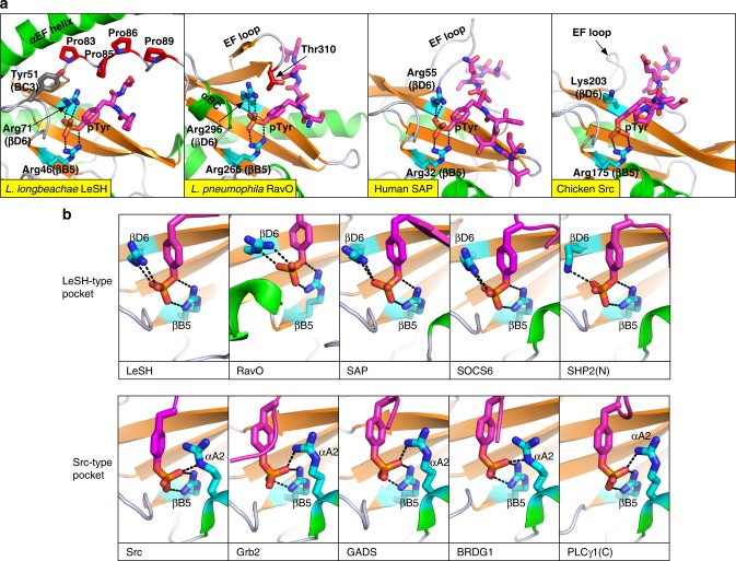
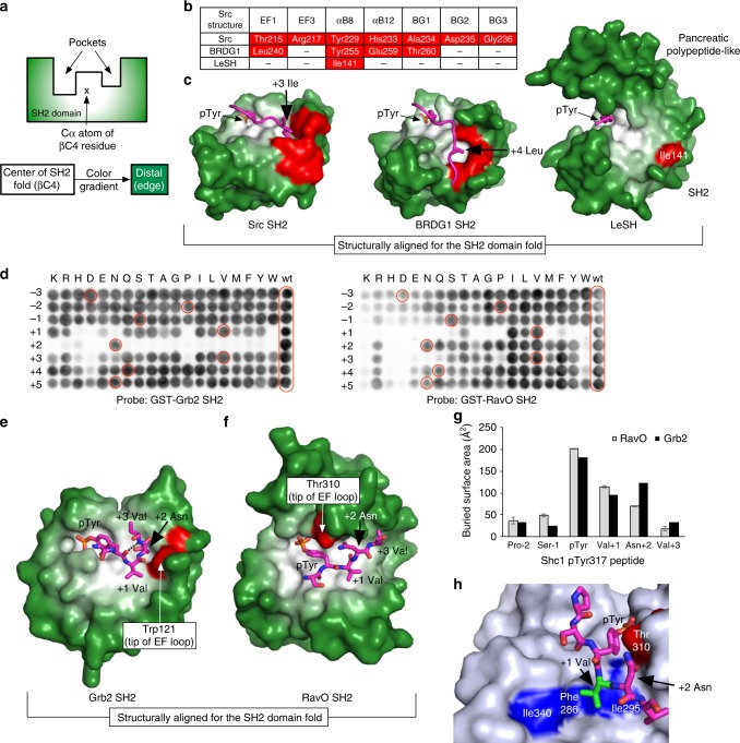
Src homology 2 (SH2) domains play a critical role in signal transduction in mammalian cells by binding to phosphorylated Tyr (pTyr). Apart from a few isolated cases in viruses, no functional SH2 domain has been identified to date in prokaryotes. Here we identify 93 SH2 domains from Legionella that are distinct in sequence and specificity from mammalian SH2 domains. The bacterial SH2 domains are not only capable of binding proteins or peptides in a Tyr phosphorylation-dependent manner, some bind pTyr itself with micromolar affinities, a property not observed for mammalian SH2 domains. The Legionella SH2 domains feature the SH2 fold and a pTyr-binding pocket, but lack a specificity pocket found in a typical mammalian SH2 domain for recognition of sequences flanking the pTyr residue. Our work expands the boundary of phosphotyrosine signalling to prokaryotes, suggesting that some bacterial effector proteins have acquired pTyr-superbinding characteristics to facilitate bacterium-host interactions.


Conflict of interest statement

The authors declare no competing interests.

Figures

Fig. 1
Fig. 1
Legionella encodes a preponderance of SH2 domains. a Structure-guided sequence alignment of the Legionella SH2 domains. Shown are the SH2 domains from the L. longbeachae LeSH and LUSH; L. dumoffii LeSH1a, LeSH2, and LeSH3; L. drancourtii LeSH4; L. pneumophila RavO and DoSH (C-terminal SH2); and L. waltersii LeSH5 (Lwal_0152). The SH2 domain structures of LeSH, RavO, and Src were used as templates for the structure-guided alignment. Secondary structure elements are based on either solved (Src, LeSH, and RavO SH2 domains) or predicted structures. The α-helices are colored green and β-strands orange. The Arg residues at the βB5 and βD6 positions (based on secondary structure nomenclature of the Src SH2 domain) are highlighted in blue. The four Pro residues in the pancreatic polypeptide-fold of LeSH are highlighted in red. b Domain organization of representative Legionella SH2 domain-containing proteins. CPD, cysteine protease domain; L22, uncharacterized Legionella effector domain
Fig. 2
Fig. 2
Legionella SH2 proteins are translocated effectors. a Distribution of SH2 domain proteins in different Legionella species and their effector probability. Included in the heat map are 40 Legionella species (including two strains from L. pneumophila that contain distinct SH2 proteins) and two species from the Coxiellaceae family (that belongs to the order Legionellales). The 28 Legionella species implicated in human disease are highlighted in yellow. The effector prediction scores derived from Burstein et al. were used to generate the heat map with a cyan-to-magenta gradient representing low-to-high effector probability for the 71 proteins listed in Burstein et al.. The scores are not available (N/A) for 13 other SH2 proteins and they are colored gray. When LeSH2 or DoSH paralogs exist for a species, the cell is divided accordingly into two or three sections. For example, L. waltersii encodes three paralogs of LeSH2 all with high effector potential. See Supplementary Table 1 for a full list of the SH2 proteins. b Type IV secretion system (T4SS)-dependent translocation of Legionella SH2 proteins from bacteria to host cells. Ten SH2 proteins from Legionella fused with the adenylate cyclase Cya were expressed in the L. pneumophila strain Lp02 (dotA+). The L. pneumophila RavO and L. longbeachae LUSH were also tested in the Lp03 strain (defect in the T4SS, or dotA−). Differentiated U937 cells were infected with bacteria expressing the different Cya-fusion protein. Translocation of the fusion proteins was monitored by the intracellular cyclic AMP (cAMP) level, which is an indicator of the Cya activity in eukaryotic host cells. The error bars indicate standard deviation from triplicate infection experiments. See Supplementary Fig. 5 for expression level of the Cya-fusion proteins and p-values that indicate statistical significance of translocation of the Cya-fusion proteins in comparison to the Cya itself (labeled no fusion). The cAMP values are derived from Supplementary Fig. 5a, except for L. dumoffii LeSH1a and LeSH2, which are taken from Supplementary Fig. 5b. L. dumoffii LeSH1a and L. anisa LeSH1b, which are deficient in pTyr binding (see Fig. 3c), are colored cyan
Fig. 3
Fig. 3
Legionella SH2 domains are bona fide phosphotyrosine superbinders. a Selective binding of L. longbeachae LeSH to the tyrosine-phosphorylated peptide GGpYGG, but not to the non-phosphorylated or pThr-containing versions. Peptides used for the fluorescence polarization assay were N-terminally labeled with fluorescein, through a GG- or a 6-aminohexanoic acid spacer. b Equilibrium binding curves (from fluorescence polarization assays) of five purified Legionella SH2 domains and the human Src SH2 triple mutant (Src superbinder) to the GGpYGG peptide. Llo, L. longbeachae; Ldr, L. drancourtii; Ldu, L. dumoffii; and Lp, L. pneumophila. c Equilibrium dissociation constants (Kd) of 13 Legionella SH2 domains for a selected group of pTyr, Tyr, or pThr-containing peptides. Except for the VCP peptide, the peptides used here were selected from the peptide arrays (Fig. 3d; Supplementary Fig. 6). The Kd values are shown in µM, with a color gradient from red (Kd = 0.2 µM) to blue (Kd = 20 µM) to denote high to low affinity. See Supplementary Table 2 for fitting statistics of the binding curves. Ahx: 6-aminohexanoic acid spacer. d A peptide array probed with the GST-tagged RavO SH2 domain. Shown here is a strip of 20 spots representing pTyr peptides taken from the EGFR, Shc1 and MidT and the artificial peptide GGpYGG. Peptides A6–A10, identified in red rectangle, are non-phosphorylated versions of peptides A1–A5. See Supplementary Fig. 6a and b, for images of the full peptide arrays. e Binding of Legionella SH2 domains to tyrosine-phosphorylated proteins. Human macrophage-like U937 cells were treated with pervanadate (+pv) to enrich protein tyrosine phosphorylation before lysate is prepared. GST-fused Legionella SH2 domains were used to pull down the phosphorylated proteins from the lysate of U937-derived macrophage. Shown are western blotting images using antibodies specific for the pTyr, Shc1, or VCP. See Supplementary Fig. 6e for data on four additional Legionella proteins
Fig. 4
Fig. 4
Structures of two Legionella SH2 domains with bound phosphopeptides. a Crystal structure of L. longbeachae LeSH in complex with the IL2Rβ pTyr387 peptide. Only three residues (Pro-pTyr-Ser) could be traced in the electron density (see Supplementary Fig. 8d). The three prolines associating with the hydrophobic side of the protruding αEF helix are shown in sticks and colored red. See Supplementary Fig. 8e for amphipathic nature of the helical extension. b The Src SH2 domain with the avian pancreatic polypeptide shown on top to mimic the LeSH structure. Pro residues in the pancreatic polypeptide that make contact with the hydrophobic side of the α-helix (αEF) are colored red. c Crystal structure of the RavO SH2 domain in complex with the Shc1 pTyr317 peptide. Panels a and c are structurally aligned to the Src SH2 domain. Helices and β strands in the SH2 domains are shown in green and orange ribbons, respectively. The conserved βB5 arginine is colored cyan. The bound phosphopeptides are in magenta sticks. d A comparison of secondary structures for the three SH2 domains. e Sequence identity between Legionella and selected eukaryotic SH2 domains based on structure-based alignments. SH2 domains from the chicken Src, human SAP, Dictyostelium STAT, and yeast Spt6 protein were included for the structure-guided sequence identity calculation. The total number of aligned residues in each pairwise structural comparison was given in the parenthesis. f Electrostatic surface potential of the LeSH, RavO, and Src SH2 domains drawn with the same scale. The bound peptides are shown as magenta sticks. The surface is colored in a gradient from red (negative) to blue (positive electronic potential)
Fig. 5
Fig. 5
Comparison of the pTyr-binding pocket between Legionella and animal SH2 domains. a The pTyr-binding pockets of the LeSH and RavO SH2 domains. The human SAP and chicken Src SH2 domains are shown for comparison. b Classification of pTyr conformation based on structural alignment of the LeSH and RavO SH2 domains with eight eukaryotic SH2 domains. Residues that contribute to salt bridges are shown in cyan sticks. The following high-resolution (resolution ≤2 Å and the crystallographic free-R <0.25) crystal structures were used here: SAP (PDB code 1D4W), SOCS6 (2VIF), SHP2 (N-terminal SH2, 3TKZ), Src (1SHA), Grb2 (1JYR), GADS (1R1Q), BRDG1 (3MAZ), and PLCγ1 (C-terminal SH2, 4K45)
Fig. 6
Fig. 6
The Legionella SH2 domains lack a specificity pocket. a A color gradient depicting the distance of the ligand-binding pockets on a typical eukaryotic SH2 domain to the center of the globular SH2 domain fold. The Cα atom of the βC4 residue of an SH2 domain was defined as the origin of the color gradient, from white to dark green, to facilitate visualization of the specificity pocket in an SH2 domain. b Residues that contribute to forming the inside lining of either the P + 3 or + 4-binding pocket in SH2 domains. The P + 3 (Ile)-binding pocket in the Src SH2 domain involves the EF1, EF3, BG2, and BG3 residues whereas the P + 4 (Leu)-binding pocket in the BRDG1 SH2 domain is formed by the EF1, αB8, αB12, and BG1 residues. A 3D structural alignment of the two SH2 domains with LeSH was used to identify structurally equivalent residues in the latter. The symbol (–) indicates absence of an equivalent residue in the structure. c Comparison of the structure of the LeSH-DnaJ-A1 peptide complex with those of the Src and BRDG1 SH2 domains. The bound peptides are pTyr-Glu-Glu-Ile for the Src SH2 domain, and pTyr-Glu-Asn-Val-Leu for the BRDG1 SH2 domain. Only the side chains of the key residues (Ile + 3 for the Src SH2 ligand and Leu + 4 for the BRDG1 SH2 ligand) are shown in magenta sticks. The pocket-forming residues listed in b are colored red. d A permutation array based on the Shc1 pTyr317 peptide (DPSpYVNVQNL) probed with either the GST-fused Grb2 (left panel) or RavO SH2 domain (right panel). The amino acid at each position between −3 and +5 was replaced by one of 19 natural amino acids (except for Cys). The pTyr residue was fixed. The peptide spots identical to the wild-type (wt) sequence are highlighted in red circles. ef Comparison of the LeSH and human Grb2 SH2 domains bound to the same peptide (Shc1 pTyr317). The human Grb2 SH2 domain (e) is structurally aligned with the L. pneumophila RavO SH2 domain (f). The Shc1 peptide Ser-pTyr-Val-Asn-Val is shown in magenta sticks. The residue located at the tip of the EF loop is colored red for each SH2 domain. g The buried surface area (in Å2) calculated for each residue of the Shc1 pTyr317 peptide bound to the SH2 domain of RavO or Grb2. The error bars for the area from the RavO complex indicate deviations between the three copies of SH2-peptide complexes in the asymmetric unit (Supplementary Fig 9e and f). h The hydrophobic patch on the RavO SH2 domain used for accommodating the +1 position of the ligand peptide

References

    1. Bartram, J., Chartier, Y., Lee, J. V., Pond, K. & Surman-Lee, S. Legionella and the Prevention of Legionellosis (World Health Organization, Geneva, 2007).
    1. Cianciotto, N. P., Hilbi, H. & Buchrieser, C. in The prokaryotes (ed. Rosenberg, E.) 147–217 (Springer, Berlin, Heidelberg, 2013).
    1. Correia AM, et al. Probable person-to-person transmission of Legionnaires’ disease. N. Engl. J. Med. 2016;374:497–498. doi: 10.1056/NEJMc1505356. - DOI - PubMed
    1. Ensminger AW, Yassin Y, Miron A, Isberg RR. Experimental evolution of Legionella pneumophila in mouse macrophages leads to strains with altered determinants of environmental survival. PLoS Pathog. 2012;8:e1002731. doi: 10.1371/journal.ppat.1002731. - DOI - PMC - PubMed
    1. Boamah DK, Zhou G, Ensminger AW, O’Connor TJ. From many hosts, one accidental pathogen: the diverse protozoan hosts of Legionella. Front. Cell Infect. Microbiol. 2017;7:21660–17. doi: 10.3389/fcimb.2017.00477. - DOI - PMC - PubMed

Publication types