. 2019 Apr;19(2):168-177.
doi: 10.1007/s12012-018-9487-6.

Airway Exposure to Modified Multi-walled Carbon Nanotubes Perturbs Cardiovascular Adenosinergic Signaling in Mice

Affiliations

Airway Exposure to Modified Multi-walled Carbon Nanotubes Perturbs Cardiovascular Adenosinergic Signaling in Mice

Leslie C Thompson et al. Cardiovasc Toxicol. 2019 Apr.

Abstract

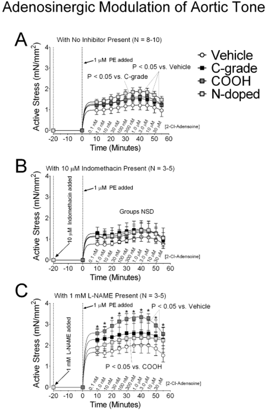
The broad list of commercial applications for multi-walled carbon nanotubes (MWCNT) can be further expanded with the addition of various surface chemistry modifications. For example, standard commercial grade MWCNT (C-grade) can be carboxylated (COOH) or nitrogen-doped (N-doped) to suite specific utilities. We previously reported dose-dependent expansions of cardiac ischemia/reperfusion (I/R) injury, 24 h after intratracheal instillation of C-grade, COOH, or N-doped MWCNT in mice. Here, we have tested the hypothesis that airway exposure to MWCNT perturbs cardiovascular adenosinergic signaling, which could contribute to exacerbation of cardiac I/R injury. 100 µL of Vehicle or identical suspension volumes containing 100 µg of C-grade, COOH, or N-doped MWCNT were instilled into the trachea of CD-1 ICR mice. 1 day later, we measured cyclic adenosine monophosphate (cAMP) concentrations in cardiac tissue and evaluated arterial adenosinergic smooth muscle signaling mechanisms related to nitric oxide synthase (NOS) and cyclooxygenase (COX) in isolated aortic tissue. We also verified cardiac I/R injury expansion and examined both lung histology and bronchoalveolar lavage fluid cellularity in MWCNT exposed mice. Myocardial cAMP concentrations were reduced (p < 0.05) in the C-grade group by 17.4% and N-doped group by 13.7% compared to the Vehicle group. Curve fits to aortic ring 2-Cl-Adenosine concentration responses were significantly greater in the MWCNT groups vs. the Vehicle group. Aortic constrictor responses were more pronounced with NOS inhibition and were abolished with COX inhibition. These findings indicate that addition of functional chemical moieties on the surface of MWCNT may alter the biological responses to exposure by influencing cardiovascular adenosinergic signaling and promoting cardiac injury.

Keywords: Adenosine; Cardiac ischemia/reperfusion injury; Multi-walled carbon nanotubes; cAMP.


Conflict of interest statement

Conflict of Interest:

The authors report no conflicts of interests related to the research reported in this manuscript. The conclusions reported in the manuscript are those of the authors and do not necessarily reflect those of the National Institute of Environment Health Sciences, East Carolina University, Bellarmine University, or the University of Colorado at Denver.

Figures

Figure 1.
Figure 1.. Lung histology.
Left lungs were collected 24 hours following Vehicle or MWCNT instillation and examined histologically by H&E staining in order to visually assess pulmonary responses to MWCNT. Images were collected using the 10× objective and inlays were collected using the 40× objective. (A) Representative images of a left lung tissue collected from mice instilled with Vehicle. (B) Representative images of lung tissue collected from mice instilled with C-grade. (C) Representative images of left lung tissue collected from mice instilled with COOH. (D) Representative images of left lung tissue collected from mice instilled with N-doped.
Figure 2.
Figure 2.. Cardiac I/R injury.
We tested the hypothesis that 24 hours after MWCNT instillation cardiac I/R injury would result in expansion of myocardial infarction. Myocardial infarct sizes from CD-1 mice instilled with Vehicle, 100 μg C-grade, COOH, or N-doped are presented as a percent of the zone at risk. *for P < 0.05 vs. Vehicle; †P < 0.05 vs. C-grade; ‡P < 05 vs. COOH as determined by ANOVA. N = 5 for all groups.
Figure 3.
Figure 3.. Heart and Lung cAMP concentrations.
Heart and right lung tissues were collected from mice 24 hours after instillation with Vehicle or MWCNT and assessed for cAMP concentrations via ELISA. (A) cAMP concentrations in heart tissue collected from mice instilled with Vehicle (n = 6), C-grade (n = 5), COOH (n = 5), or N-doped (n = 6) normalized to total protein. (B) cAMP concentrations in lung tissue collected from mice instilled with Vehicle (n = 8), 100 μg C-grade (n = 4), COOH (n = 4), and N-doped (n = 8) normalized to total protein. *P < 0.05 vs. Vehicle.
Figure 4.
Figure 4.. Adenosinergic modulation of aortic tone.
Isolated rings of thoracic aorta were stimulated with 1 μM phenylepherine (PE) for 5 minutes followed by additions of 2-Cl-adenosine every 5 minutes to generate cumulative concentration-responses from 0.1 nM to 30 μM. (A) Concentration-response profiles with NO Inhibitor present (n = 8-10). (B) Concentration-response profiles with 10 μM general cyclooxygenase (COX) inhibitor indomethacin present (n = 3-5). (C) Concentration-response profiles with 1 mM nitric oxide synthase (NOS) inhibitor L-NAME present (n = 3-5). P < 0.05 for comparisons of the nonlinear fits of the treatment response curve was determined by best fit regression analysis [24]. *indicates P < 0.05 vs. Vehicle for mean data reported as done by Repeated Measures ANOVA with Bonferroni post-test. (n = 3-10).
Figure 5.
Figure 5.. Aortic contraction with Phenylephrine (PE) and relaxation with 2-Cl-adenosine.
(A) Maximal aortic stress generated 5 minutes following stimulation with 1 μM PE, just prior to the start of 2-Cl-adenosine protocols (n = 3-10). (B) The maximal aortic vasorelaxation generated by stimulation with 30 μM 2-Cl-adenosine taken as a function of peak stress generated during experimental protocols with PE + 2-Cl-adenosine (n = 3-10). *for P < 0.05 by Two-way ANOVA with Bonferroni post-test.

References

    1. Kuijpers E, Bekker C, Fransman W, Brouwer D, Tromp P, Vlaanderen J, Godderis L, Hoet P, Lan Q, Silverman D, Vermeulen R and Pronk A (2016). Occupational Exposure to Multi-Walled Carbon Nanotubes During Commercial Production Synthesis and Handling. Ann Occup Hyg 60: 305–317. - PMC - PubMed
    1. Poulsen SS, Knudsen KB, Jackson P, Weydahl IE, Saber AT, Wallin H and Vogel U (2017). Multi-walled carbon nanotube-physicochemical properties predict the systemic acute phase response following pulmonary exposure in mice. PloS One 12: e0174167. - PMC - PubMed
    1. Ayala P, Arenal R, Rummeli M, Rubio A and Pichler T (2010). The doping of carbon nanotubes with nitrogen and their potential applications. Carbon 48: 575–586.
    1. Min C, Zhang Q, Shen C, Liu D, Shen X, Song H, Li S, Xu D, Lin X and Zhang K (2017). Graphene oxide/carboxyl-functionalized multi-walled carbon nanotube hybrids: powerful additives for water-based lubrication. RSC Advances 7: 32574–32580.
    1. Urankar RN, Lust RM, Mann E, Katwa P, Wang X, Podila R, Hilderbrand SC, Harrison BS, Chen P, Ke PC, Rao AM, Brown JM and Wingard CJ (2012). Expansion of cardiac ischemia/reperfusion injury after instillation of three forms of multi-walled carbon nanotubes. Part Fibre Toxicol 9: 38. - PMC - PubMed

Publication types

MeSH terms