. 2018 Oct 31;239(3):339-350.
doi: 10.1530/JOE-18-0449.

Stress rapidly suppresses in vivo LH pulses and increases activation of RFRP-3 neurons in male mice

Affiliations

Stress rapidly suppresses in vivo LH pulses and increases activation of RFRP-3 neurons in male mice

Jennifer A Yang et al. J Endocrinol. .

Abstract

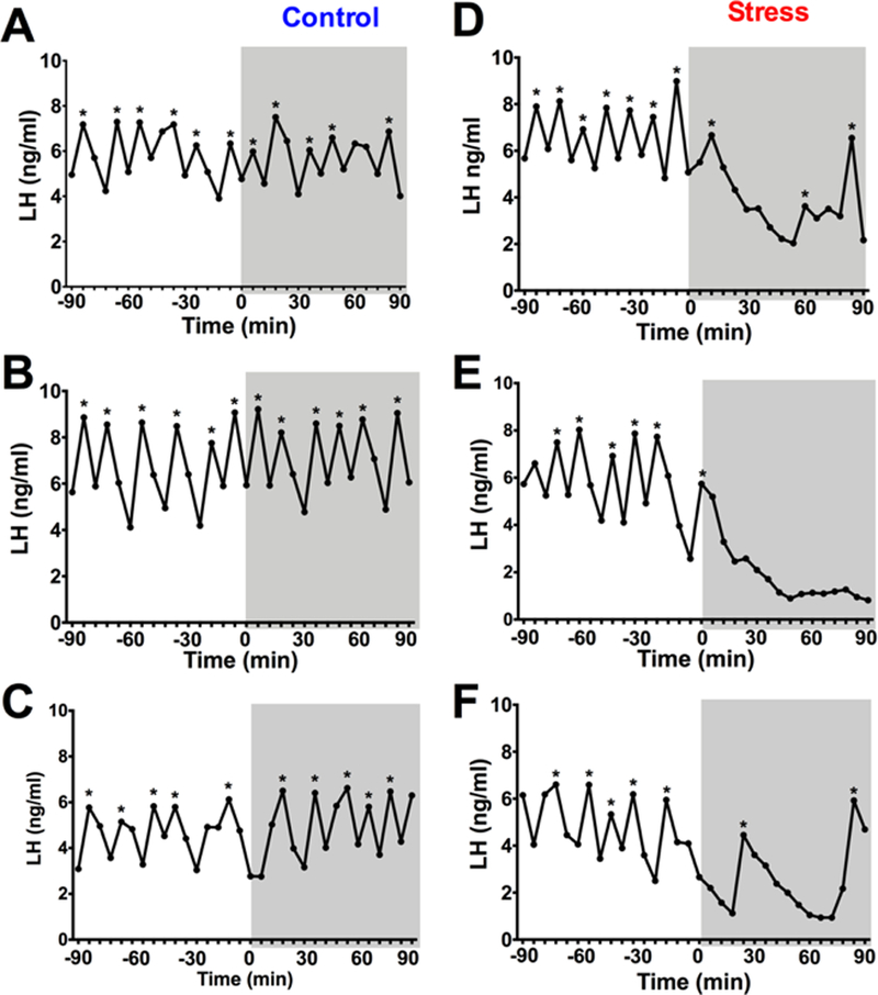
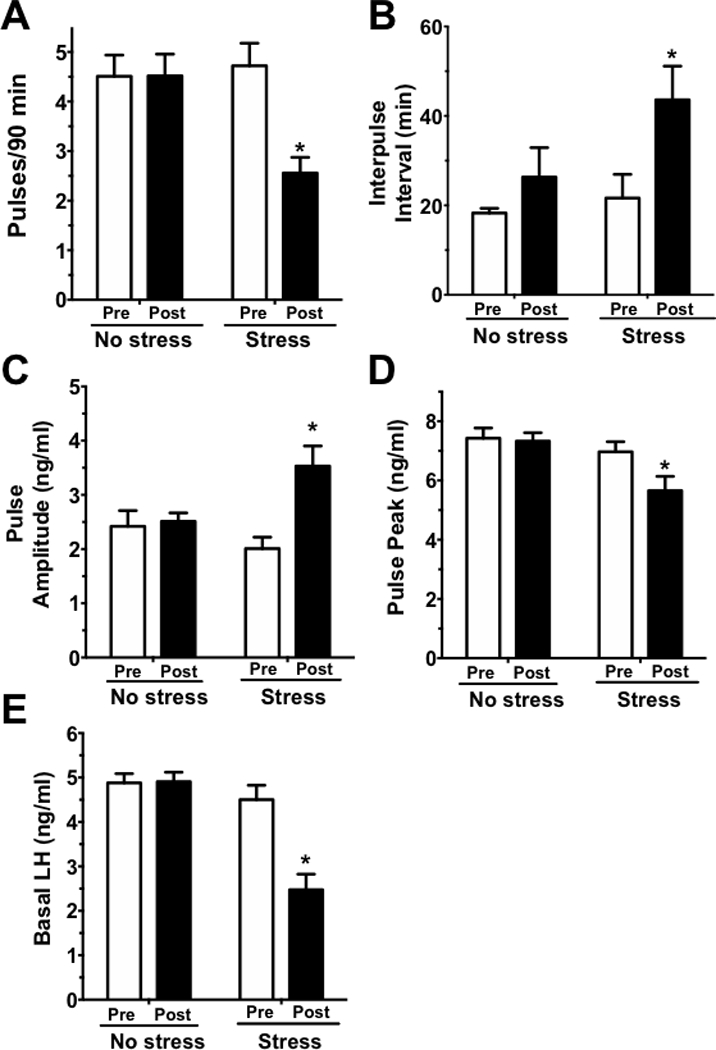
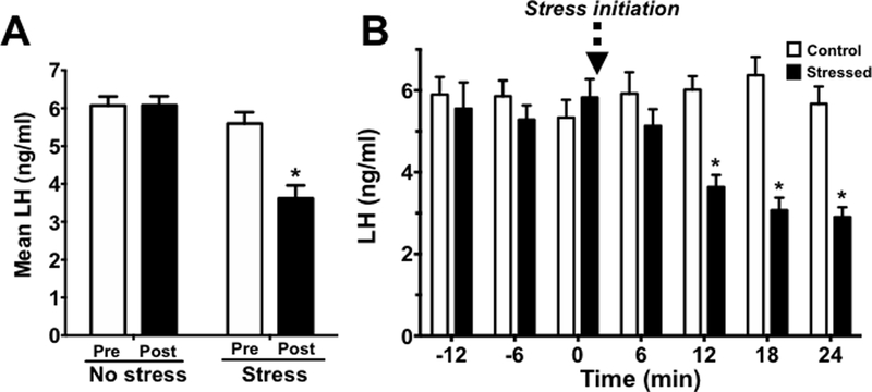
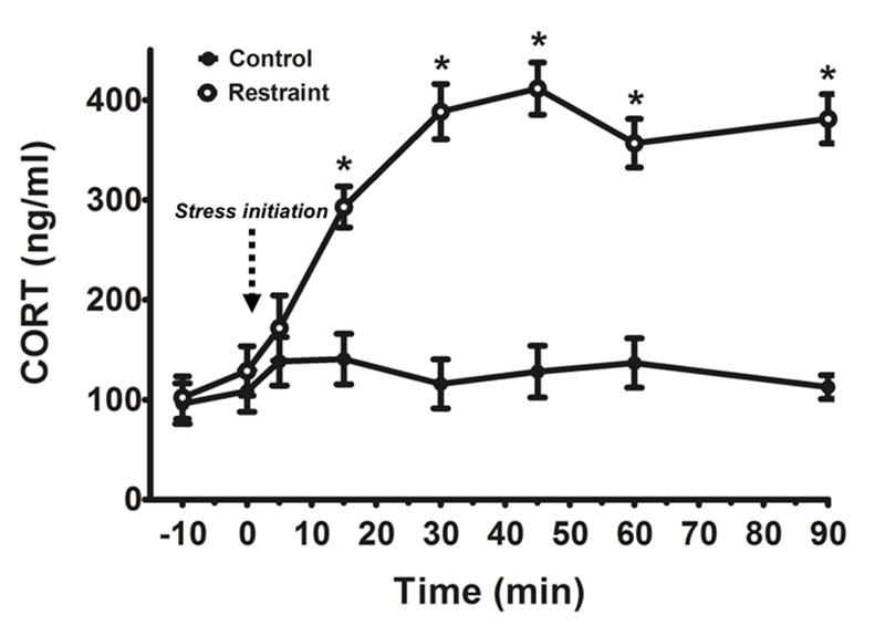
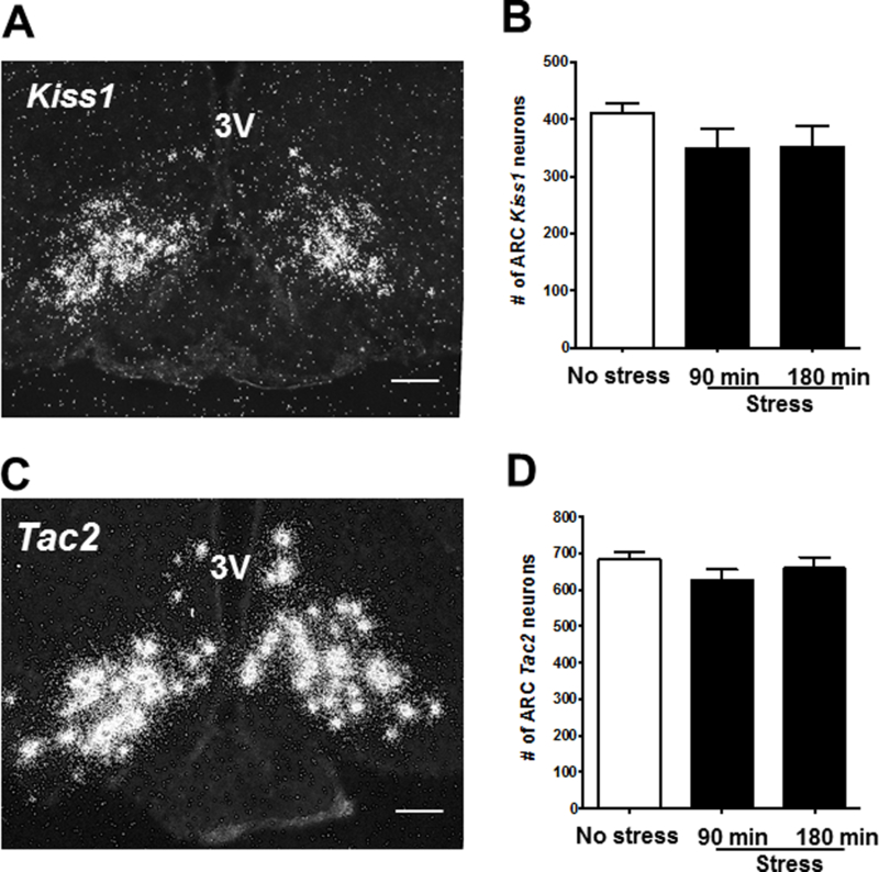
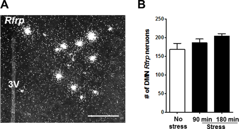
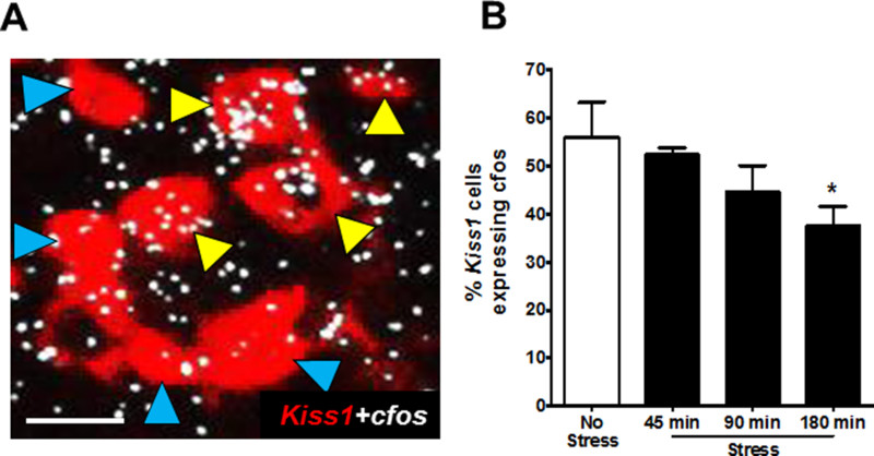
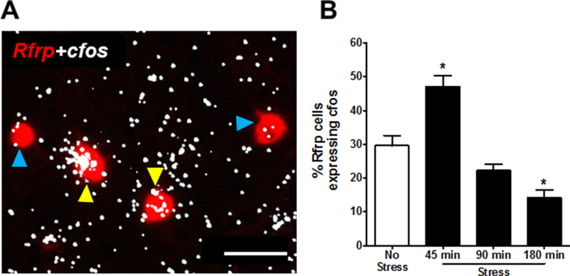
Restraint stress is a psychosocial stressor that suppresses reproductive status, including LH pulsatile secretion, but the neuroendocrine mechanisms underlying this inhibition remains unclear. Reproductive neural populations upstream of gonadotropin-releasing hormone (GnRH) neurons, such as kisspeptin, neurokinin B and RFRP-3 (GnIH) neurons, are possible targets for psychosocial stress to inhibit LH pulses, but this has not been well examined, especially in mice in which prior technical limitations prevented assessment of in vivo LH pulse secretion dynamics. Here, we examined whether one-time acute restraint stress alters in vivo LH pulsatility and reproductive neural populations in male mice, and what the time-course is for such alterations. We found that endogenous LH pulses in castrated male mice are robustly and rapidly suppressed by one-time, acute restraint stress, with suppression observed as quickly as 12–18 min. This rapid LH suppression parallels with increased in vivo corticosterone levels within 15 min of restraint stress. Although Kiss1, Tac2 and Rfrp gene expression in the hypothalamus did not significantly change after 90 or 180 min restraint stress, arcuate Kiss1 neural activation was significantly decreased after 180 min. Interestingly, hypothalamic Rfrp neuronal activation was strongly increased at early times after restraint stress initiation, but was attenuated to levels lower than controls by 180 min of restraint stress. Thus, the male neuroendocrine reproductive axis is quite sensitive to short-term stress exposure, with significantly decreased pulsatile LH secretion and increased hypothalamic Rfrp neuronal activation occurring rapidly, within minutes, and decreased Kiss1 neuronal activation also occurring after longer stress durations.


Conflict of interest statement

Declaration of interest: The authors have no conflict of interest to disclose.

Figures

Figure 1.
Figure 1.
Restraint stress inhibits pulsatile LH in gonadectomized (GDX) male mice. (A-C) represent three individual control mice and (D-F) represent three individual stress mice. Each graph represents an individual male mouse with blood collected every 6 min for 180 min. The initial 90 min period (−90 to 0 min) represent the pre-stress (baseline) period and the gray boxes (time 0 to 90 min) represent the post (stress) period. Pulse peaks are indicated by *.
Figure 2.
Figure 2.
LH pulse parameters in GDX no stress (control) and stress male mice. The following LH parameters were analyzed: (A) pulse frequency, shown as pulses per 90 min, (B) interpulse interval, (C) pulse amplitude, (D) pulse peak levels, and (E) basal LH levels. White bars are the pre-period (baseline) of blood collection (−90 to 0 min) and the black bars are the post-period (stress) of blood collection (0 to 90 min). *, p<0.05 versus baseline (pre-stress) value.
Figure 3.
Figure 3.
(A) Mean LH measured pre-stress (baseline; white bars) and post-stress initiation (black bars) in no stress (control) and stressed male mice. (B) Detailed time-course of LH changes after stress exposure, measured at 6 min intervals to determine how soon LH decreased following initiation of restraint stress. Mean blood LH levels before (−12 to 0) and after (0 to 24) restraint stress. White bars are control mice and black bars are restraint stressed mice. *p<0.05.
Figure 4.
Figure 4.
CORT secretion time-course in non-stressed control (closed, black circles) and acutely restrained male mice (open, white circles). Time 0 min represents time of restraint stress initiation in the restraint mice. *p<0.05 control vs. restraint mice, within time point.
Figure 5.
Figure 5.
Acute restraint stress has no effect on arcuate Kiss1 or Tac2 cell number in male mice. (A) Representative image of single-label ISH for arcuate Kiss1 in a no stress control mouse. (B) Mean (± SEM) Kiss1 cell number in no-stress control (white bar) and stressed groups (90 and 180 min; black bars). (C) Representative images of single-label ISH for Tac2 in a no stress control mouse. (D) Mean (± SEM) Tac2 cell number in no stress control (white bar) and stressed groups (90 and 180 min; black bars). *p<0.05 versus no stress controls. 3V, third ventricle; ARC, arcuate. Scale bar = 100μm.
Figure 6.
Figure 6.
Restraint stress does not change Rfrp coexpression in male mice. (A) Representative image of single-label ISH for Rfrp in the DMN of a no stress control male. (B) Mean (± SEM) Rfrp cell number in no-stress control (white bar) and stressed groups (90 and 180 min; black bars). *p<0.05 versus no stress controls. 3V, third ventricle; DMN, dorsal medial nucleus. Scale bar = 100μm.
Figure 7.
Figure 7.
Arcuate Kiss1 neural activation in stress vs no stress control male mice. (A) Representative images of Kiss1 (red fluorescence) and cfos mRNA (silver grains) gene expression in a no stress control male. Yellow arrowheads represent coexpression of Kiss1+cfos; blue arrowheads denote Kiss1 neurons not coexpressing cfos mRNA. (B) Quantification of coexpression of Kiss1 and cfos mRNA in the arcuate of male mice. *p<0.05, compared to no stress controls. Scale bar = 25μm.
Figure 8.
Figure 8.
DMN Rfrp neural activation in stress vs no stress control male mice. (A) Representative images of Rfrp (red fluorescence) and cfos mRNA (silver grains) gene expression in a no stress control male. Yellow arrowheads represent coexpression of Rfrp+cfos mRNA; blue arrowheads denote Rfrp neurons not coexpressing cfos mRNA. (B) Quantification of coexpression of Rfrp and cfos mRNA in the DMN of male mice. *p<0.05, compared to no stress controls. Scale bar = 50μm.

Similar articles

Cited by

References

    1. Anderson GM, Relf HL, Rizwan MZ & Evans JJ 2009. Central and peripheral effects of RFamide-related peptide-3 on luteinizing hormone and prolactin secretion in rats. Endocrinology 150 1834–1840. - PubMed
    1. Chen MD, O’Byrne KT, Chiappini SE, Hotchkiss J & Knobil E 1992. Hypoglycemic ‘stress’ and gonadotropin-releasing hormone pulse generator activity in the rhesus monkey: role of the ovary. Neuroendocrinology 56 666–673. - PubMed
    1. Chen MD, Ordog T, O’Byrne KT, Goldsmith JR, Connaughton MA & Knobil E 1996. The insulin hypoglycemia-induced inhibition of gonadotropin-releasing hormone pulse generator activity in the rhesus monkey: roles of vasopressin and corticotropin-releasing factor. Endocrinology 137 2012–2021. - PubMed
    1. Clarke IJ, Sari IP, Qi Y, Smith JT, Parkington HC, Ubuka T, Iqbal J, Li Q, Tilbrook A, Morgan K, et al. 2008. Potent action of RFamide-related peptide-3 on pituitary gonadotropes indicative of a hypophysiotropic role in the negative regulation of gonadotropin secretion. Endocrinology 149 5811–5821. - PubMed
    1. Clarkson J, Han SY, Piet R, McLennan T, Kane GM, Ng J, Porteous RW, Kim JS, Colledge WH, Iremonger KJ, et al. 2017. Definition of the hypothalamic GnRH pulse generator in mice. Proc Natl Acad Sci U S A 114 E10216–E10223. - PMC - PubMed

Publication types