. 2019 Feb 23;219(6):856-866.
doi: 10.1093/infdis/jiy585.

Characterization of the Genital Mucosa Immune Profile to Distinguish Phases of the Menstrual Cycle: Implications for HIV Susceptibility

Affiliations

Characterization of the Genital Mucosa Immune Profile to Distinguish Phases of the Menstrual Cycle: Implications for HIV Susceptibility

Geneviève Boily-Larouche et al. J Infect Dis. .

Abstract

Background: Inflammation and immune activation are key factors in sexual transmission of human immunodeficiency virus (HIV). We sought to define the impact of hormonal cycling on the mucosal immune environment and HIV risk in sex workers with a natural menstrual cycle.

Methods: We compared soluble mucosal immune factors and cervical mononuclear cells during hormone titer-defined phases of the menstrual cycle among 37 sex workers from Nairobi, Kenya. Systemic and mucosal samples were collected 14 days apart to distinguish the follicular and luteal phases of the menstrual cycle, and phases were confirmed by hormone measurements. Vaginal concentrations of 19 immune modulators and cervical T-cell activation markers were measured.

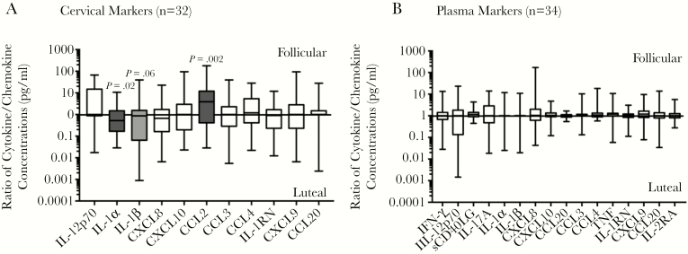
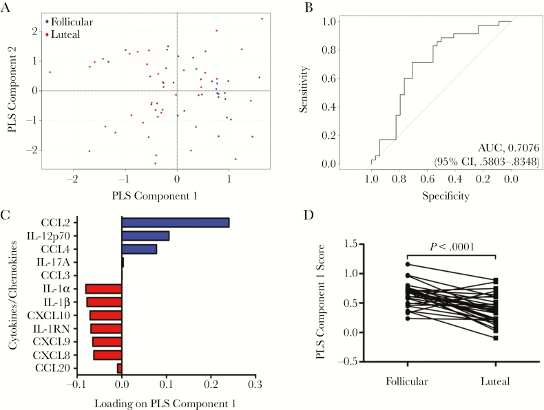
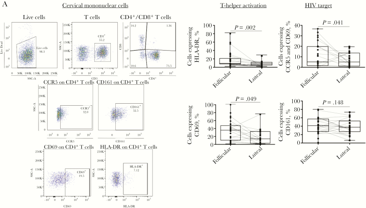
Results: The follicular phase signature was characterized by an elevated CCL2 level, decreased interleukin 1α and interleukin 1β cervical concentrations, and a significant increase in the proportion of CD4+ T cells that expressed CD69. The genital concentration of CCL2 was the best marker to distinguish the follicular from the luteal phase in univariate and multivariate analyses and remained independent of elevated genital inflammation and bacterial vaginosis.

Conclusion: The follicular phase of the menstrual cycle was associated with an elevated CCL2 level and retention of resident memory CD4+ T cells, which has implications for increased susceptibility to HIV infection.

Keywords: Genital inflammation; HIV; immune activation; menstrual cycle.


Figures

Figure 1.
Figure 1.
Ratios of cytokine/chemokine concentrations in picograms/milliliter during the follicular phase to those during the luteal phase (ie, fold change) in paired cervicovaginal lavage and plasma specimens from women who underwent sampling during both phases of the menstrual cycle (1, no change in level; >1, elevated level during the follicular phase; and <1, elevated level during the luteal phase). Only ratios of markers for which concentrations were detectable in at least 30% of the participants are presented in this figure. The ratios are presented as box and whisker plots showing the medians (middle lines), interquartile ranges (upper and lower limits of boxes), and minimum and maximum values (upper and lower whiskers). The parametric paired t test was used for comparing the log10-transformed cytokine/chemokine concentrations between phases. Levels of cytokines/chemokines that were significantly altered by the menstrual cycle are indicated by shaded bars. IFN-γ, interferon γ; IL-1RN, interleukin 1 receptor antagonist; IL-1α, interleukin 1α; IL-1β, interleukin 1β; IL-2RA, interleukin 2 receptor agonist; IL-12p70, interleukin 12p70; IL-17A, interleukin 17A; TNF, tumor necrosis factor.
Figure 2.
Figure 2.
Identification of multivariate cytokine profiles associated with distinct phases of the menstrual cycle. A, Partial least squares discriminant analysis (PLS-DA) model of all 12 cytokines classified in individuals (blue, follicular phase; red, luteal phase). B, The area under the curve (AUC) was 0.79 (95% confidence interval [CI], .68–.90), with a sensitivity of 0.74 and a specificity of 0.74, for the autopredictive model; the AUC was 0.71 (95% CI, .58–.83), with a sensitivity of 0.71 and a specificity of 0.71, for the cross-validated model. C, Loading plots of the 12 cytokines/chemokines measured. PLS component 1 best separated the follicular phase from the luteal phase of the menstrual cycle. The follicular phase clustered in the positive region of PLS component 1 (A). Levels of cytokines positively loaded on PLS component 1 (CCL2, interleukin 12p70 [IL-12p70], CCL4, interleukin 17A [IL-17A], and CCL3) are elevated in the follicular phase profile, whereas levels of cytokines negatively loaded (interleukin 1α [IL-1α], interleukin 1β [IL-1β], CXCL10, interleukin 1 receptor antagonist [IL-1RN], CXCL9, CXCL8, and CCL20) were comparatively reduced during the luteal phase. D, PLS component 1 scores were significantly higher in the follicular phase of the menstrual cycle as compared to the luteal phase (P < .0001). Model-fitted values calculated were compared by the parametric paired t test (n = 32).
Figure 3.
Figure 3.
Proportion of activated T-helper cells in cervical mononuclear cells. A, Gating strategy to characterize expression of mucosal T-helper cell activation markers. For each cervical cell sample, 100000 events were collected and gated to identify lymphocytes (forward scatter [FSC-A] vs side scatter [SSC-A]). Live CD3 cells were selected and subdivided into CD4+ and CD8+ T-cell subset, and those expressing CD161, CCR5 (depicted as an example), CD69, and HLA-DR were identified. B, Comparison of the proportion of T-helper (CD4+) cells in cervical cells expressing activation markers CD69, HLA-DR, Th17 marker CD161, and human immunodeficiency virus (HIV) coreceptor CCR5 between the follicular and luteal phases of the menstrual cycle. Results are presented as box plots showing the medians (middle lines), interquartile ranges (upper and lower limits of boxes), and minimum and maximum values (upper and lower whiskers). Differences between phases were assessed using the Wilcoxon signed rank test. P values of <.05 were considered statistically significant.

References

    1. Joint United Nations Programme on HIV. UNAIDS data 2017. Geneva: UNAIDS, 2017:1–246.
    1. Masson L, Passmore JA, Liebenberg LJ, et al. . Genital inflammation and the risk of HIV acquisition in women. Clin Infect Dis 2015; 61:260–9. - PMC - PubMed
    1. Haase AT. Early events in sexual transmission of HIV and SIV and opportunities for interventions. Annu Rev Med 2011; 62:127–39. - PubMed
    1. Naranbhai V, Abdool Karim SS, Altfeld M, et al. ; CAPRISA004 TRAPS team Innate immune activation enhances hiv acquisition in women, diminishing the effectiveness of tenofovir microbicide gel. J Infect Dis 2012; 206:993–1001. - PMC - PubMed
    1. Lajoie J, Juno J, Burgener A, et al. . A distinct cytokine and chemokine profile at the genital mucosa is associated with HIV-1 protection among HIV-exposed seronegative commercial sex workers. Mucosal Immunol 2012; 5:277–87. - PubMed

Publication types

Grants and funding