Review
. 2019 Feb;92(1094):20180505.
doi: 10.1259/bjr.20180505. Epub 2018 Nov 30.

Role and future of MRI in radiation oncology

Affiliations
Review

Role and future of MRI in radiation oncology

Indra J Das et al. Br J Radiol. 2019 Feb.

Abstract

Technical innovations and developments in areas such as disease localization, dose calculation algorithms, motion management and dose delivery technologies have revolutionized radiation therapy resulting in improved patient care with superior outcomes. A consequence of the ability to design and accurately deliver complex radiation fields is the need for improved target visualization through imaging. While CT imaging has been the standard of care for more than three decades, the superior soft tissue contrast afforded by MR has resulted in the adoption of this technology in radiation therapy. With the development of real time MR imaging techniques, the problem of real time motion management is enticing. Currently, the integration of an MR imaging and megavoltage radiation therapy treatment delivery system (MR-linac or MRL) is a reality that has the potential to provide improved target localization and real time motion management during treatment. Higher magnetic field strengths provide improved image quality potentially providing the backbone for future work related to image texture analysis-a field known as Radiomics-thereby providing meaningful information on the selection of future patients for radiation dose escalation, motion-managed treatment techniques and ultimately better patient care. On-going advances in MRL technologies promise improved real time soft tissue visualization, treatment margin reductions, beam optimization, inhomogeneity corrected dose calculation, fast multileaf collimators and volumetric arc radiation therapy. This review article provides rationale, advantages and disadvantages as well as ideas for future research in MRI related to radiation therapy mainly in adoption of MRL.


Figures

Figure 1.
Figure 1.
(a) A small brain lesion invisible in CT images, but can be clearly visible in MR images, (b) another patient with dental filling where CT images are difficult to provide volume delineation. The MR images can provide structural information. The CT-MR fusion is used for target and normal tissue delineation.
Figure 2.
Figure 2.
(a) ViewRay MRIdian system, (b) Elekta 1.5 T MRI-Linac, (c) Alberta’s Aurora RT system and (d) Australian (Sydney) MRI System, image courtesy of Paul Keall and Brad Oburn.
Figure 3.
Figure 3.
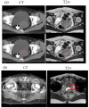
MR simulation example cases: (a) Axial CT + MR fusion of a Gynaecological case. GTV boundary, red colour (bottom panel) is clearly visible on the T2w MRI acquired in treatment position, (b) Axial CT + MR fusion of a prostate patient with bilateral hip implants. Prostate and rectal spacer is clearly visible on the axial T2w MRI as compared to CT (arrow).
Figure 4.
Figure 4.
MR only approach to patient treatment. (a) T1/T2 image acquired on a flat table top used for contouring, (b) sCT generation, (c) treatment planning (d) two images (bone and soft tissue) of sCT DRR for treatment verification. Similar approaches and detailed DRR images are also provided by several other investigators.
Figure 5.
Figure 5.
Morphological assessment of tumour burden based on longitudinal T2 weighted MRI of rectal cancer patients undergoing neoadjuvant chemoradiation treatment. Please note changes in tumor burden over time with tumour location indicated by the arrow.
Figure 6.
Figure 6.
Schema for the image post-processing on contrast-enhanced T1 and T2 images. The tumour areas were contoured on all MRI slices. Radiomics features are extracted that is correlative with clinical data for survival prediction. Adopted from Ouyang et al with open access source.

References

    1. Citrin DE. Recent developments in radiotherapy. N Engl J Med Overseas Ed 2017; 377: 1065–75. doi: 10.1056/NEJMra1608986 - DOI - PubMed
    1. Srivastava SP, Cheng C-W, Das IJ. Image guidance-based target volume margin expansion in IMRT of head and neck cancer. Technol Cancer Res Treat 2016; 15: 107–13. doi: 10.1177/1533034614561162 - DOI - PubMed
    1. Martinez AA, Yan D, Lockman D, Brabbins D, Kota K, Sharpe M, et al. . Improvement in dose escalation using the process of adaptive radiotherapy combined with three-dimensional conformal or intensity-modulated beams for prostate cancer. Int J Radiat Oncol Biol Phys 2001; 50: 1226–34. doi: 10.1016/S0360-3016(01)01552-8 - DOI - PubMed
    1. Das IJ, Cheng CW, Cao M, Johnstone PA. Computed tomography imaging parameters for inhomogeneity correction in radiation treatment planning. J Med Phys 2016; 41: 3–11. doi: 10.4103/0971-6203.177277 - DOI - PMC - PubMed
    1. Mah D, Steckner M, Palacio E, Mitra R, Richardson T, Hanks GE. Characteristics and quality assurance of a dedicated open 0.23 T MRI for radiation therapy simulation. Med Phys 2002; 29: 2541–7. doi: 10.1118/1.1513991 - DOI - PubMed