. 2018 Nov 1;13(11):e0206951.
doi: 10.1371/journal.pone.0206951. eCollection 2018.

Pathology of experimentally induced mouthrot caused by Tenacibaculum maritimum in Atlantic salmon smolts

Affiliations

Pathology of experimentally induced mouthrot caused by Tenacibaculum maritimum in Atlantic salmon smolts

Kathleen Frisch et al. PLoS One. .

Abstract

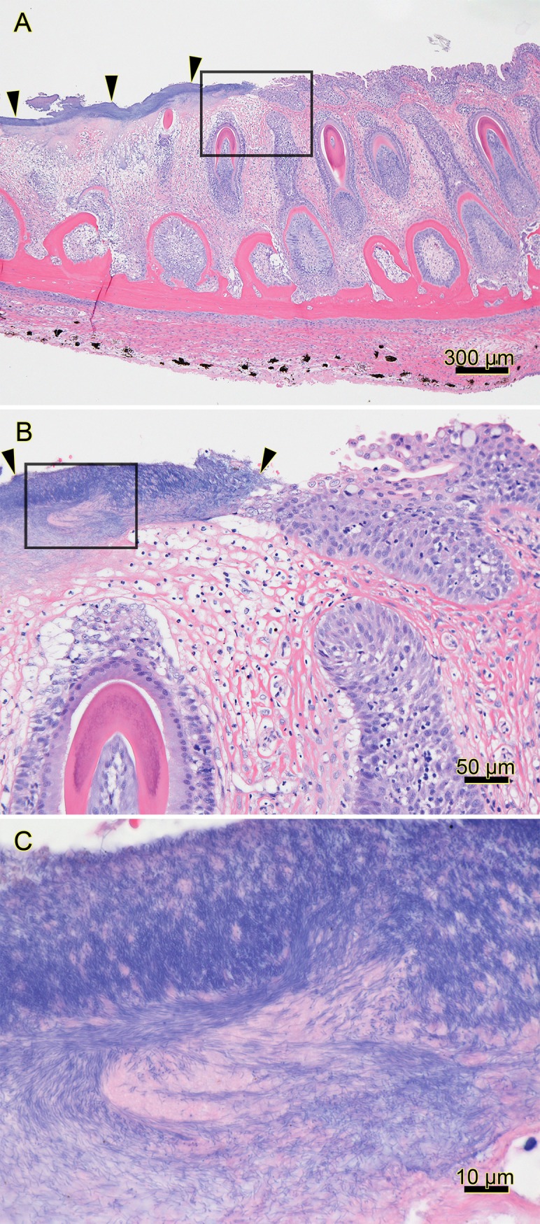
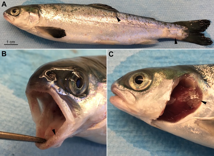
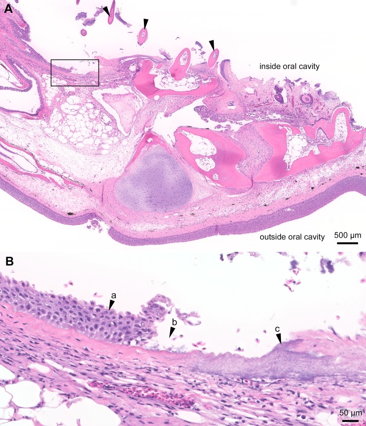
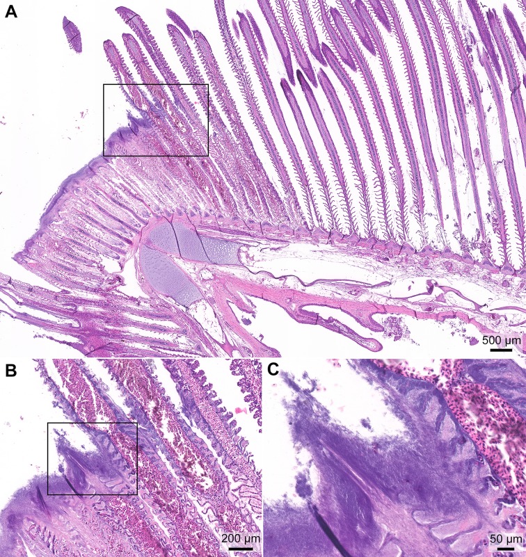
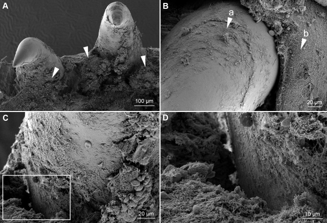
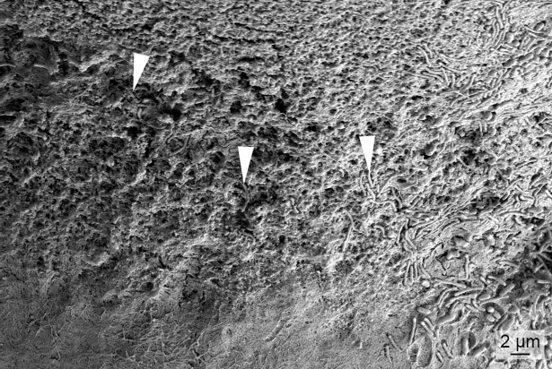
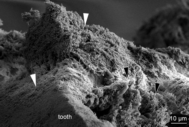
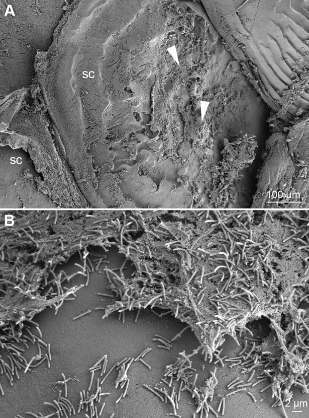
Mouthrot, caused by Tenacibaculum maritimum is a significant disease of farmed Atlantic salmon, Salmo salar on the West Coast of North America. Smolts recently transferred into saltwater are the most susceptible and affected fish die with little internal or external clinical signs other than the characteristic small (usually < 5 mm) yellow plaques that are present inside the mouth. The mechanism by which these smolts die is unknown. This study investigated the microscopic pathology (histology and scanning electron microscopy) of bath infected smolts with Western Canadian T. maritimum isolates TmarCan15-1, TmarCan16-1 and TmarCan16-5 and compared the findings to what is seen in a natural outbreak of mouthrot. A real-time RT-PCR assay based on the outer membrane protein A specific for T. maritimum was designed and used to investigate the tissue tropism of the bacteria. The results from this showed that T. maritimum is detectable internally by real-time RT-PCR. This combined with the fact that the bacteria can be isolated from the kidney suggests that T. maritimum becomes systemic. The pathology in the infected smolts is primarily mouth lesions, including damaged tissues surrounding the teeth; the disease is similar to periodontal disease in mammals. The pathological changes are focal, severe, and occur very rapidly with little associated inflammation. Skin lesions are more common in experimentally infected smolts than in natural outbreaks, but this could be an artefact of the challenge dose, handling and tank used during the experiments.


Conflict of interest statement

We have the following interests: Kathleen Frisch, Sverre Bang Småge, Henrik Duesund and Øyvind Jakobsen Brevik are employed by Cermaq Group AS, who planned and performed this study, and Renate Johansen is employed by Pharmaq Analytiq, who helped analyse the data. There are no patents, products in development or marketed products to declare. This does not alter our adherence to all the PLOS ONE policies on sharing data and materials.

Figures

Fig 1
Fig 1. Histopathology of the jaw of a smolt from a natural outbreak of mouthrot.
Histopathology of the jaw from a farmed Atlantic salmon that died 2 months after it was transferred from freshwater into a saltwater net-pen in BC; H&E stain. (A) The mucosal epithelium on the left side of the section is ulcerated and covered by a layer of deeply basophilic bacteria (arrowheads). The black box surrounds the transition from the bacteria-covered ulcer (left) to intact epithelium (right), and it outlines the area included in B. (B) Higher magnification of the transition between the ulcer covered by filamentous bacteria (arrowheads) and intact epithelium (right of right arrow); black box outlines the area included in C. (C) Higher magnification of abundant filamentous bacteria streaming in a proteinaceous matrix. (Optimization of photomicrograph illumination and color balance followed published methods [25]).
Fig 2
Fig 2. Gross clinical signs of an experimentally infected smolt.
A moribund Atlantic salmon smolt that was bath infected with T. maritimum strain TmarCan16-1. Gross lesion scoring [11]: mouth = 2 out of 3, skin = 1 out of 3, gills = 1 out of 2. (A) Very few clinical signs are on the body surface other than some scale loss at the base of the peduncle and dorsal-lateral surface (arrows). (B) The gingiva is swollen (arrow). (C) A gill lesion (arrow).
Fig 3
Fig 3. Histopathology of the jaw from an experimentally infected smolt.
Histopathology of the gills from a moribund Atlantic salmon smolt experimentally bath infected with T. maritimum strain TmarCan15-1; H&E stain. (A) Oblique section of the jaw with mouthrot and loose teeth (arrowheads) with only a few of them connected to the jaw. The top is the inside of the oral cavity and the bottom the outside. The epidermis on the outside is intact. The black box outlines the area included in B and represents the transition at the edge of the ulcer. (B) The distance between intact mucosal epithelium (arrow "a") and the ulcer (arrow "b") is very short. Large quantities of bacteria with T. maritimum morphology are within the ulcer (arrow "c"). No signs of inflammation at the edge of the ulcer.
Fig 4
Fig 4. Histopathology of the jaw from an experimentally infected smolt.
Histopathology of the jaw from the smolt in Fig 2; H&E stain. (A) Oblique section of the jaw. The epidermis is completely missing and the outer surface is covered with a thick mat of long thin rod-shaped T. maritimum-like bacteria that have infiltrated the submucosa (arrow "a"). Only one tooth (arrow "b") remains and holes are present where there used to be more teeth (arrow "c"). The black boxes labelled "B" and "C" outline the areas included in Fig 4B and 4C. (B) A mat of bacteria with T. maritimum morphology is on the outer surface (arrow "d") and the bacteria have infiltrated the underlying submucosa. (C) Large quantities of bacteria with T. maritimum morphology are within the destructed submucosa surrounding the tooth (arrow "e"). Some intact red blood cells (arrow "f") are within the mass of bacteria and remnants of tissue.
Fig 5
Fig 5. Histopathology of the gills from an experimentally infected smolt.
Histopathology of the gills from the smolt in Fig 2; H&E stain. (A) Section of the gills with a distinct lesion on the top of the curve of the gill arch. The tips of the filaments are missing in the center of the lesion, and the remaining distal end of the filament is necrotic. The tissue is replaced by a thick layer of bacteria with T. maritimum morphology. The black box includes the transition between the lesion and normal tissue and outlines the area included in B. (B) The distance between the lesion and normal gill filaments is very short. In the damaged area, only the blood vessels remain in some of the lamellae. The black box outlines the area included in C. (C) Abundant bacteria with T. maritimum morphology cover the destroyed region of the gills. Only remnants of the lamellae are within the ulcer.
Fig 6
Fig 6. SEM of teeth from an experimentally infected smolt.
Micrographs of teeth and the surrounding tissue from the mouth of a diseased smolt bath infected with TmarCan15-1 in the cohabitation experiment. (A) Teeth and surrounding gingiva are covered by mats of bacteria with T. maritimum morphology (arrowheads) and the associated tissue is damaged. (B) Zoomed in view of a tooth showing bacterial growth on the surface of the tooth (arrow "a") as well as the surrounding gingival tissue (arrow "b"). (C) The dentin-enameloid interface with associated tissue destruction. White box indicates area in D. (D) Cellular debris within the bacterial mats.
Fig 7
Fig 7. SEM of a tooth surface from an experimentally infected smolt.
Micrograph of a tooth surface from a diseased smolt bath infected with TmarCan15-1 in the cohabitation experiment. Bacteria with T. maritimum morphology are within the enameloid of the tooth (arrowheads).
Fig 8
Fig 8. SEM of a fractured tooth from an experimentally infected smolt.
Micrograph of a fractured tooth from a diseased smolt bath infected with TmarCan15-1 in the cohabitation experiment. Large aggregates of bacteria with T. maritimum morphology are on the outside of the tooth (white arrowheads) as well as within the exposed pulp (black arrowheads) of the tooth.
Fig 9
Fig 9. SEM of a skin lesion from an experimentally infected smolt.
Micrographs of a skin lesion on the dorsal-lateral surface of a diseased smolt bath infected with TmarCan15-1 in the cohabitation experiment. (A) Mats of bacteria with T. maritimum morphology (arrowheads) are associated with epithelial damage exposing the scales (sc). (B) Cellular debris with aggregates of bacteria with T. maritimum morphology.

References

    1. Toranzo AE, Magariños B, Romalde JL. A review of the main bacterial fish diseases in mariculture systems. Aquaculture. 2005;246: 37–61. 10.1016/j.aquaculture.2005.01.002 - DOI
    1. Avendaño-Herrera R, Toranzo AE, Magariños B. Tenacibaculosis infection in marine fish caused by Tenacibaculum maritimum: a review. Diseases of Aquatic Organisms. 2006;71: 255–266. 10.3354/dao071255 - DOI - PubMed
    1. Avendaño-Herrera R, Irgang R, Sandoval C, Moreno-Lira P, Houel A, Duchaud E, et al. Isolation, characterization and virulence potential of Tenacibaculum dicentrarchi in salmonid cultures in Chile. Transboundary and Emerging Diseases. 2016;63: 121–126. 10.1111/tbed.12464 - DOI - PubMed
    1. Småge SB, Brevik ØJ, Duesund H, Ottem KF, Watanabe K, Nylund A. Tenacibaculum finnmarkense sp. nov., a fish pathogenic bacterium of the family Flavobacteriaceae isolated from Atlantic salmon. Antonie van Leeuwenhoek. 2015;109: 273–285. 10.1007/s10482-015-0630-0 - DOI - PMC - PubMed
    1. Småge SB, Brevik ØJ, Frisch K, Watanabe K, Duesund H, Nylund A. Concurrent jellyfish blooms and tenacibaculosis outbreaks in Northern Norwegian Atlantic salmon (Salmo salar) farms. PLOS ONE. 2017;12: e0187476–21. 10.1371/journal.pone.0187476 - DOI - PMC - PubMed

Publication types