. 2019 Jan 2:259:46-53.
doi: 10.1016/j.virusres.2018.10.016. Epub 2018 Oct 30.

Identification of gammaherpesvirus infection in free-ranging black bears (Ursus americanus)

Affiliations

Identification of gammaherpesvirus infection in free-ranging black bears (Ursus americanus)

Wendy Black et al. Virus Res. .

Abstract

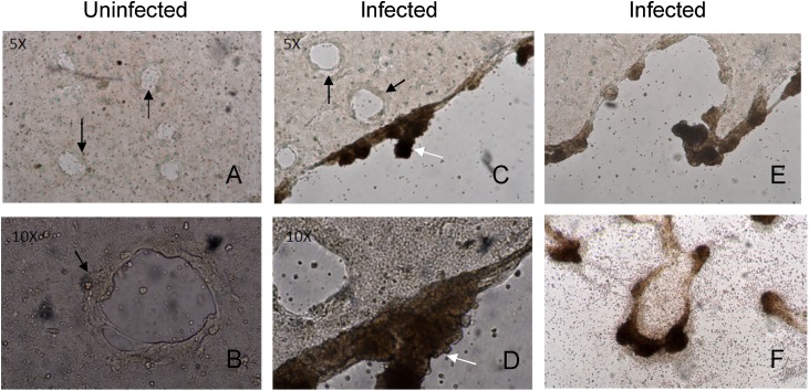
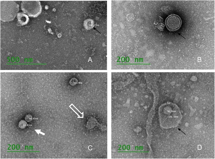
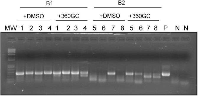
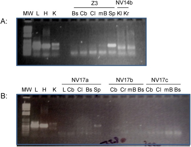
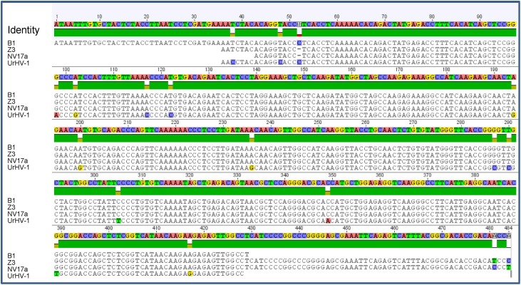
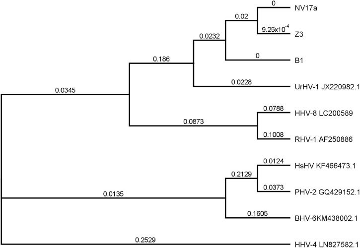
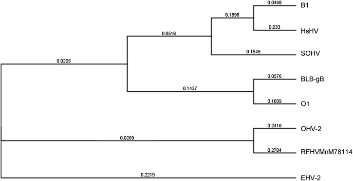
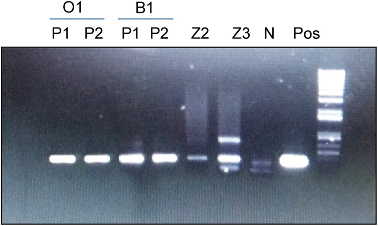
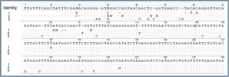
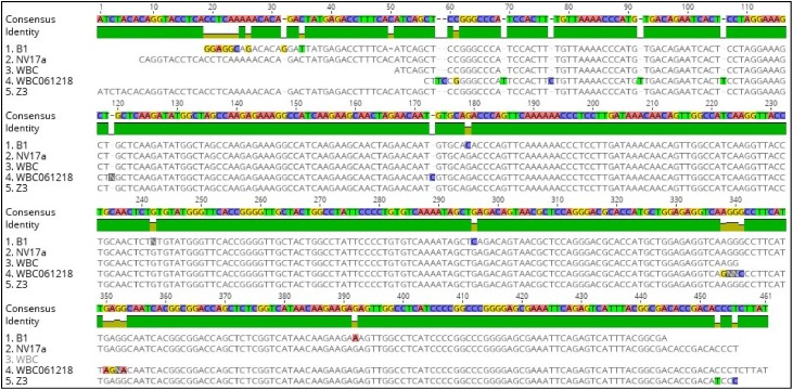
Herpesvirus infection was investigated in black bears (Ursus americanus) with neurological signs and brain lesions of nonsuppurative encephalitis of unknown cause. Visible cytopathic effects (CPE) could only be observed on days 3-5 post-infection in HrT-18G cell line inoculated with bear tissue extracts. The observed CPE in HrT-18G cells included syncytia, intranuclear inclusions, and cell detachments seen in herpesvirus infection in vitro. Herpesvirus-like particles were observed in viral culture supernatant under the electron microscope, however, capsids ranging from 60 nm to 100 nm in size were often observed in viral cultures within the first two passages of propagation. Herpesvirus infection in the bear tissues and tissue cultures were detected by PCR using degenerate primers specific to the DNA polymerase gene (DPOL) and glycoprotein B gene (gB). DNA sequencing of the amplicon revealed that the detected herpesvirus has 94-95% identity to Ursid gammaherpesvirus 1 (UrHV-1) DNA sequences of DPOL. Phylogenetic analysis of DPOL sequences indicates that black bear herpesviruses and UrHV-1 are closely related and have small distances to members of Rhadinovirus. Interestingly, black bear herpesvirus infections were also found in bears without neurological signs. The DPOL DNA sequence of black bear herpesviruses detected in neurological bears were similar to the those detected in the non-neurological bears. However, the gB DNA sequence detected from the neurological bear is different from non-neurological bear and has only 64.5%-70% identity to each other. It is possible that at least two different types of gammaherpesviruses are present in the U. americanus population or several gammaherpesviruses exist in ursine species.

Keywords: Black bears; DNA sequencing; Degenerative PCR; PCR; UrHV-1; Virus isolation.


Figures

Fig. 1
Fig. 1
Light microscope images of HrT18G cells with or without exposure to bear tissue extracts. A (5X) & B (10X): mock infected HrT-18G cells at day 4 post-infection. C (5X) &D (10X): HrT-18G cells infected with B1 bear tissue extracts at day 4 post-infection. E &F: HrT-18G cells exposed to O1 tissue extract at day 4 post-infection (E), and at day 8 post-infection (F).
Fig. 2
Fig. 2
Electron micrograph images of virions purified from infected HrT-18G cell culture supernatant. A: Purified virions from tissue isolation with B1 bear showing neurological signs; B: Negative staining of purified virions from tissue culture infected with pooled tissues from O1 bear (B), without neurological signs. C and D: Negative staining of purified virions from tissue culture infected with fecal samples of CA bear Z3 and OR bear 1, without neurological signs. The black arrow indicates the herpesvirus like virion. The open arrow indicates coronavirus like virion. The solid white arrow indicates capsids with sizes between 40-60nm: Scale bar = 0.2 μm or 0.5 μm.
Fig. 3
Fig. 3
Panherpesvirus PCR analysis of tissue total DNA isolated from B1 and B2 bears diagnosed with neurological diseases. The PCR reaction was performed similarly as described in Supplemental Fig. 1 with either DMSO or 360 GC Enhancer. The predicted product is >400 bp in size. Template DNA was as follows: 1&5: brain, 2&6: kidney; 3&7: liver; 4&8: lymph node; P: HHV-1; MW: 1 kb-plus DNA ladder (Invitrogen).
Fig. 4
Fig. 4
Panherpesvirus PCR analysis of tissue total DNA isolated from bears without clinical signs. The reaction was performed as described in Supplemental Fig. 1 with 360 GC Enhancer included in the PCR. A: Z3 is bear from California, NV14b: bear collected in 2015 from Nevada. B: NV17a-c: bears collected in 2017 from Nevada. The abbreviations of tissues are as follows: Bs: Brain stem, Cb: cerebrum, Cl: cerebellum, mB: mid-brain, Sp: spleen, Kl: left kidney, Kr: right kidney, L: liver; Pos: L: LHV-4, H: HHV-1, K: KHV; MW: 1 kb-plus DNA ladder.
Fig. 5
Fig. 5
DNA sequence alignments of amplicons from panherpesvirus PCR analysis with UrHV-1 DNA sequence available in the Genbank. PCR amplicons are produced by panherpesvirus PCR using primers POLdegF1 and POLdegR1 in the first round of reaction, POLdegF2 and POLdegR2 in second round of reaction. B1, Z3, NV17a: PCR products amplified from total DNA from lymph node of bear B1, spleen of bear Z3, spleen od bear NV17a, respectively. UrHV-1: DNA sequence detected in the sun bear (accession number JX220982). Green bar stands for identical sequences.
Fig. 6
Fig. 6
Phylogenetic analysis of the amplicons from panherpesvirus PCR. Phylogenetic tree of the DNA sequences of the DNA polymerase subunit. The scale bar represents genetic distance (nucleotide substitutions per site), and branch lengths are given above the branches. The abbreviations in the virus names are as follows: HHV-4: human herpesvirus type; BHV-6: bovine gammaherpesvirus type 6; CPV-2: Caprine herpesvirus 2; HsHV: Harp seal herpesvirus isolate; PHV-2: Phocid herpesvirus 2; EHV-2: Equid gammaherpesvirus 2; HHV-8: human herpesvirus type 8; RHV-1: Gorilla rhadinovirus 1 isolate; UrHV-1: Ursid gammaherpesvirus 1; B1: amplicon from Nevada bear B1lymph node; NV17a: amplicon from Nevada bear NV17a spleen; Z3: amplicon from California bear Z3 spleen. The accession numbers used for DNA polymerase coding sequence analysis were given following each virus name.
Fig. 7
Fig. 7
Phylogenetic tree of the gB DNA sequences. The scale bar represents genetic distance (nucleotide substitutions per site), and branch lengths are given above the branches. The abbreviations in the virus names are as follows: HsHV: Harp seal herpesvirus isolate FMV04-1493874 (KP136799); SOHV: Sea otter herpesvirus (KX024493.1); OHV-2: Ovine herpesvirus 2 (AF385442.1); RFHVMnM78114: the Macaque Homolog of Kaposi's Sarcoma (KS)-Associated Herpesvirus (KF703446); EHV-2: Equid herpesvirus 2 (HQ247756.1); BLB-gB (MK089801) : gB DNA amplified from non-neurological black bear. B1: gB amplicon from Nevada bear B1 lymph node; O1: gB amplicon from Oregon bear spleen.
Fig. 8
Fig. 8
Detection of UrHV-1 like DNA using UrHV-1 specific primers. Total DNA samples were analyzed by PCR using UrHV-1 specific primers UG341F and UG341R. The abbreviations of tissues are as follows: P: total DNA of lymph node from bear B1; N: total DNA of HrT-18G cells; Z2: total DNA of spleen from Z2; Z3: total DNA of spleen from Z3; P1: total DNA of viral culture passage 1, P2: total DNA of viral culture passage 2. O1: Oregon bear without neurological signs, B1: Nevada bear with neurological signs. MW, 1 kb-plus DNA ladder.
Fig. 9
Fig. 9
DNA sequence alignments of UG341 PCR product with UrHV-1 sequence detected in the sun bear (accession number JX220982). 1: amplicon of P2 from Nevada bear B1 virus isolation shown in Fig. 8. 2: amplicon of P2 from Oregon bear O1 virus isolation shown in Fig. 8. 3: JX220982, UrHV-1 detected in sun bear.
Fig. 10
Fig. 10
DNA sequence alignments of UG341 PCR product amplified from white blood cell (WBC) total DNA. 1: amplicon of lymph node total DNA from bear B1; 2: amplicon of spleen total DNA from bear NV17a. 3: amplicon of WBC total DNA from a new Oregon bear submitted to OVDL in 2018. 4: amplicon of WBC total DNA from a new Nevada bear submitted to OVDL in 2018. 5: amplicon of spleen total DNA from bear Z3.

References

    1. Ali K., Lawthom C. Epstein-Barr virus-associated cerebellar ataxia. BMJ Case Rep. 2013;2013 - PMC - PubMed
    1. Altschul S.F., Gish W., Miller W., Myers E.W., Lipman D.J. Basic local alignment search tool. J. Mol. Biol. 1990;215:403–410. - PubMed
    1. Axthelm M.K., Bourdette D.N., Marracci G.H., Su W., Mullaney E.T., Manoharan M., Kohama S.G., Pollaro J., Witkowski E., Wang P., Rooney W.D., Sherman L.S., Wong S.W. Japanese macaque encephalomyelitis: a spontaneous multiple sclerosis-like disease in a nonhuman primate. Annals Neurol. 2011;70:362–373. - PMC - PubMed
    1. Brenner J., Perl S., Lahav D., Garazi S., Oved Z., Shlosberg A., David D. An unusual outbreak of malignant catarrhal fever in a beef herd in Israel. J. Vet. Med. B Infect. Dis. Vet. Public Health. 2002;49:304–307. - PubMed
    1. Cho H.J., Song M.J. A gammaherpesvirus establishes persistent infection in neuroblastoma cells. Mol. Cells. 2014;37:518–525. - PMC - PubMed

Publication types