

Interaction of Developmental Venous Anomalies with Resting-State Functional MRI Measures
- PMID: 30385467
- PMCID: PMC6476411
- DOI: 10.3174/ajnr.A5847
Interaction of Developmental Venous Anomalies with Resting-State Functional MRI Measures
Abstract
Background and purpose: Functional MR imaging of the brain, used for both clinical and neuroscientific applications, relies on measuring fluctuations in blood oxygenation. Such measurements are susceptible to noise of vascular origin. The purpose of this study was to assess whether developmental venous anomalies, which are frequently observed normal variants, can bias fMRI measures by appearing as true neural signal.
Materials and methods: Large developmental venous anomalies (1 in each of 14 participants) were identified from a large neuroimaging cohort (n = 814). Resting-state fMRI data were decomposed using independent component analysis, a data-driven technique that creates distinct component maps representing aspects of either structured noise or true neural activity. We searched all independent components for maps that exhibited a spatial distribution of their signals following the topography of developmental venous anomalies.
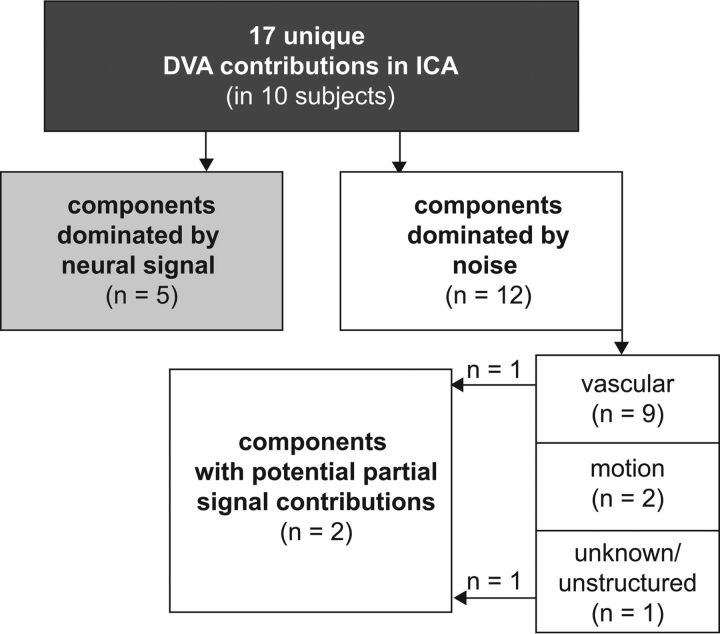
Results: Of the 14 developmental venous anomalies identified, 10 were clearly present in 17 fMRI independent components in total. While 9 (52.9%) of these 17 independent components were dominated by venous contributions and 2 (11.8%) by motion artifacts, 2 independent components (11.8%) showed partial neural signal contributions and 5 independent components (29.4%) unambiguously exhibited typical neural signal patterns.
Conclusions: Developmental venous anomalies can strongly resemble neural signal as measured by fMRI. They are thus a potential source of bias in fMRI analyses, especially when present in the cortex. This could impede interpretation of local activity in patients, such as in presurgical mapping. In scientific studies with large samples, developmental venous anomaly confounds could be mainly addressed using independent component analysis-based denoising.
© 2018 by American Journal of Neuroradiology.
Figures


References
Publication types
MeSH terms
Grants and funding
LinkOut - more resources
Full Text Sources
Medical