

MR Imaging for Differentiating Contrast Staining from Hemorrhagic Transformation after Endovascular Thrombectomy in Acute Ischemic Stroke: Phantom and Patient Study
- PMID: 30385471
- PMCID: PMC7655397
- DOI: 10.3174/ajnr.A5848
MR Imaging for Differentiating Contrast Staining from Hemorrhagic Transformation after Endovascular Thrombectomy in Acute Ischemic Stroke: Phantom and Patient Study
Abstract
Background and purpose: Early differentiation of contrast staining from hemorrhagic transformation in patients with acute ischemic stroke who have undergone endovascular treatment is critical in preventing the delayed administration of antiplatelet agents. We aimed to demonstrate the usefulness of an immediate postinterventional DWI protocol including B0 and gradient recalled-echo sequences to discriminate those 2 conditions through phantom and preliminary retrospective patient studies.
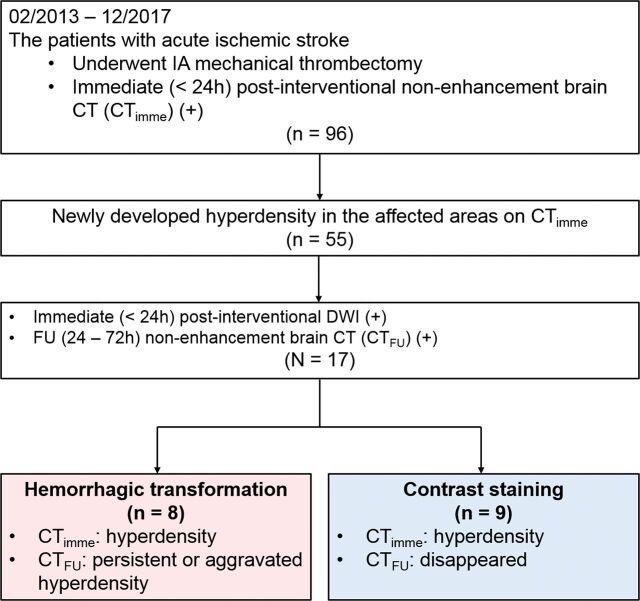
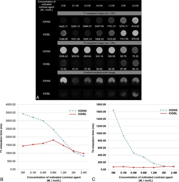
Materials and methods: On 3T MR imaging, the signal intensities of the phantom models consisting of iodinated contrast agents diluted with normal saline and arterial blood were compared using T1WI, T2WI, and gradient recalled-echo sequences. A total 17 patients (8 with hemorrhagic transformation and 9 with contrast staining; 8 men and 9 women; mean age, 72.00 ± 10.91 years; range, 52-90 years) who underwent mechanical thrombectomy for acute ischemic stroke and showed newly appearing hyperdense lesions on immediate (<24 hours) postinterventional nonenhanced CT scans were included in this study. Immediate postinterventional DWI of patients were compared.
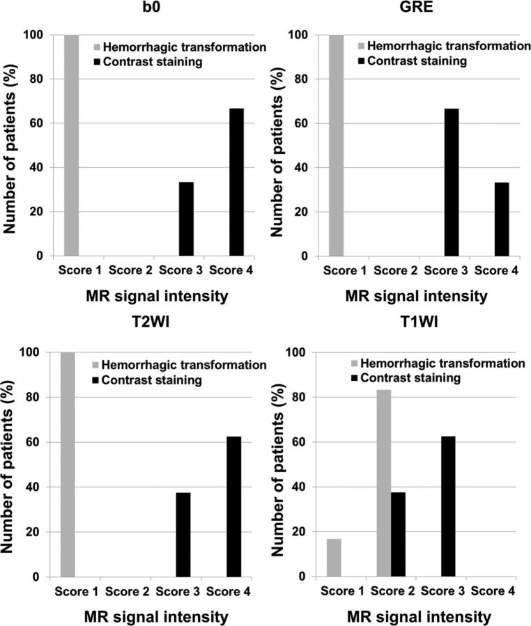
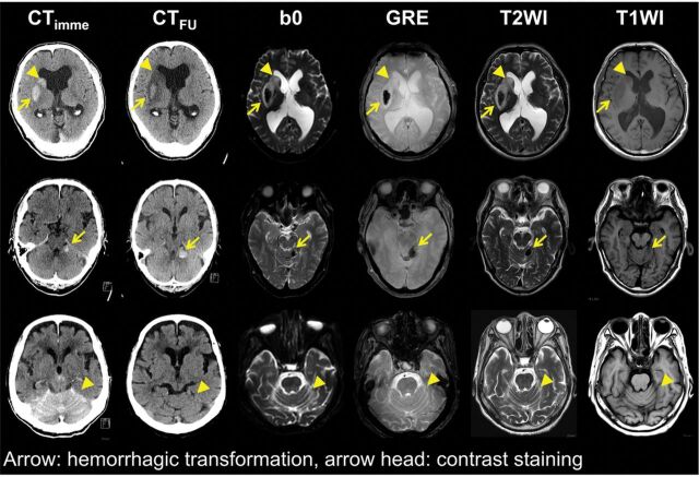
Results: In the phantom study, iodinated contrast agents diluted with normal saline showed minimal signal drop, while those diluted with arterial blood demonstrated dark signal intensity in the T2WI and gradient recalled-echo sequences. In the patient study, all hemorrhagic transformations and none of the contrast staining demonstrated dark or low signal (<gray matter) intensities similar to those of the vessel in the B0-DWI and gradient recalled-echo images.
Conclusions: According to our preliminary results, contrast staining might be differentiated from hemorrhagic transformation using an immediate postinterventional DWI protocol including gradient recalled-echo sequences. It might be possible to expedite establishment of postinterventional medical treatment strategy.
© 2018 by American Journal of Neuroradiology.
Figures
Similar articles
-
Immediate postinterventional flat-panel CT: Differentiation of hemorrhagic transformation from contrast exudation of acute ischemic stroke patients after thrombectomy.Acta Radiol. 2023 Apr;64(4):1600-1607. doi: 10.1177/02841851221122429. Epub 2022 Aug 28. Acta Radiol. 2023. PMID: 36036263
-
A Novel Dual-Energy CT Method for Detection and Differentiation of Intracerebral Hemorrhage From Contrast Extravasation in Stroke Patients After Endovascular Thrombectomy : Feasibility and First Results.Clin Neuroradiol. 2023 Mar;33(1):171-177. doi: 10.1007/s00062-022-01198-3. Epub 2022 Aug 12. Clin Neuroradiol. 2023. PMID: 35960327 Free PMC article.
-
Flat Panel CT Scanning Is Helpful in Predicting Hemorrhagic Transformation in Acute Ischemic Stroke Patients Undergoing Endovascular Thrombectomy.Biomed Res Int. 2021 Apr 13;2021:5527101. doi: 10.1155/2021/5527101. eCollection 2021. Biomed Res Int. 2021. Retraction in: Biomed Res Int. 2024 Mar 20;2024:9823857. doi: 10.1155/2024/9823857. PMID: 33954174 Free PMC article. Retracted.
-
CT Hyperdense Lesions after Endovascular Therapy in Acute Ischemic Stroke: Imaging Findings and Clinical Significance.Cerebrovasc Dis. 2024;53(5):607-617. doi: 10.1159/000535369. Epub 2023 Nov 21. Cerebrovasc Dis. 2024. PMID: 37989121 Review.
-
Acute ischemic stroke patients with diffusion-weighted imaging-Alberta Stroke Program Early Computed Tomography Score ≤ 5 can benefit from endovascular treatment: a single-center experience and literature review.Neuroradiology. 2019 Apr;61(4):451-459. doi: 10.1007/s00234-019-02177-1. Epub 2019 Feb 6. Neuroradiology. 2019. PMID: 30725121 Free PMC article. Review.
Cited by
-
Contrast extravasation mimicking intracerebral and intraventricular hemorrhage after intravenous thrombolytic treatment of ischemic stroke: a case report.BMC Neurol. 2024 Apr 19;24(1):134. doi: 10.1186/s12883-024-03618-y. BMC Neurol. 2024. PMID: 38641592 Free PMC article.
-
Development of estimation method for T1 and T2 values using the relaxivity of contrast agent and coagulant for a magnetic resonance imaging phantom.Radiol Phys Technol. 2025 Jun;18(2):469-476. doi: 10.1007/s12194-025-00900-7. Epub 2025 Mar 23. Radiol Phys Technol. 2025. PMID: 40122940
-
Contrast Extravasation Post Thrombectomy in Patients With Acute Cerebral Stroke: A Review and Recommendations for Future Studies.Cureus. 2020 Sep 23;12(9):e10616. doi: 10.7759/cureus.10616. Cureus. 2020. PMID: 33123430 Free PMC article. Review.
-
Comparison of diffusion weighted imaging b0 with T2*-weighted gradient echo or susceptibility weighted imaging for intracranial hemorrhage detection after reperfusion therapy for ischemic stroke.Neuroradiology. 2023 Nov;65(11):1649-1655. doi: 10.1007/s00234-023-03180-3. Epub 2023 Jun 29. Neuroradiology. 2023. PMID: 37380891 Free PMC article.
-
Intracranial Bleeding After Reperfusion Therapy in Acute Ischemic Stroke.Front Neurol. 2021 Feb 9;11:629920. doi: 10.3389/fneur.2020.629920. eCollection 2020. Front Neurol. 2021. PMID: 33633661 Free PMC article. Review.
References
MeSH terms
LinkOut - more resources
Full Text Sources
Medical