. 2018 Nov 2;362(6414):eaat0572.
doi: 10.1126/science.aat0572.

Defining the human C2H2 zinc finger degrome targeted by thalidomide analogs through CRBN

Affiliations

Defining the human C2H2 zinc finger degrome targeted by thalidomide analogs through CRBN

Quinlan L Sievers et al. Science. .

Abstract

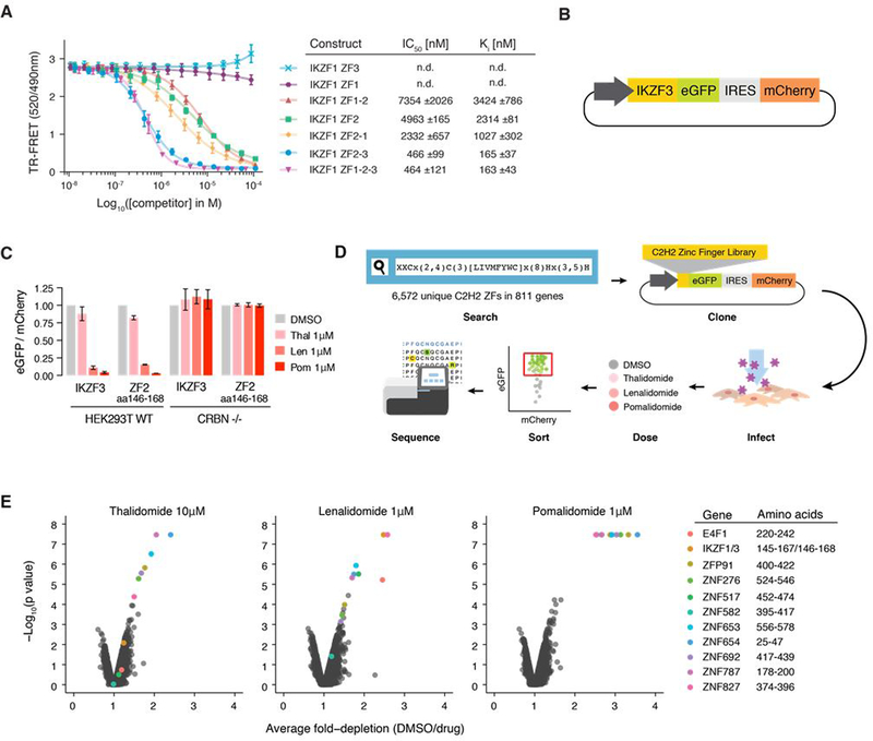
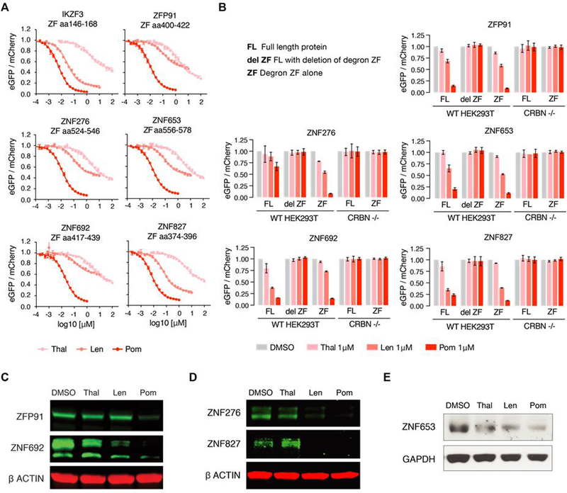
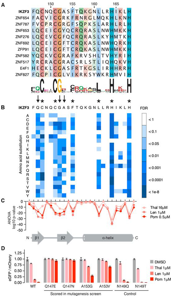
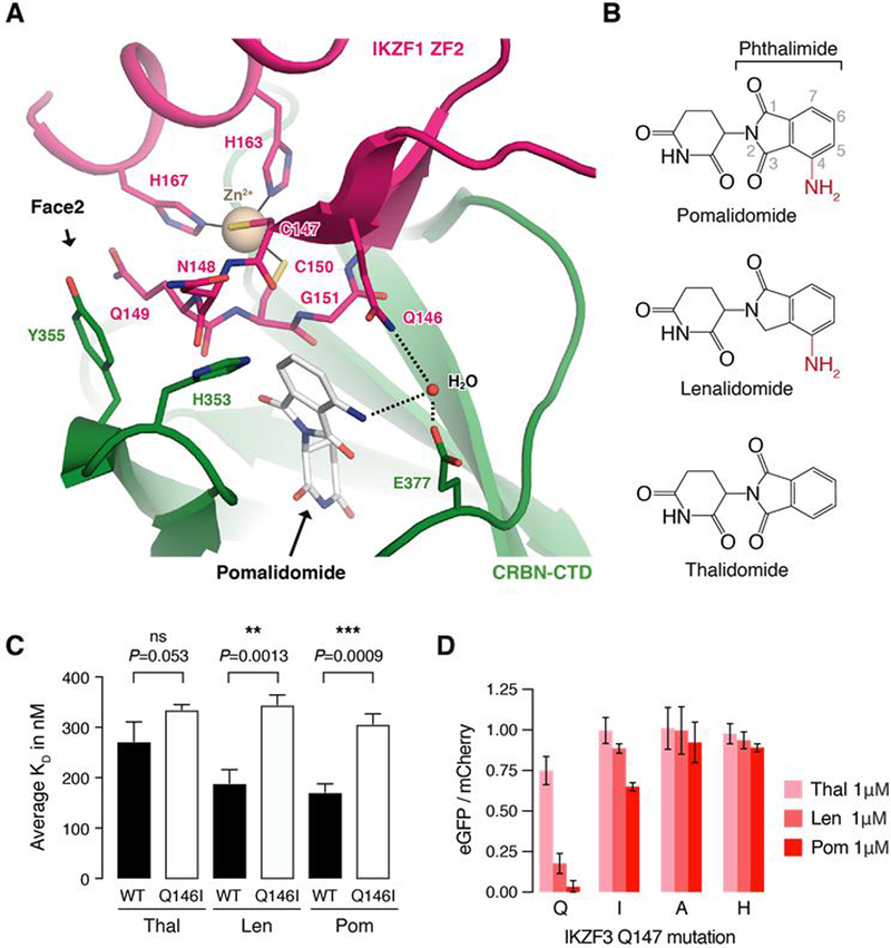
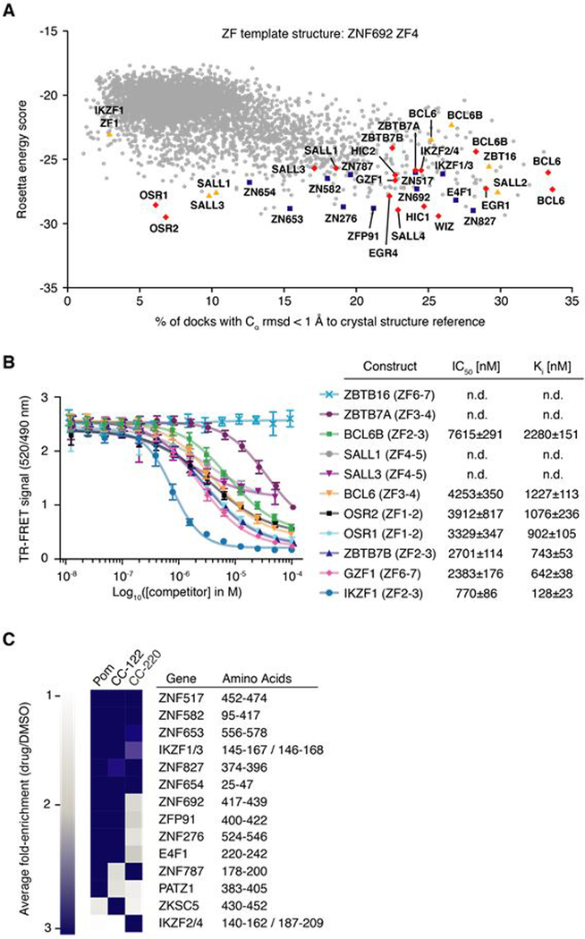
The small molecules thalidomide, lenalidomide, and pomalidomide induce the ubiquitination and proteasomal degradation of the transcription factors Ikaros (IKZF1) and Aiolos (IKZF3) by recruiting a Cys2-His2 (C2H2) zinc finger domain to Cereblon (CRBN), the substrate receptor of the CRL4CRBN E3 ubiquitin ligase. We screened the human C2H2 zinc finger proteome for degradation in the presence of thalidomide analogs, identifying 11 zinc finger degrons. Structural and functional characterization of the C2H2 zinc finger degrons demonstrates how diverse zinc finger domains bind the permissive drug-CRBN interface. Computational zinc finger docking and biochemical analysis predict that more than 150 zinc fingers bind the drug-CRBN complex in vitro, and we show that selective zinc finger degradation can be achieved through compound modifications. Our results provide a rationale for therapeutically targeting transcription factors that were previously considered undruggable.


Conflict of interest statement

Competing interests: Authors declare no competing interests.

Figures

Fig. 1.
Fig. 1.. C2H2 ZF library screen identifies 11 C2H2 ZFs degraded in the presence of thalidomide, lenalidomide, or pomalidomide.
(A) TR-FRET counter titration. Unlabeled IKZF1 constructs (0.01–100 μM) titrated to preassembled Alexa488DDB1ΔB-CRBN-pomalidomide-biotinIKZF1ZF1−2−3 (200 nM Alexa488DDB1ΔB-CRBN, 100nM biotinIKZF1ZF1−2−3, 5μM pomalidomide). (B) Schematic of the protein degradation reporter vector (IRES: Internal ribosome entry site). (C) HEK293T WT and CRBN−/− cells expressing IKZF3 constructs in the degradation reporter were treated for 20 hours with DMSO or drug then analyzed by flow cytometry to quantify the DMSO-normalized ratio of eGFP/mCherry fluorescence (exp. rep. = 3, tech. rep. = 3, bar heights indicate mean of experimental replicates, error bars indicate 95% CI). (D) Schematic of the human C2H2 ZF library screen. (E) Average fold-depletion of sequencing read counts (DMSO/drug) and corresponding p values (empirical rank-sum test-statistic) for the 5,611 C2H2 ZFs with raw read count >200 in all three control replicates (exp. rep. = 3, labeled data points possess FDR<0.01 in at least one of the three drugs).
Fig. 2.
Fig. 2.. Six proteins are degraded by thalidomide, lenalidomide, or pomalidomide conditional on the presence of a C2H2 ZF degron.
(A) HEK239T WT and CRBN−/− cells expressing the 11 ZFs identified in the screen in the degradation reporter were treated for 20 hours with drug then analyzed by flow cytometry to measure the DMSO-normalized ratio of eGFP/mCherry fluorescence (exp. rep. = 3, tech. rep. =3, dots represent average of experimental replicates, error bars indicate range). ZFs not shown are in fig S2A. (B) HEK293T WT or CRBN−/− cells expressing full-length (FL), FL with ZF deleted (del ZF), or ZF alone cDNA constructs were treated for 20 hours with DMSO or 1μM drug then analyzed using flow cytometry to measure the DMSO-normalized ratio of eGFP/mCherry fluorescence (exp. rep. = 3, tech. rep. = 3, bar heights indicate mean of exp. rep., error bars indicate 95% CI). Proteins not shown are in fig. S2B. (C) KG1 (human acute myeloid leukemia), (D) WM266–4 (human melanoma), and (E) MOLM-16 cells (human acute myeloid leukemia) were treated with DMSO or 1μM drug for 20 or 24 hours after which protein lysates were harvested, run on a polyacrylamide gel, and immunoblotted for the specified targets (images representative of 3 experimental replicates).
Fig. 3.
Fig. 3.. Identification of amino acids required for drug-induced degradation of a C2H2 ZF degron.
(A) Sequence alignment of the 11 C2H2 ZFs with FDR<0.01 in at least one drug condition (amino acids colored by property). (B) Saturation mutagenesis screen of IKZF3 aa 130–189 in presence of lenalidomide displayed as heat map of the FDR for mutant amino acids (unpaired, one-sided t-test, FDR correction performed within each column, tech. rep. = 3). Asterisks indicate amino acids required for the ZF fold and arrows indicate non-structural IKZF3 residues required for degradation. Complete results for all 60 amino acids are located in fig. S3C (C) ANOVA p values for difference in frequency of mutant amino acids at each position in IKZF3 aa 130–189 (DMSO vs drug). Complete results for all 60 aa are located in fig. S3D. (D) HEK293T cells expressing IKZF3 ZF2 constructs in the degradation reporter were treated for 20 hours with DMSO or 1μM drug after which flow cytometry was used to measure the DMSO-normalized ratio of eGFP/mCherry fluorescence (exp. rep. = 1, tech. rep. = 3, bar height is the average of tech. rep, error bars denote 95% CI).
Fig. 4.
Fig. 4.. The pomalidomide-CRBN interface accommodates C2H2 ZF degrons with distinct amino acid sequences and properties.
(A) Crystal structure of DDB1ΔB-CRBNΔN40 bound to pomalidomide and IKZF1ZF2. Zinc ions are shown as beige spheres. (B) Crystal structure of DDB1ΔB-CRBNΔN40 bound to pomalidomide and ZNF692ZF4. CTD, carboxy-terminal domain; HBD, helical bundle domain; NTD, amino-terminal domain. BPA, BPC: β-propeller A and B. (C) Superposition of the CRBN-CTDs bound to IKZF1ZF2 and ZNF692ZF4. A dashed line indicates the complementary groove. (D) Side chain interactions between IKZF1ZF2, CRBN and pomalidomide (dashed lines indicate hydrogen bonds) and sequence alignment of IKZF1ZF2 and ZNF692ZF4 (amino acids colored by property). Green dots or open circles indicate side chain interactions with CRBN or pomalidomide, respectively. A black line indicates ZF residues involved in backbone interactions with CRBN and/or pomalidomide. (E) Superposition of both CRBN-CTDs shows differences in IKZF1/ZNF692 ZF orientation.
Fig. 5.
Fig. 5.. IKZF3 Q147 interacts with the amino group on pomalidomide.
(A) Side chain interactions between IKZF1 ZF2, CRBN, and pomalidomide. Dashed lines indicate hydrogen bonds. (B) Chemical structures of pomalidomide, lenalidomide, and thalidomide with differences highlighted in red. (C) Inhibitory constants (Ki) for wild-type (WT) and Q146I mutant IKZF1ZF2-ZF3 in the presence of thalidomide, lenalidomide, and pomalidomide in TR-FRET counter titration experiments. (D) HEK293T cells expressing IKZF3 ZF2 Q147 mutants in the degradation reporter were treated for 20 hours with DMSO or drug and then analyzed by flow cytometry to measure the DMSO-normalized ratio of eGFP/mCherry fluorescence (exp. rep. = 3, tech. rep. = 3, bar height is average of exp. rep, error bars indicate 95% CI). The complete set of Q147 amino acid mutants is located in fig. S5D.
Fig. 6.
Fig. 6.. Thalidomide analogs with chemical modifications at the ZF-interface target different sets of C2H2 ZF degrons.
(A) ZF library docking results using RosettaDock with the CRBN C-terminal domain bound to pomalidomide and ZNF692ZF4 as a crystal structure reference. Blue squares indicate degraded ZFs from the library screen, red diamonds highlight ZFs that bind pomalidomide-CRBN in vitro, and yellow triangles mark ZFs for which CRBN-binding could not be confirmed in vitro or in vivo (B) TR-FRET counter titration. Unlabeled dual ZF candidate constructs (0.01–100 μM) titrated to preassembled Alexa488DDB1ΔB-CRBN-pomalidomide-biotinIKZF1ZF1−2−3 (500 nM Alexa488DDB1ΔB-CRBN, 50 nM biotinIKZF1ZF1−2−3, 5 μM pomalidomide). (C) Average fold-enrichment of sequencing read counts (drug/DMSO) for the 11 previously identified ZF degrons and 3 novel ZF hits.

Comment in

References

    1. Fink EC, Ebert BL, The novel mechanism of lenalidomide activity. Blood. 126, 2366–2369 (2015). - PMC - PubMed
    1. Lu G et al., The Myeloma Drug Lenalidomide Promotes the Cereblon-Dependent Destruction of Ikaros Proteins. Science. 343, 305–309 (2014). - PMC - PubMed
    1. Krönke J et al., Lenalidomide Causes Selective Degradation of IKZF1 and IKZF3 in Multiple Myeloma Cells. Science. 343, 301–305 (2014). - PMC - PubMed
    1. Krönke J et al., Lenalidomide induces ubiquitination and degradation of CK1α in del(5q) MDS. Nature. 523, 183–188 (2015). - PMC - PubMed
    1. Matyskiela ME et al., A novel cereblon modulator recruits GSPT1 to the CRL4CRBN ubiquitin ligase. Nature, 1–24 (2016). - PubMed

Publication types

MeSH terms