. 2018 Nov 20;115(47):E11138-E11147.
doi: 10.1073/pnas.1801156115. Epub 2018 Nov 1.

Vorinostat, a pan-HDAC inhibitor, abrogates productive HPV-18 DNA amplification

Affiliations

Vorinostat, a pan-HDAC inhibitor, abrogates productive HPV-18 DNA amplification

N Sanjib Banerjee et al. Proc Natl Acad Sci U S A. .

Abstract

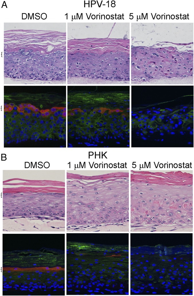
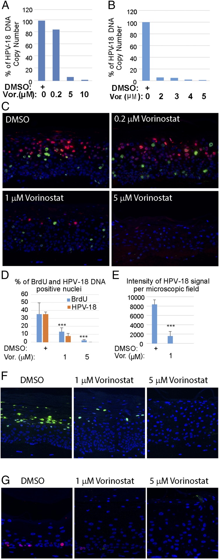
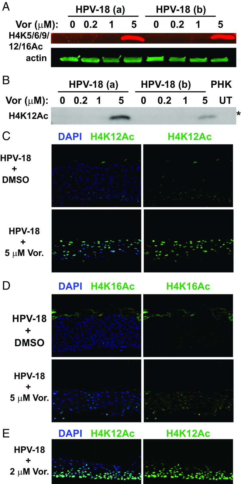
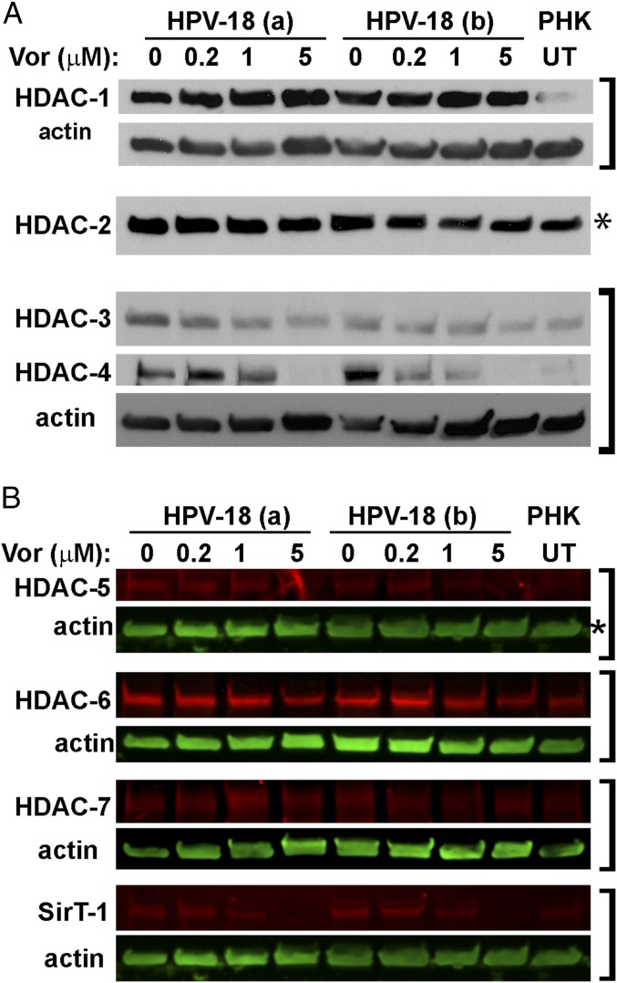
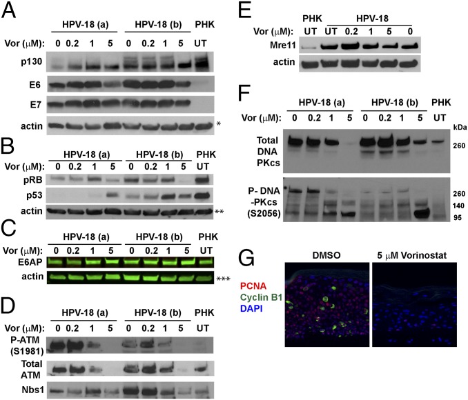
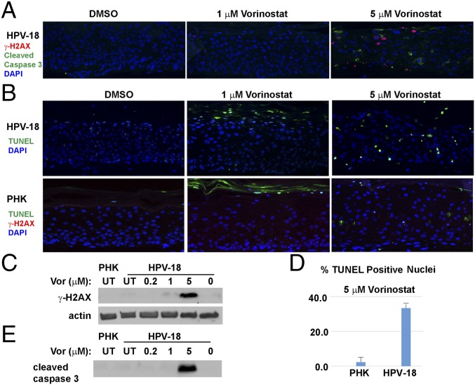
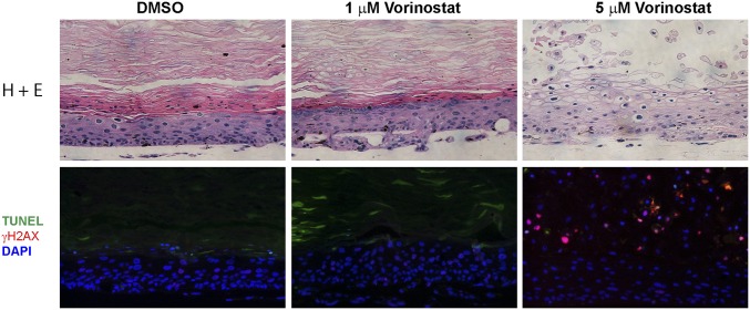
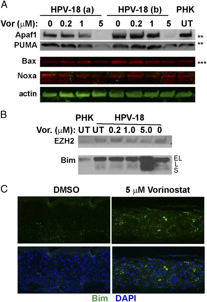
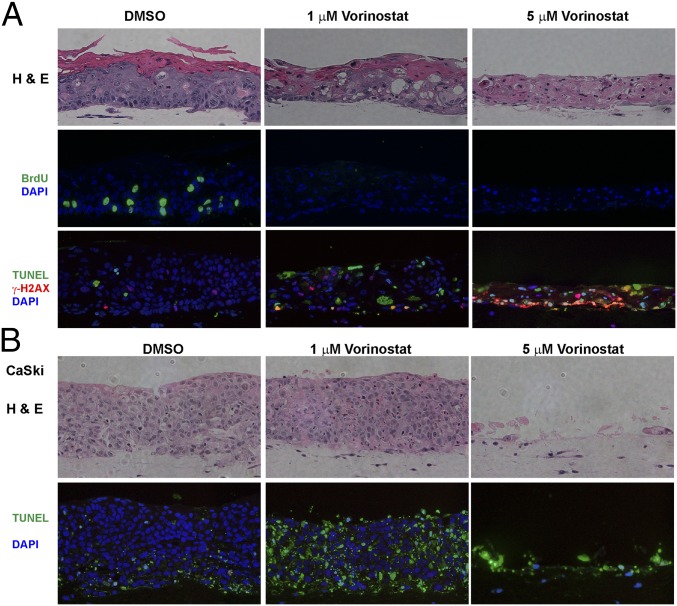
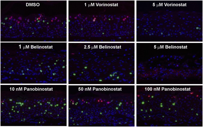
Human papillomaviruses (HPVs) cause epithelial proliferative diseases. Persistent infection of the mucosal epithelia by the high-risk genotypes can progress to high-grade dysplasia and cancers. Viral transcription and protein activities are intimately linked to regulation by histone acetyltransferases and histone deacetylases (HDACs) that remodel chromatin and regulate gene expression. HDACs are also essential to remodel and repair replicating chromatin to enable the progression of replication forks. As such, Vorinostat (suberoylanilide hydroximic acid), and other pan-HDAC inhibitors, are used to treat lymphomas. Here, we investigated the effects of Vorinostat on productive infection of the high-risk HPV-18 in organotypic cultures of primary human keratinocytes. HPV DNA amplifies in the postmitotic, differentiated cells of squamous epithelia, in which the viral oncoproteins E7 and E6 establish a permissive milieu by destabilizing major tumor suppressors, the pRB family proteins and p53, respectively. We showed that Vorinostat significantly reduced these E6 and E7 activities, abrogated viral DNA amplification, and inhibited host DNA replication. The E7-induced DNA damage response, which is critical for both events, was also compromised. Consequently, Vorinostat exposure led to DNA damage and triggered apoptosis in HPV-infected, differentiated cells, whereas uninfected tissues were spared. Apoptosis was attributed to highly elevated proapoptotic Bim isoforms that are known to be repressed by EZH2 in a repressor complex containing HDACs. Two other HDAC inhibitors, Belinostat and Panobinostat, also inhibited viral DNA amplification and cause apoptosis. We suggest that HDAC inhibitors are promising therapeutic agents to treat benign HPV infections, abrogate progeny virus production, and hence interrupt transmission.

Keywords: HDAC inhibitors; HPV DNA amplification; HPV E6 and E7 activities; apoptosis; organotypic cultures.


Conflict of interest statement

The authors declare no conflict of interest.

Figures

Fig. 1.
Fig. 1.
Histology and differentiation markers of representative raft cultures. HPV-18 (A) infected and (B) uninfected day 13 PHK raft cultures were exposed, from days 6 to 13, to DMSO and 1 or 5 μM Vorinostat. Four-micrometer sections of FFPE tissues were stained with hematoxylin and eosin (Upper). Indirect IF detection of loricrin (red) and keratin 10 (green) (Lower). Brackets denote stratum granulosum. Microphotographic images here and in later figures were captured with a 20× objective, and DAPI staining (blue) revealed all nuclei.
Fig. 2.
Fig. 2.
Effects of Vorinostat on HPV-18 DNA amplification and cellular DNA synthesis. Relative HPV-18 DNA copy number/cell was computed from qPCR of total DNA, extracted from fresh day 13 raft cultures following exposure (A) to DMSO (as 100%) (0) or 0.2 (0.2), 5 (5), or 10 μM (10) of Vorinostat, or (B) to DMSO (0), 2 (2), 3 (3), 4 (4), or 5 (5) μM Vorinostat from days 6 to 13. (C) In situ detection of HPV-18 DNA (red/cy3) and BrdU incorporation (green/FitC) in FFPE tissue sections of HPV-18 raft cultures exposed to DMSO or Vorinostat. Nuclei were detected with DAPI. (D) Average percentage of BrdU-positive and HPV-18−positive nuclei from four nonoverlapping microscopic fields per slide from the above experiment (C: DMSO, 1 or 5 μM Vorinostat) using the Count And Measure Application of cellSens software (Olympus). (E) Average intensity of HPV-18 DNA signals from the same microscopic fields as in D. Error bars in D and E represent SD. (F) Indirect IF detection of the major capsid protein L1 (green) in day 14 HPV-18 raft cultures treated with DMSO or Vorinostat from days 6 to 14. (G) Indirect IF probing for BrdU incorporation (red) in day 13 uninfected cultures exposed to DMSO or to Vorinostat from days 6 to 13. ***P < 0.001.
Fig. 3.
Fig. 3.
Detection of acetylated histone 4 in the presence or absence of Vorinostat. (A) Acetylated histone 4 was detected with antibody reactive to H4 K5/6/9/12/16 (red signals) in IBs of lysates from two independent sets of day 13 HPV-18−infected cultures (a and b). The cultures were exposed to DMSO (0) or to 0.2, 1, or 5 μM Vorinostat from days 6 to 13. Acetylated H4 (n red) and actin (in green) were recorded with the LI-COR CLx system. (B) Acetylated H4K12 was detected in the IB of the above lysates using the enhanced chemiluminescence (ECL) method. Lysate from UT PHK was one of the controls. This IB (marked with an asterisk) was derived from the same gel as depicted for HDAC-2 in Fig. 4A and for HDAC-5 and actin loading reference in Fig. 4B. Indirect IF detection of elevated (C) H4K12Ac and (D) H4K16Ac in day 13 HPV-18 raft cultures in the DMSO-treated control (Upper) or exposed to 5 μM Vorinostat (Lower) from days 6 to 13. (E) Indirect IF detection of H4K12Ac (green) in day 13 HPV-18 raft cultures treated with 2 μM Vorinostat from days 6 to 13. In CE, merged images are shown in which nuclei were detected with DAPI and acetylated H4K in green (Left), while only the acetylated H4K signals were shown (Right).
Fig. 4.
Fig. 4.
IBs to detect HDACs in control and Vorinostat-treated day 13 raft cultures. (A) IBs of lysates of HPV-18 and PHK raft cultures reveal steady-state levels of HDAC-1, HDAC-2, HDAC-3, and HDAC-4. The cultures were exposed to vehicle (0) or Vorinostat at three concentrations, as indicated, from days 6 to 13. Lysates from UT PHK raft cultures served as one of the controls. The signals were detected with ECL. (B) HDACs-5, HDAC -6, HDAC -7, and SirT-1 (in red) and actin (in green) from the same raft cultures were documented with the LI-COR CLx system. Brackets indicate blots from the same gel. Panels showing bands for H4K12Ac in Fig. 3B as well as bands of HDAC2 in A and HDAC5 in B (each marked with an asterisk) were detected in strips from the same gel.
Fig. 5.
Fig. 5.
IBs and IF to detect steady-state levels of HPV-18 E6 and E7 and target host proteins. (A and B) Two sets of independent day 13 HPV-18 cultures were exposed to the indicated Vorinostat concentrations from days 6 to 13. UT PHK or HPV-18−infected raft cultures exposed to vehicle (0) served as references. Actin was loading control. The ECL detection system was used to detect (A) p130, HPV-18 E6 and E7, and actin, (B) pRB, p53, and actin, (D) total ATM, phosphorylated ATM S1981, and Nbs1, (E) Mre-11 and actin, and (F) total (Upper) and phosphorylated (Lower) DNA-PKcs. (C) E6AP and actin were detected and documented with LI-COR CLx system. Images in AC and E each represent blots from a separate gel. Protein bands identified in D and F are from the same gel as those in A and B, respectively. (G) PCNA (red) and cyclin B1 (green) were detected by IF in FFPE sections from one set of the infected cultures, exposed to DMSO (Left) or 5 μM Vorinostat (Right). A and F are from the same gel and had same actin loading control, indicated by a single asterisk (*) in A. ** and *** indicate actin loading controls of B and C IBs.
Fig. 6.
Fig. 6.
Assays to detect DNA damage and apoptosis in day 13 PHK and HPV-18−infected raft cultures. (A) Detection of cleaved caspase 3 (green) and γ-H2AX (S139) (red) in HPV-18 raft cultures following exposure to DMSO or Vorinostat (1 and 5 μM) from days 6 to 13. (B) HPV-18 raft cultures were probed for TUNEL (green) (Upper), whereas PHK raft cultures were probed for TUNEL (green) and γ-H2AX (red) (Lower). (C) IB detection of γ-H2AX S139 in parallel HPV-18 raft cultures. Lysates from UT PHK or infected raft culture as well as infected culture exposed to vehicle (0) were used as references. Actin served as a loading control. (D) Percentage of TUNEL-positive nuclei in A and B were averaged from four microscopic fields under a 20× objective. (E) IB detection of cleaved caspase 3 in parallel Vorinostat-treated HPV-18 raft and untreated PHK raft cultures. This strip of IB was derived from the same gel as depicted in Fig. 5E, and the actin loading control was presented in Fig. 5E.
Fig. 7.
Fig. 7.
Prolonged exposure of HPV-18−infected raft culture to Vorinostat. The day 22 cultures were exposed to DMSO or 1 or 5 μM Vorinostat from days 6 to 22. Histology was revealed by staining with hematoxylin and eosin (Upper). DNA damage and apoptosis were detected by indirect immunofluorescent detection of γ-H2AX and TUNEL (Lower).
Fig. 8.
Fig. 8.
IBs and IF to detect steady-state levels of the proapoptotic proteins. (A) Apaf1, PUMA, Bax, and Noxa. IBs marked with ** and *** are from the same gels as shown in Fig. 5 B and C, which included the actin loading control. (B) IBs to detect EZH2 (Upper) and Bim (Lower) in day 13 lysates from UT PHK and HPV-18 cultures as well as HPV-18 raft cultures exposed to Vorinostat from days 6 to 13 at the indicated concentrations. EL, L, and S indicate extralong, long, and small forms of Bim, respectively. The blots are from the same gels as presented in Figs. 5E and 6C, where the actin loading controls are shown. (C) Indirect IF detection of Bim protein (green) in tissue sections of HPV-18 raft cultures.
Fig. 9.
Fig. 9.
Effects of Vorinostat on raft cultures of W12-E cells (A) and CaSki cells (B). Day 14 cultures were exposed to DMSO (Left) or 1 (Middle) or 5 μM (Right) Vorinostat from days (A) 6 to 14 or (B) 5 to 14. Rows are as follows: (A) histology (Upper), BrdU incorporation (Middle), and γ-H2AX and TUNEL (Lower) signals; (B) histology (Upper) and TUNEL (Lower) signals.
Fig. 10.
Fig. 10.
HPV-18−infected raft cultures exposed to Vorinostat, Belinostat, or Panobinostat. Parallel cultures were exposed to these HDAC inhibitors at the specified concentration from days 6 to 14. Day 14 cultures were probed for viral DNA amplification (red) and BrdU incorporation (green). Portions of the cultures exposed to 5 μM Belinostat had significant cell death, causing trapping of in situ probe signals (red). Histology, L1, and TUNEL assays are shown in SI Appendix, Figs. S3 and S4.

References

    1. de Martel C, Plummer M, Vignat J, Franceschi S. Worldwide burden of cancer attributable to HPV by site, country and HPV type. Int J Cancer. 2017;141:664–670. - PMC - PubMed
    1. Stoler MH, Wolinsky SM, Whitbeck A, Broker TR, Chow LT. Differentiation-linked human papillomavirus types 6 and 11 transcription in genital condylomata revealed by in situ hybridization with message-specific RNA probes. Virology. 1989;172:331–340. - PubMed
    1. Wang HK, Duffy AA, Broker TR, Chow LT. Robust production and passaging of infectious HPV in squamous epithelium of primary human keratinocytes. Genes Dev. 2009;23:181–194. - PMC - PubMed
    1. McLaughlin-Drubin ME, Münger K. The human papillomavirus E7 oncoprotein. Virology. 2009;384:335–344. - PMC - PubMed
    1. Genovese NJ, Banerjee NS, Broker TR, Chow LT. Casein kinase II motif-dependent phosphorylation of human papillomavirus E7 protein promotes p130 degradation and S-phase induction in differentiated human keratinocytes. J Virol. 2008;82:4862–4873. - PMC - PubMed

Publication types

MeSH terms