

Single-Cell Transcriptomics of Pancreatic Cancer Precursors Demonstrates Epithelial and Microenvironmental Heterogeneity as an Early Event in Neoplastic Progression
- PMID: 30385653
- PMCID: PMC6445737
- DOI: 10.1158/1078-0432.CCR-18-1955
Single-Cell Transcriptomics of Pancreatic Cancer Precursors Demonstrates Epithelial and Microenvironmental Heterogeneity as an Early Event in Neoplastic Progression
Abstract
Purpose: Early detection of pancreatic ductal adenocarcinoma (PDAC) remains elusive. Precursor lesions of PDAC, specifically intraductal papillary mucinous neoplasms (IPMNs), represent a bona fide pathway to invasive neoplasia, although the molecular correlates of progression remain to be fully elucidated. Single-cell transcriptomics provides a unique avenue for dissecting both the epithelial and microenvironmental heterogeneities that accompany multistep progression from noninvasive IPMNs to PDAC.
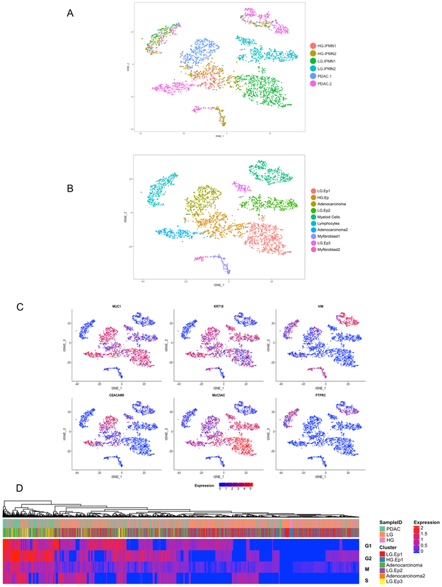
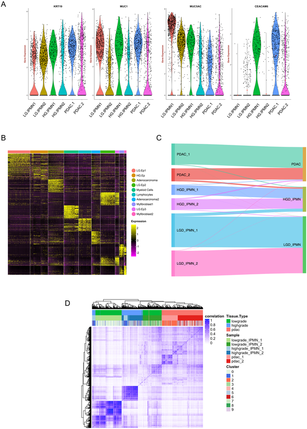
Experimental design: Single-cell RNA sequencing was performed through droplet-based sequencing on 5,403 cells from 2 low-grade IPMNs (LGD-IPMNs), 2 high-grade IPMNs (HGD-IPMN), and 2 PDACs (all surgically resected).
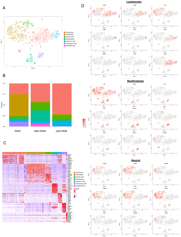
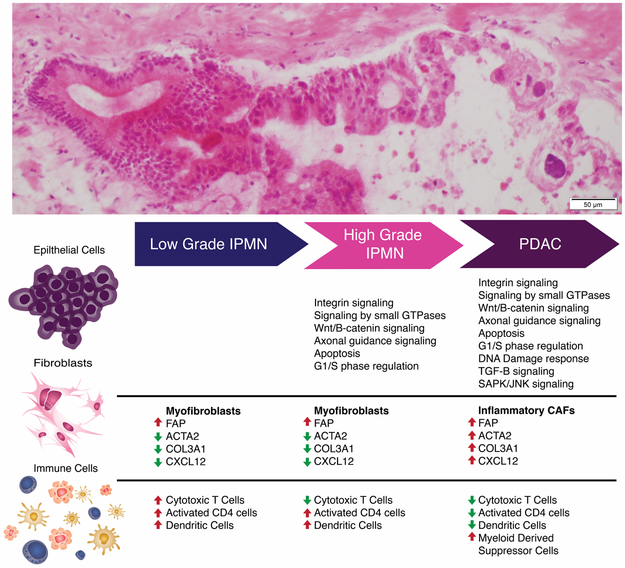
Results: Analysis of single-cell transcriptomes revealed heterogeneous alterations within the epithelium and the tumor microenvironment during the progression of noninvasive dysplasia to invasive cancer. Although HGD-IPMNs expressed many core signaling pathways described in PDAC, LGD-IPMNs harbored subsets of single cells with a transcriptomic profile that overlapped with invasive cancer. Notably, a proinflammatory immune component was readily seen in low-grade IPMNs, composed of cytotoxic T cells, activated T-helper cells, and dendritic cells, which was progressively depleted during neoplastic progression, accompanied by infiltration of myeloid-derived suppressor cells. Finally, stromal myofibroblast populations were heterogeneous and acquired a previously described tumor-promoting and immune-evading phenotype during invasive carcinogenesis.
Conclusions: This study demonstrates the ability to perform high-resolution profiling of the transcriptomic changes that occur during multistep progression of cystic PDAC precursors to cancer. Notably, single-cell analysis provides an unparalleled insight into both the epithelial and microenvironmental heterogeneities that accompany early cancer pathogenesis and might be a useful substrate to identify targets for cancer interception.See related commentary by Hernandez-Barco et al., p. 2027.
©2018 American Association for Cancer Research.
Conflict of interest statement
Figures
Comment in
-
No Cell Left Unturned: Intraductal Papillary Mucinous Neoplasm Heterogeneity.Clin Cancer Res. 2019 Apr 1;25(7):2027-2029. doi: 10.1158/1078-0432.CCR-18-3877. Epub 2019 Jan 14. Clin Cancer Res. 2019. PMID: 30642914 Free PMC article.
References
-
- Rezaee N, Barbon C, Zaki A, He J, Salman B, Hruban RH, et al. Intraductal papillary mucinous neoplasm (IPMN) with high-grade dysplasia is a risk factor for the subsequent development of pancreatic ductal adenocarcinoma. HPB (Oxford) 2016;18(3):236–46 doi 10.1016/j.hpb.2015.10.010. - DOI - PMC - PubMed
MeSH terms
Grants and funding
LinkOut - more resources
Full Text Sources
Medical
