. 2019 Jan;74(1):69-82.
doi: 10.1136/thoraxjnl-2018-211535. Epub 2018 Nov 1.

Fas activation alters tight junction proteins in acute lung injury

Affiliations

Fas activation alters tight junction proteins in acute lung injury

Raquel Herrero et al. Thorax. 2019 Jan.

Abstract

Background:The acute respiratory distress syndrome (ARDS) is characterized by protein-rich oedema in the alveolar spaces, a feature in which Fas-mediated apoptosis of the alveolar epithelium has been involved. Objective:To determine whether Fas activation increases protein permeability by mechanisms involving disruption of the paracellular tight junction (TJ) proteins in the pulmonary alveoli. Methods: Protein permeability and the expression of TJ proteins were assessed in vivo in wild-type and Fas-deficient lpr mice 16 hours after the intratracheal instillation of recombinant human soluble Fas ligand (rh-sFasL), and at different time points in vitro in human pulmonary alveolar epithelial cells (HPAEpiC) exposed to rh-sFasL Results:Activation of the Fas pathway increased protein permeability in mouse lungs and altered the expression of the TJ proteins occludin and zonula occludens-1 in the alveolar-capillary membrane in vivo and in human alveolar epithelial cell monolayers in vitro. Blockade of caspase-3, but not inhibition of tyrosine kinase dependent pathways, prevented the alterations in TJ protein expression and permeability induced by the Fas/FasL system in human alveolar cell monolayers in vitro. We also observed that both the Fas-induced increase of protein permeability and disruption of TJ proteins occurred before cell death could be detected in the cell monolayers in vitro. Conclusion:Targeting caspase pathways could prevent the disruption of TJs and reduce the formation of lung oedema in the early stages of ARDS.

Keywords: ards; innate immunity; pulmonary oedema.


Conflict of interest statement

Competing interests: None declared.

Figures

Fig. 1.
Fig. 1.. Effects of FasL on pulmonary protein permeability (A), cellular inflammatory responses (B, C) and left lung weight (D) in wild-type and Fas-deficient lpr mice in vivo.
Mice were treated with intratracheal instillation of recombinant human soluble FasL (rh-sFasL, 25 ng/g body wt), or PBS (as control), and then studied 16 h later. The graphs show the effect of rh-sFasL on (A) the concentration of IgM (a plasma protein of large size, 900 kDa), (B) the number of polymorphonuclear (PMN) leukocytes, and (C) the number of macrophages in bronchoalveolar lavage (BAL) fluid, and on (D) the left lung wt-to-body wt ratio. In the graphs, each dot represents an individual mouse (n=10 per group). Horizontal bars represent means. *P < 0.05 vs WT-PBS group.
Fig. 2.
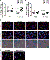
Fig. 2.. Human soluble FasL alters the expression of occludin in the lungs of wild-type, but not in the lungs of lpr Fas-deficient mice.
Mice were treated with intratracheal instillation of recombinant human soluble FasL (rh-sFasL, 25 ng/g body wt), and then studied 16 h later. As control, wild-type (WT) and lpr Fas-deficient (lpr) mice were treated with PBS via intratracheal instillation. We measured the concentration of occludin in the membrane and cytosolic fractions in mouse lungs by ELISA (A and B, respectively). The merged images of fluorescence microscopy and light microscopy with differential interference contrast (DIC) show occludin protein signal (red) and cell nuclei (DAPI staining, blue) over the structure of the alveolar walls. Original image magnification X200. (C). Representative images at larger magnification (x400) show the decreased expression of occludin protein (red signal) in the lung of WT mice treated with rh-sFasL compared with mice treated with PBS (D). In the graphs, each dot represents an individual mouse (n=10 per group). n of 5 mice per group for the immunofluorescence analyses. Horizontal bars represent means. *P < 0.05 vs WT-PBS group
Fig. 3.
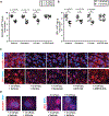
Fig. 3.. Human soluble FasL alters the expression of ZO1 in the lungs of wild-type, but not in the lungs of lpr Fas-deficient mice.
Wild-type (WT) and lpr Fas-deficient (lpr) mice were treated with intratracheal instillation of recombinant human soluble FasL (rh-sFasL, 25 ng/g body wt), and then studied 16 h later. As control, WT and lpr mice were intratracheally instilled with PBS. We measured the concentration of ZO1 in the membrane and cytosolic fractions in mouse lungs by ELISA (A and B, respectively). The merged images of fluorescence microscopy and light microscopy with differential interference contrast (DIC) show occludin protein signal (red) and cell nuclei (DAPI staining, blue) over the structure of the alveolar walls. Original image magnification X200 (C). Representative images at larger magnification (x400) show the decreased expression of ZO1 protein (red signal) in the lung of WT mice treated with rh-sFasL compared with mice treated with PBS (D). In the graphs, each dot represents an individual mouse (n=10 per group). n of 5 mice per group for the immunofluorescence analyses. Horizontal bars represent means. *P < 0.05 vs WT-PBS group
Fig. 4.
Fig. 4.. Fas-induced apoptosis is locally associated with altered expression of occludin and ZO1 in mouse lungs in vivo.
Wild-type (WT) and Fas deficient (lpr) mice were treated with one intratracheal instillation of recombinant human soluble FasL (rh-sFasL, 25 ng/g body wt), or PBS (as control). At 16 h post-instillation, we measured (A) the number of nuclei containing DNA strand breaks (TUNEL-positive signal) in lung tissue sections, and (B) caspase-3 activity in the lung homogenates. Merged images of representative lung tissue sections from PBS or FasL-treated WT mice (C), showed that occludin and ZO1 protein signals (red) were diminished in the alveolar walls only in areas with TUNEL positive cells (green). Original image magnification X400. n of 5 mice per group for the immunofluorescence analyses. In the graphs, each dot represents an individual mouse (n= 10 mice per group). Horizontal bars represent means. *P < 0.05 vs WT-PBS group
Fig. 5.
Fig. 5.. Human soluble FasL increases alveolar epithelial cell permeability prior to cell death in vitro.
The permeability to FITC-albumin (A) and the percentage of cell death (B) were measured in human primary alveolar epithelial cell (HAEpiC) monolayers after 2 h of incubation with increasing concentrations of a cytotoxic long form of human recombinant sFasL from 33 to 600 ng/mL. Cells with medium only (MO) or with a non-cytotoxic short form of human recombinant soluble FasL (300 ng/mL) were used as controls. Results from 3 separate experiments performed in duplicate. Each dot of the graphs represents a single data point. Horizontal bars represent means. *P < 0.05 vs medium only (MO).
Fig.6.
Fig.6.. Human sFasL increases protein permeability in human alveolar epithelial cell monolayers by mechanisms dependent on Caspase-3, but not on tyrosine-kinase signaling-dependent cytokine production.
HPAEpiC monolayers were pre-incubated with caspase-3 inhibitor zDEVD.fmk (or zFA.fmk or vehicle as controls) and exposed for 2 h to rh-sFasL (100 ng/mL or 300 ng/mL) or medium only (MO) (A-C). After treatment, we measured (A) caspase 3-activity, (B) percentage of cell death and (C) FITC-albumin permeability in these cell monolayers. HPAEpiC monolayers were also pre-incubated with the tyrosine-kinase specific inhibitor genistein (or vehicle as control) (D-H) and exposed for 2 h to rh-sFasL (100 ng/mL or 300 ng/mL) or medium only (MO). After treatment, we measured IL-8 concentration in cell supernatant by ELISA (D), percentage of cell death by PrestoBlue (E), permeability to FITC-albumin (F), IL-6 concentration in cell supernatant by ELISA (G), and tyrosine kinase activity in the cell monolayers (H). Cell monolayers treated with medium only were used to determine 100% survival. Results from 4 separate experiments performed in duplicate. Each dot of the graphs represents a single data point. Horizontal bars represent means. *P<0.05 vs all conditions with MO; # P< 0.05.
Fig. 7.
Fig. 7.. Human sFasL alters the levels and distribution of occludin and ZO1 proteins in human lung alveolar epithelial cells in vitro by mechanisms dependent on caspase-3, but independent of tyrosine kinase-mediated cytokine production.
After 2 h exposure of rh-sFasL (+FasL) at the dose of 100 ng/mL or medium only (-FasL), the concentrations of occludin and ZO1 proteins were measured by ELISA in HAECpiC monolayers pre-incubated with the caspase-3 inhibitor zDEVD.fmk, its inactive analog (zFA.fmk), or the tyrosine kinase inhibitor genistein or vehicle (DMSO). Compared to control cells, rh-sFasL decreased the concentrations of occludin and ZO1 in cell protein extracts (A and B, respectively). Representative immunofluorescence images (original image magnification X400) (C) show the expression of occludin and ZO1 proteins (red signals) localized along the cytoplasmic membrane of control cells in HAECpiC monolayers (−rhFasL/+vehicle). Exposure to rh-sFasL resulted in a global decrease of occludin and ZO1 fluorescence signals that was particularly profound in the cytoplasmic extensions, while some signal remained close to the cell nuclei (DAPI staining, blue signal). Only pre-incubation with the caspase-3 inhibitor zDEVD.fmk prevented the sFasL-induced decrease of occludin and ZO1 proteins evaluated both by ELISA (A and B) and by immunocytochemistry (C). Representative immunofluorescence images at larger magnification (x1000) show the decreased expression of occludin and ZO1 proteins (red signals) in HAECpiC monolayers treated with rh-sFasL (+rh-sFasL/+vehicle) compared with control cells (−rh-sFasL/+vehicle) (D) Results from 4 separate experiments performed in duplicate. Each dot of the graphs represents a single data point. Horizontal bars represent means. *P<0.05 vs their corresponding-rh-sFasL conditions.
Fig. 8.
Fig. 8.. FasL-mediated changes in the levels and distribution of occludin and ZO1 proteins occur before nuclear DNA fragmentation in human lung alveolar epithelial cells in vitro.
Representative fluorescence images of HAECpiC monolayers exposed to rh-sFasL (100 ng/mL) or medium only (MO) for 1, 2, 3 or 5 h. The cells were labeled with the TUNEL method for the detection of DNA fragmentation (green signal), followed by labelling with an antibody to occludin or ZO1 proteins (red signal). The merged image shows a decrease of occludin and ZO1 fluorescence signals after exposure to FasL in a time-dependent manner. Despite this loss of occludin and ZO1 fluorescence signal, no TUNEL positive signal was observed during the first 3 h of FasL exposure. However, TUNEL positive cells were observed at 5 h-exposure of FasL. Original image magnification X400.

Similar articles

Cited by

References

    1. Matthay MA, and Wiener-Kronish JP 1990. Intact epithelial barrier function is critical for the resolution of alveolar edema in humans. Am Rev Respir Dis 142:1250–1257. - PubMed
    1. Berthiaume Y, Lesur O, and Dagenais A 1999. Treatment of adult respiratory distress syndrome: plea for rescue therapy of the alveolar epithelium. Thorax 54:150–160. - PMC - PubMed
    1. Ware LB, and Matthay MA 2001. Alveolar fluid clearance is impaired in the majority of patients with acute lung injury and the acute respiratory distress syndrome. American journal of respiratory and critical care medicine 163:1376–1383. - PubMed
    1. Hartsock A, and Nelson WJ 2008. Adherens and tight junctions: structure, function and connections to the actin cytoskeleton. Biochimica et biophysica acta 1778:660–669. - PMC - PubMed
    1. Schneeberger EE, and Lynch RD 1992. Structure, function, and regulation of cellular tight junctions. The American journal of physiology 262:L647–661. - PubMed

Publication types

MeSH terms