Observational Study
. 2018 Nov 2;3(21):e120694.
doi: 10.1172/jci.insight.120694.

Heme scavenging reduces pulmonary endoplasmic reticulum stress, fibrosis, and emphysema

Affiliations
Observational Study

Heme scavenging reduces pulmonary endoplasmic reticulum stress, fibrosis, and emphysema

Saurabh Aggarwal et al. JCI Insight. .

Abstract

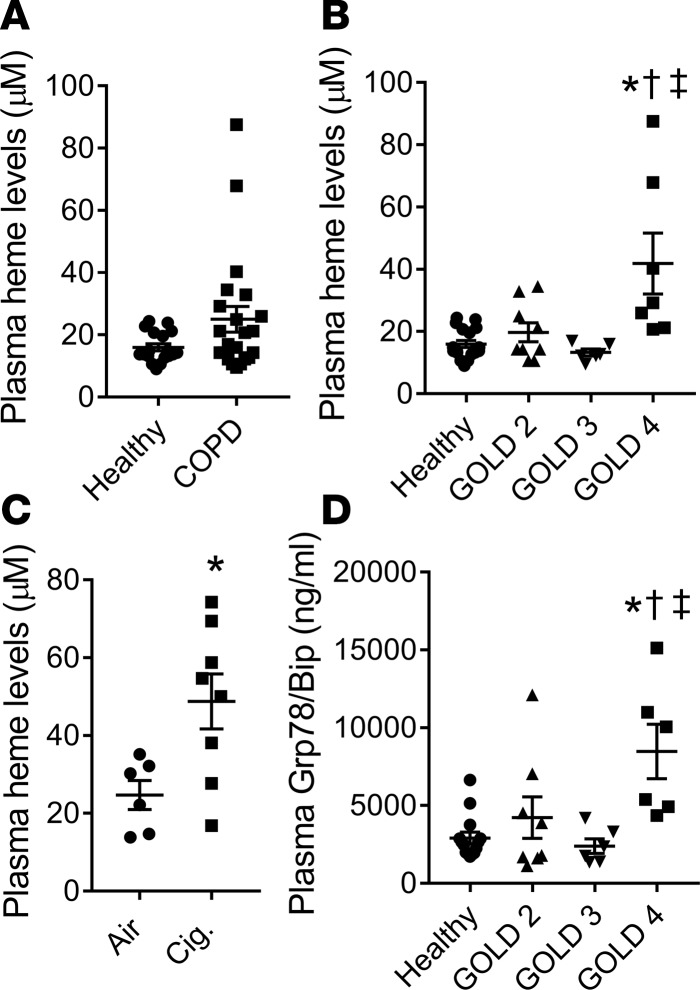
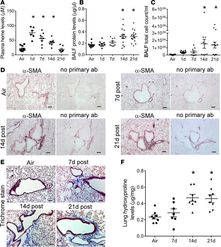
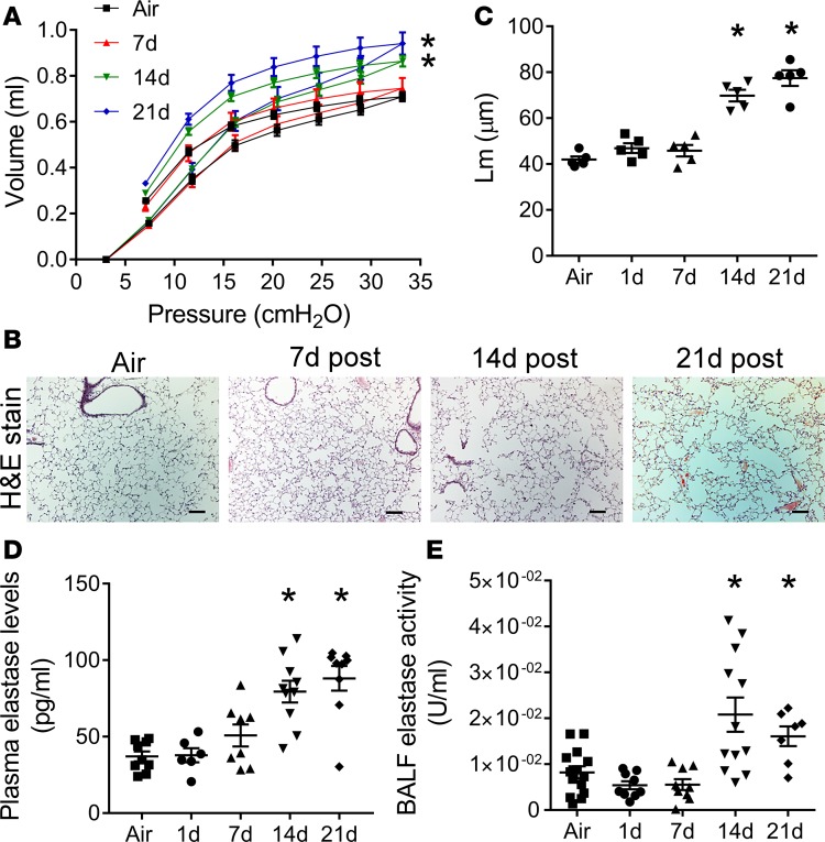
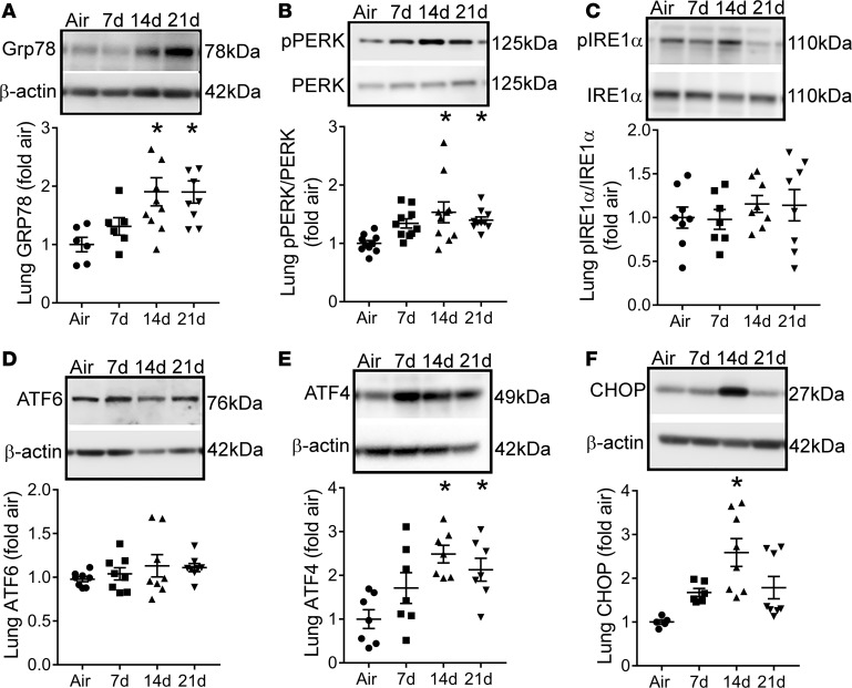
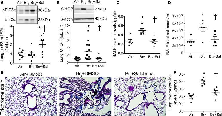
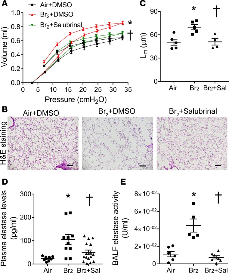
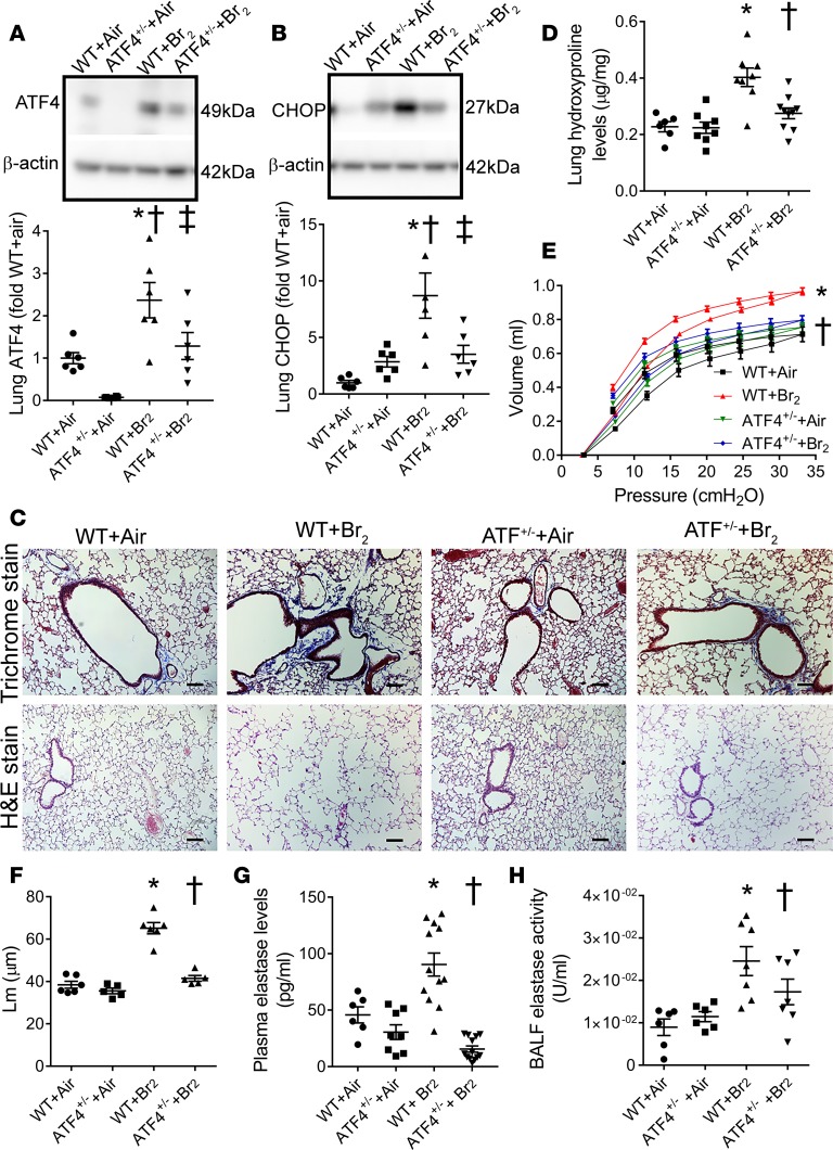
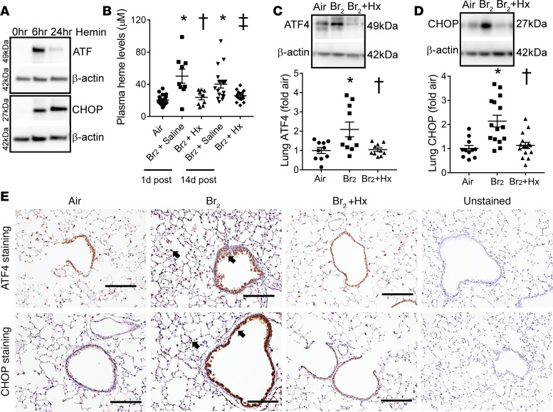
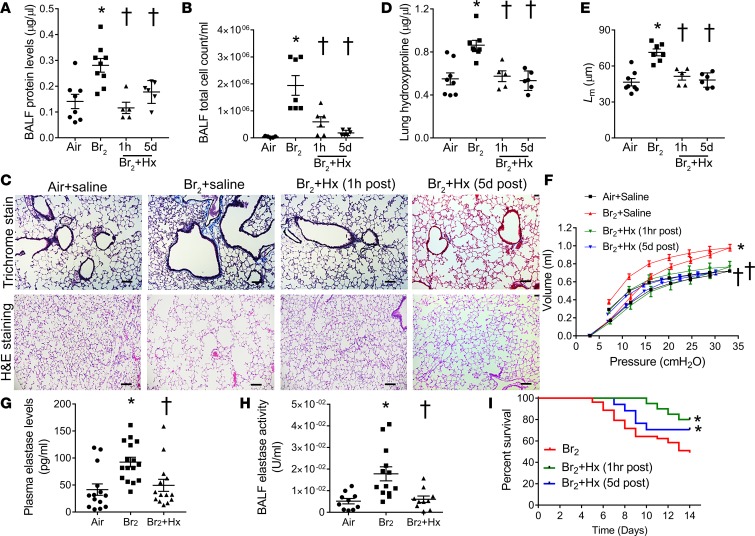
Pulmonary fibrosis and emphysema are irreversible chronic events after inhalation injury. However, the mechanism(s) involved in their development remain poorly understood. Higher levels of plasma and lung heme have been recorded in acute lung injury associated with several insults. Here, we provide the molecular basis for heme-induced chronic lung injury. We found elevated plasma heme in chronic obstructive pulmonary disease (COPD) (GOLD stage 4) patients and also in a ferret model of COPD secondary to chronic cigarette smoke inhalation. Next, we developed a rodent model of chronic lung injury, where we exposed C57BL/6 mice to the halogen gas, bromine (Br2) (400 ppm, 30 minutes), and returned them to room air resulting in combined airway fibrosis and emphysematous phenotype, as indicated by high collagen deposition in the peribronchial spaces, increased lung hydroxyproline concentrations, and alveolar septal damage. These mice also had elevated pulmonary endoplasmic reticulum (ER) stress as seen in COPD patients; the pharmacological or genetic diminution of ER stress in mice attenuated Br2-induced lung changes. Finally, treating mice with the heme-scavenging protein, hemopexin, reduced plasma heme, ER stress, airway fibrosis, and emphysema. This is the first study to our knowledge to report elevated heme in COPD patients and establishes heme scavenging as a potential therapy after inhalation injury.

Keywords: COPD; Fibrosis; Pulmonology.


Conflict of interest statement

Conflict of interest: The authors have declared that no conflict of interest exists.

Figures

Figure 1
Figure 1. Plasma heme and ER stress levels are elevated in patients with very severe COPD and in ferrets exposed to cigarette smoke.
Total heme levels were measured in the plasma of COPD patients and their healthy counterparts. Although total heme levels were not significantly higher in COPD patients compared with the healthy individuals (P = 0.06) (n = 18–22) (A), the stratification of patients according to the severity of the disease revealed significantly elevated plasma heme in COPD patients with GOLD stage 4 disease compared with the healthy individuals or patients with the mild disease (n = 6–18) (B). Plasma heme levels were also significantly higher in ferrets exposed to cigarette smoke for 6 months, which induced emphysema and attenuated lung function (n = 6–8) (C). ELISA showed that GOLD stage 4 COPD patients had significantly higher levels of ER stress marker Grp78/Bip (n = 6–14) (D). Values are means ± SEM. *P < 0.05 versus healthy patients or air-exposed ferrets; P < 0.05 versus COPD GOLD stage 2; P < 0.05 versus COPD GOLD stage 3, by unpaired t test for 2 groups or 1-way ANOVA followed by Tukey’s post hoc testing for more than 2 groups.
Figure 2
Figure 2. Lung injury and fibrosis in rodent model of inhalation injury.
Male C57BL/6 mice were exposed to air or Br2 gas (400 ppm, 30 minutes) and returned to room air. Plasma heme and acute and chronic lung injury parameters were measured in mice on days 1, 7, 14, or 21 after Br2 exposure. Plasma levels of total heme were elevated in mice until 14 days after Br2 inhalation (n = 6–11) (A). Bronchoalveolar lavage fluid (BALF) showed a significant increase in protein levels (n = 9–14) (B) and total cell count (n = 9–17) (C) on days 14 and 21 after Br2 inhalation. Peripheral lung tissue staining for α-smooth muscle actin (α-SMA) (n = 5) (D) and with Masson’s trichrome stain (n = 5) (E) demonstrated an increased accumulation of α-SMA and thickening of the smooth muscle layer and collagen deposition (blue stain) primarily around airways on days 14 and 21 after Br2. Characteristic images were obtained from the indicated number of lungs for each condition. Similarly, the quantification of collagen by measuring lung hydroxyproline levels showed significant increases at 14 and 21 days after Br2 inhalation (F). Values are means ± SEM. *P < 0.05 versus air-exposed C57BL/6 mice by 1-way ANOVA followed by Tukey’s post hoc test. Scale bars are 100 µm.
Figure 3
Figure 3. Acute exposure to Br2 induces lung emphysematous changes.
Male C57BL/6 mice were exposed to air or Br2 gas (400 ppm, 30 minutes) and then returned to room air. On days 7, 14, or 21 after Br2 exposure, mouse lung compliance was assessed by the slope of the deflation limbs of pressure-volume (PV) curves. Br2 inhalation increased lung volumes as indicated by shifting up and left of PV curves on days 14 and 21 after exposure (n = 5–9) (A). The staining of peripheral lung tissue with hematoxylin and eosin (H&E) showed airspace enlargement (B) and increased alveolar mean linear intercept (Lm) (n = 5). Scale bars are 100 µm. (C) on days 14 and 21 after exposure. Characteristic images are shown. Finally, plasma elastase levels (n = 6–10) (D) and BALF elastase activity (n = 7–14) (E) were elevated in Br2-exposed mice on days 14 and 21 after exposure. Values are means ± SEM. *P < 0.05 versus air-exposed C57BL/6 mice by 1-way ANOVA followed by Tukey’s post hoc test. PV curves were analyzed by 2-way ANOVA with Bonferroni’s post hoc test.
Figure 4
Figure 4. ER stress is increased in Br2-exposed mice.
Male C57BL/6 mice were exposed to air or Br2 gas (400 ppm, 30 minutes) and then returned to room air. On days 7, 14, or 21 after exposure, lungs were harvested and immunoblotted for ER stress markers Grp78/Bip (n = 6–10) (A), phospho-PERK (n = 10) (B), phospho-IRE1α (n = 8) (C), and ATF6 (n = 8) (D). Br2 exposure increased the lung expression of Grp78, phopho-PERK and the downstream transcriptional effectors of the PERK pathway ATF4 (n = 7) (E) and CHOP (n = 5–8) (F), 14 days after exposure. Values are means ± SEM. *P < 0.05 versus air-exposed C57BL/6 mice by 1-way ANOVA followed by Tukey’s post hoc test.
Figure 5
Figure 5. Attenuation of ER stress abrogates lung injury and fibrosis.
Male C57BL/6 mice were exposed to air or Br2 gas (400 ppm, 30 minutes) and then returned to room air. Some Br2-exposed mice received an intraperitoneal injection of the ER stress inhibitor salubrinal (1 mg/kg BW), starting at 1 hour after Br2 exposure, and then daily for 13 consecutive days. Air-exposed and some Br2-exposed mice received DMSO (vehicle) as a control. Fourteen days after Br2 exposure, immunoblot of lung tissue showed that salubrinal increased phospho-eIF2α levels (n = 7–8) (A) but decreased lung CHOP levels (n = 12–13) (B) after Br2 exposure. Salubrinal also attenuated BALF protein (n = 5–6) (C) and total cell count (n = 5–6) (D) in Br2-exposed mice. Masson’s trichrome staining (n = 5) (E) and quantification of lung hydroxyproline levels (n = 5–6). Scale bars are 100 µm. (F) showed a decrease in collagen levels and lung fibrotic changes 14 days after Br2 exposure in salubrinal-treated mice. Values are means ± SEM. *P < 0.05 versus air + DMSO–treated mice and P < 0.05 versus Br2 + DMSO–treated mice by 1-way ANOVA followed by Tukey’s post hoc test.
Figure 6
Figure 6. Attenuation of ER stress reduces lung emphysematous changes.
Male C57BL/6 mice were exposed to air or Br2 gas (400 ppm, 30 minutes) and then returned to room air. Some Br2-exposed mice were given an intraperitoneal injection of the ER stress inhibitor salubrinal (1 mg/kg BW), starting at 1 hour after Br2 exposure, and then daily for 13 consecutive days. Air-exposed and some Br2-exposed mice received DMSO (vehicle) as a control. Fourteen days after Br2 exposure, the lung volumes were increased in mice as indicated by the shifting up and left of the pressure-volume (PV) curves. (n = 5–6) (A). Staining of peripheral lung tissue with hematoxylin and eosin (H&E) showed airspace enlargement (n = 5) (B), which was quantified by measuring alveolar mean linear intercept (Lm) (n = 5). Scale bars 100 µm. (C). Treatment with salubrinal attenuated these Br2-induced emphysematous changes in mouse lung. Salubrinal also reduced plasma elastase levels (n = 10–16) (D) and BALF elastase activity (n = 5–7) (E) in Br2-exposed mice. Values are means ± SEM. All animals were males. *P < 0.05 versus air + DMSO–treated mice and P < 0.05 versus Br2 + DMSO–treated mice by 1-way ANOVA followed by Tukey’s post hoc test. PV curves were analyzed by 2-way ANOVA with Bonferroni’s post hoc test.
Figure 7
Figure 7. ATF4-haplodeficient mice (ATF4+/–) mice are protected against inhalation injury.
Male wild-type (WT, C57BL/6) and ATF4+/– mice were exposed to air or Br2 gas (400 ppm, 30 minutes) and then returned to room air. Fourteen days later, mouse lungs were harvested. Immunoblot analyses showed that ATF4+/– mice had lower lung ATF4 (n = 6) (A) and CHOP levels (n = 6) (B) compared with WT mice exposed to Br2. Masson’s trichrome staining (n = 5) (C) and quantification of lung hydroxyproline levels (n = 6–10). Scale bars are 100 µm. (D) showed increased collagen deposition primarily around airways in the WT mice compared with the ATF4+/– mice. Assessment of lung pressure-volume (PV) curves demonstrated that Br2 exposure increased lung volumes as indicated by shifting up and left of PV curve in the WT but not the ATF4+/– mice (n = 4 for WT + air and n = 5 for others) (E). Staining of peripheral lung tissue with hematoxylin and eosin (H&E) showed airspace enlargement after Br2 exposure (n = 5–6) in WT but not ATF4+/– mice (C), which was quantified by measuring alveolar mean linear intercept (Lm) (n = 5–6) (F). Plasma elastase levels (n = 6–13) (G) and BALF elastase activity (n = 6–7) (H) were significantly higher in the WT compared with the ATF4+/– mice after Br2 exposure. Values are means ± SEM. *P < 0.05 versus WT + air, P < 0.05 versus ATF+/– + air, and ‡P < 0.05 versus WT + Br2 for A and B; P < 0.05 versus WT + Br2 for DH by 1-way ANOVA followed by Tukey’s post hoc test. PV curves were analyzed by 2-way ANOVA with Bonferroni’s post hoc test.
Figure 8
Figure 8. Heme scavenging attenuates ER stress.
Immunoblot analysis showed that the incubation of the human bronchial epithelial cells with hemin (a form of heme, 25 μM), increased the ER stress markers ATF4 and CHOP (n = 3) (A) at 6 and 24 hours after hemin challenge. In addition, male C57BL/6 mice were exposed to air or Br2 gas (400 ppm, 30 minutes) and then returned to room air. Some Br2-exposed mice were given an intraperitoneal injection of purified human hemopexin (Hx) (4 μg/g BW) 1 hour after Br2 exposure. All air-exposed mice and some Br2-exposed mice received saline injection as an appropriate control. Hx attenuated plasma total heme levels in Br2-exposed mice (n = 9–24) (B). Immunoblotting showed that Hx lowered Br2-induced ER stress markers, ATF4 (n = 10) (C) and CHOP (n = 11–15) (D), in mouse lungs 14 days after Br2 exposure. Similarly, immunohistochemical staining of lung sections showed an increased accumulation of ATF4 and CHOP (n = 4–5) (E) (arrows showing brown stain) lining bronchioles and in the lung parenchyma in Br2-exposed mice 14 days after exposure. Hx lowered ATF4 and CHOP levels. Values are mean ± SEM. All animals were males. Scale bars are 200 µm. For A, *P < 0.05 versus air + saline, P < 0.05 versus Br2 + saline (1 day after), and P < 0.05 versus Br2 + saline (14 days after); for B and C, P < 0.05 versus Br2 + saline (14 days after) by 1-way ANOVA followed by Tukey’s post hoc test.
Figure 9
Figure 9. Hemopexin attenuates lung injury, airway fibrosis, and lung emphysema.
Male C57BL/6 mice were exposed to air or Br2 gas (400 ppm, 30 minutes) and then returned to room air. Some Br2-exposed mice were given an intraperitoneal injection of purified human hemopexin (Hx) (4 μg/g BW) 1 hour or 5 days after Br2 exposure. All air-exposed mice and some Br2-exposed mice received saline injection as an appropriate control. Fourteen days after Br2 exposure, mouse BALF protein levels (n = 5–9) (A) and total cell count (n = 5–7) (B) were elevated in saline-treated mice but were significantly lower in Hx-treated mice. Hx-treated mice had decreased lung deposition of collagen on Masson’s trichrome staining (n = 5–8) (C) and lower lung hydroxyproline levels (n = 5–8) (D) compared with saline-treated mice, 14 days after Br2 inhalation. Assessment of mouse lung pressure-volume (P-V) curves demonstrated that Br2 exposure increased lung volumes, as indicated by the shifting up and left of PV curve in the saline-treated mice but not in the Hx-treated mice (n = 5–10) (F). Hematoxylin and eosin (H&E) staining of lungs showed that Hx prevented Br2-induced alveolar septa damage (n = 5–8) (C) and reduced alveolar Lm (n = 5–8). Scale bars 100 µm. (E). In addition, Hx lowered plasma elastase levels (n = 14–16) (G) and BALF elastase activity (n = 10–13) (H) in Br2-exposed mice. The Kaplan-Meier curve demonstrated that Hx reduced mortality after Br2 exposure (n = 42 for Br2 + saline; n = 20 for Br2 + Hx [1 hour after]; n = 17 for Br2 + Hx [5 days after]) (I). *P < 0.05 versus air + saline and P < 0.05 versus Br2 + saline by 1-way ANOVA followed by Tukey’s post hoc test. PV curves were analyzed by 2-way ANOVA with Bonferroni’s post hoc test. Overall survival was analyzed by the Kaplan-Meier method. Differences in survival were tested for statistical significance by the log-rank test.

References

    1. Auerbach O, Garfinkel L, Hammond EC. Relation of smoking and age to findings in lung parenchyma: a microscopic study. Chest. 1974;65(1):29–35. doi: 10.1378/chest.65.1.29. - DOI - PubMed
    1. Cottin V, et al. Pulmonary hypertension in patients with combined pulmonary fibrosis and emphysema syndrome. Eur Respir J. 2010;35(1):105–111. doi: 10.1183/09031936.00038709. - DOI - PubMed
    1. Cottin V, et al. Combined pulmonary fibrosis and emphysema: a distinct underrecognised entity. Eur Respir J. 2005;26(4):586–593. doi: 10.1183/09031936.05.00021005. - DOI - PubMed
    1. Grubstein A, Bendayan D, Schactman I, Cohen M, Shitrit D, Kramer MR. Concomitant upper-lobe bullous emphysema, lower-lobe interstitial fibrosis and pulmonary hypertension in heavy smokers: report of eight cases and review of the literature. Respir Med. 2005;99(8):948–954. doi: 10.1016/j.rmed.2004.12.010. - DOI - PubMed
    1. Kitaguchi Y, Fujimoto K, Hanaoka M, Kawakami S, Honda T, Kubo K. Clinical characteristics of combined pulmonary fibrosis and emphysema. Respirology. 2010;15(2):265–271. doi: 10.1111/j.1440-1843.2009.01676.x. - DOI - PubMed

Publication types

MeSH terms