Comparative Study
. 2018 Nov 2;3(21):e123468.
doi: 10.1172/jci.insight.123468.

Transcriptomic heterogeneity in multifocal prostate cancer

Affiliations
Comparative Study

Transcriptomic heterogeneity in multifocal prostate cancer

Simpa S Salami et al. JCI Insight. .

Abstract

Background: Commercial gene expression assays are guiding clinical decision making in patients with prostate cancer, particularly when considering active surveillance. Given heterogeneity and multifocality of primary prostate cancer, such assays should ideally be robust to the coexistence of unsampled higher grade disease elsewhere in the prostate in order to have clinical utility. Herein, we comprehensively evaluated transcriptomic profiles of primary multifocal prostate cancer to assess robustness to clinically relevant multifocality.

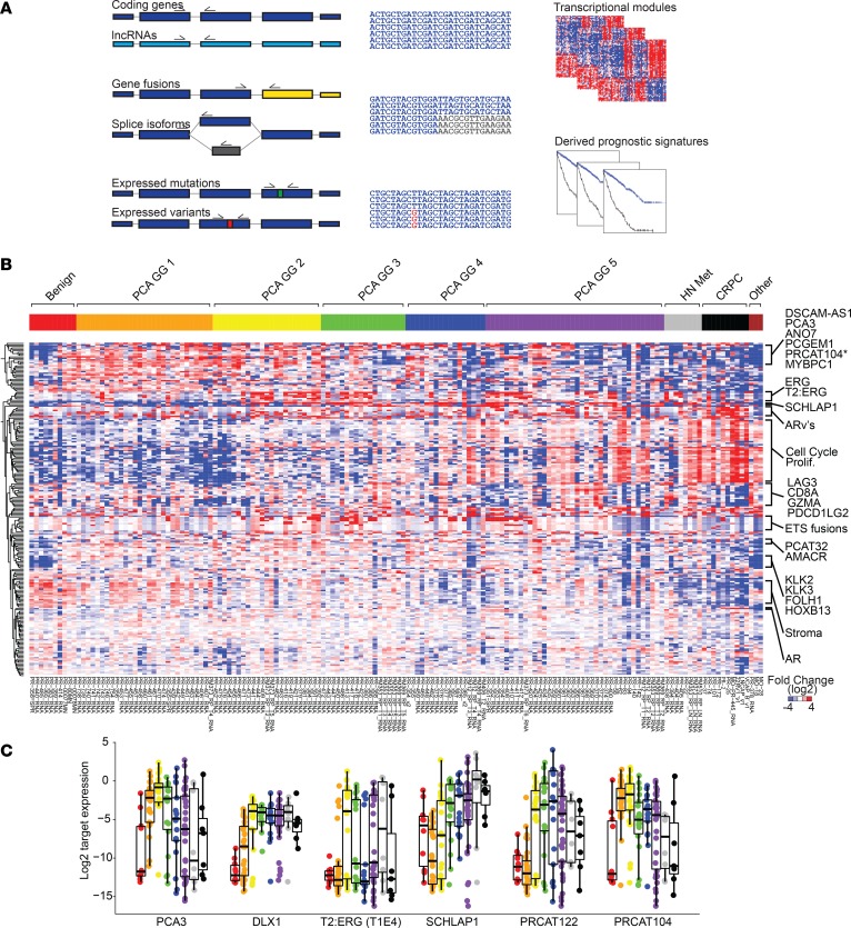
Methods: We designed a comprehensive, multiplexed targeted RNA-sequencing assay capable of assessing multiple transcriptional classes and deriving commercially available prognostic signatures, including the Myriad Prolaris Cell Cycle Progression score, the Oncotype DX Genomic Prostate Score, and the GenomeDX Decipher Genomic Classifier. We applied this assay to a retrospective, multi-institutional cohort of 156 prostate cancer samples. Derived commercial biomarker scores for 120 informative primary prostate cancer samples from 44 cases were determined and compared.

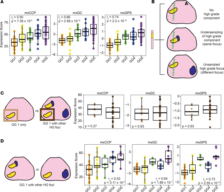
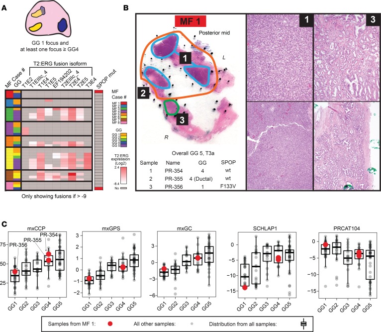
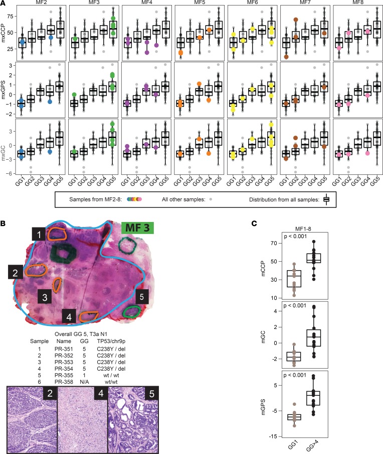
Results: Derived expression scores were positively correlated with tumor grade (rS = 0.53-0.73; all P < 0.001), both within the same case and across the entire cohort. In cases of extreme grade-discordant multifocality (co-occurrence of grade group 1 [GG1] and ≥GG4 foci], gene expression scores were significantly lower in low- (GG1) versus high-grade (≥GG4) foci (all P < 0.001). No significant differences in expression scores, however, were observed between GG1 foci from prostates with and without coexisting higher grade cancer (all P > 0.05).

Conclusions: Multifocal, low-grade and high-grade prostate cancer foci exhibit distinct prognostic expression signatures. These findings demonstrate that prognostic RNA expression assays performed on low-grade prostate cancer biopsy tissue may not provide meaningful information on the presence of coexisting unsampled aggressive disease.

Funding: Prostate Cancer Foundation, National Institutes of Health (U01 CA214170, R01 CA183857, University of Michigan Prostate Specialized Program of Research Excellence [S.P.O.R.E.] P50 CA186786-05, Weill Cornell Medicine S.P.O.R.E. P50 CA211024-01A1), Men of Michigan Prostate Cancer Research Fund, University of Michigan Comprehensive Cancer Center core grant (2-P30-CA-046592-24), A. Alfred Taubman Biomedical Research Institute, and Department of Defense.

Keywords: Cancer; Molecular diagnosis; Oncology; Prostate cancer.


Conflict of interest statement

Conflict of interest: SAT has received travel support from and had a sponsored research agreement with Compendia Bioscience/Life Technologies/Thermo Fisher Scientific, which provided access to a DNA-sequencing panel used herein. The University of Michigan and Brigham and Women’s Hospital have been issued patents (10,041,123; 9,745,635; 9,719,143; 9,303,291; 9,284,609; 8,969,527; 8,580,509; 8,211,645; and 7,718,369) on ETS gene fusions in prostate cancer, on which AMC, MAR, and SAT are coinventors. The diagnostic field of use was licensed to Hologic/Gen-Probe Inc., which has sublicensed rights to Roche/Ventana Medical Systems. SAT has served as a consultant for and received honoraria from Janssen, AbbVie, Sanofi, Almac Diagnostics, and Astellas/Medivation. SAT has sponsored research agreements with Astellas/Medivation and GenomeDX. SAT is a cofounder and consultant for Strata Oncology. TMM has received research funding from MDxHealth, Myriad Genetics, and GenomeDX. TMM has served as a consultant for Myriad Genetics.

Figures

Figure 1
Figure 1. Targeted multiplexed RNA sequencing enables robust transcriptional profiling of prostate cancer.
(A) Prostate cancer multiplexed RNA-sequencing (mxRNAseq) panel. To interrogate multiple classes of prostate cancer relevant transcriptional biomarkers, we designed a custom 306-amplicon mxRNAseq assay targeting coding genes, lncRNAs, gene fusions (e.g., TMPRSS2:ERG [T2:ERG]), splice isoforms (e.g., AR variants), expressed somatic mutations (e.g., SPOP hotspot mutations), and expressed germline risk variants (e.g., HOXB13 p.G84E). Targets to derive commercially available prognostic assays, including Prolaris Cell Cycle Progression (CCP) score, Oncotype Dx Genomic Prostate Score (GPS), and Decipher Genomic Classifier (GC) score were also included. (B) mxRNAseq confirms expected transcriptional deregulation across prostate cancer progression. Unsupervised, centroid linkage hierarchical clustering of log2-normalized expression for 223 informative amplicons from our cohort of FFPE samples representing the full spectrum of prostate cancer progression (n = 156) is shown. Samples are sorted from benign prostate tissue, ascending grade group (GG) of localized prostate cancer (GG1–5, equivalent to Gleason scores 6, 3+4 = 7, 4+3 = 7, 8, and >8), hormone therapy naive metastases (HN Met), castration-resistant prostate cancer (CRPC), and nonprostate cancer specimens included as assay controls (far right). Individual transcripts and modules (e.g., cell cycle/proliferation [Prolif.], stroma, etc.) are indicated to the right. (C) Transcriptional deregulation of individual prostate cancer transcriptional biomarker. Log2-normalized expression for prostate cancer–specific markers (PCA3, DLX1, T2:ERG [isoform T1E4]), overexpressed in aggressive prostate cancer–specific markers (SCHLaP1 and PRCAT122) and underexpressed in aggressive prostate cancer–specific markers (PRCAT104), is shown across prostate cancer progression (same order/color legend as in B).
Figure 2
Figure 2. Derivation and assessment of clinically available prognostic signatures.
(A) mxRNAseq-derived CCP (mxCCP), Decipher Genomic Classifier (mxGC), and Oncotype Dx GPS (mxGPS) scores are plotted stratified by grade group (GG1–5) for 125 FFPE primary prostate cancer tissue samples. Derived scores significantly increase with ascending GG (Spearman rank [rs] correlation, and 1-sided ANOVA P values are shown). (B) Due to both intratumoral grade heterogeneity as well as true multifocality, diagnostic prostate biopsy sampling of only low-grade prostate cancer (GG1, yellow) may reflect a lack of high-grade (HG) tumor component (top), undersampling of a high-grade (blue) component (GG2 or GG3 tumor focus, middle), or unsampling of a separate high-grade prostate cancer focus (bottom). As the most concerning scenario for a patient considering active surveillance (AS) is the latter, we tested the robustness of derived prognostic scores from GG1 to multifocality through several analyses. (C) Derived prognostic scores do not differ between GG1 prostate cancer when present in isolation or when other HG tumor foci are present. Derived prognostic mxCCP, mxGC, and mxGPS scores were plotted from samples of pT2 GG1 prostate cancer without HG foci (clinically indolent, light brown, n = 8 from 6 cases) versus scores from GG1 prostate cancer foci (n = 21 from 15 cases) where other HG foci were present. No significant differences between the 2 groups for any derived prognostic score were observed (2-sample, unpaired 2-sided t test P values are shown). (D) Derived prognostic scores differ between GG1 prostate cancer foci and concurrent HG foci. Derived prognostic scores were plotted for all samples from the 15 cases from C where samples were taken from both GG1 prostate cancer foci as well as other concurrent higher grade foci. Samples were stratified by the GG of the profiled component. Spearman rank (rs) correlation and 1-sided ANOVA P values for scores stratified by GG are shown, demonstrating significantly increased derived prognostic scores by GG of the profiled component (similar to findings from our entire cohort, including these samples as shown in Supplemental Figure 7C).
Figure 3
Figure 3. Derived prognostic signatures in the context of prostate cancer multifocality with extreme grade differences.
(A) Our cohort included 8 informative multifocal cases (MF cases 1–8) with “extreme” grade differences (e.g., at least 1 low-grade [GG1] focus and at least 1 spatially independent high-grade [≥GG4] focus), as shown in the diagram on top. An integrative heatmap summarizes mxRNAseq profiling data supporting multifocality of these cases below. Individual T2:ERG fusion isoform expression and SPOP mutation status are shown for the 35 samples from these cases (case and GG of each sample shown according to the legend). T2:ERG/SPOP status (and unique T2:ERG isoform expression) support multifocality in these cases as described in the text). (B) Histology and mxRNAseq support extreme multifocality in case MF1, which had a large pT3a GG5 index tumor focus (orange). The posterior capsule section additionally contained a small, nearby but spatially distinct GG1 focus (green). Two samples were taken from the GG5 index focus (cyan 1 and 2), and 1 sample was taken from the GG1 focus (green, 3). Sample name, profiled morphologic GG, and SPOP mutation status is shown, along with high- and low-power histopathology of the indicated samples (original magnification, ×10 [top]; ×4 [bottom]). The morphology and SPOP p.F133V mutation only in the GG1 focus support clear multifocality. (C) Derived prognostic scores (mxCCP, mxGPS, mxGC), as well as expression levels of candidate prognostic long noncoding RNA (lncRNA) biomarkers (SCHLAP1 and PRCAT104) from MF 1 samples, are indicated as red points (stratified by profiled GG) overlying the distribution of all 125 localized prostate cancer samples from our cohort (see Figure 2A).
Figure 4
Figure 4. Derived prognostic scores are not robust to multifocal prostate cancer with extreme grade differences.
(A) Derived prognostic scores (mxCCP, mxGPS, mxGC) from all profiled multifocal cases (MF2–8; MF1 in Figure 3) with extreme grade differences (i.e., at least 1 sample each from GG1 and ≥GG4 tumor foci). For each case, profiled samples are indicated by colored points overlying the overall cohort distribution, as in Figure 3C. (B) Histology and mxRNAseq support extreme multifocality in case MF3, which had a large pT3a GG5 index tumor focus (cyan) and a spatially distinct small GG1 focus (focus 5, green). Informative samples from the GG5 (orange) and GG1 (focus 5, green) foci are indicated (as well as a sampled area of normal prostate stroma in gray), with the chart showing sample name, profiled morphologic GG, and TP53 mutation/chromosome [chr] 9p deletion status, along with histopathology of the indicated samples (original magnification, ×4). The morphology and distinct TP53 p.C238Y/9p deletion status support clear multifocality, in addition to the unique T2:ERG isoform expression seen only in the GG1 focus (Figure 3A). (C) Derived prognostic scores are not robust to multifocal prostate cancer with extreme grade differences. Derived prognostic scores from all GG1 samples versus ≥GG4 tumor foci from the 8 multifocal cases with extreme grade group differences shown in Figure 3, A and C, are plotted (2-sample, unpaired 2-sided t test P values are shown).

Comment in

References

    1. Draisma G, et al. Lead times and overdetection due to prostate-specific antigen screening: estimates from the European Randomized Study of Screening for Prostate Cancer. J Natl Cancer Inst. 2003;95(12):868–878. doi: 10.1093/jnci/95.12.868. - DOI - PubMed
    1. Andriole GL, et al. Mortality results from a randomized prostate-cancer screening trial. N Engl J Med. 2009;360(13):1310–1319. doi: 10.1056/NEJMoa0810696. - DOI - PMC - PubMed
    1. Schröder FH, et al. Prostate-cancer mortality at 11 years of follow-up. N Engl J Med. 2012;366(11):981–990. doi: 10.1056/NEJMoa1113135. - DOI - PMC - PubMed
    1. Cuzick J, et al. Prognostic value of a cell cycle progression signature for prostate cancer death in a conservatively managed needle biopsy cohort. Br J Cancer. 2012;106(6):1095–1099. doi: 10.1038/bjc.2012.39. - DOI - PMC - PubMed
    1. Cuzick J, et al. Prognostic value of an RNA expression signature derived from cell cycle proliferation genes in patients with prostate cancer: a retrospective study. Lancet Oncol. 2011;12(3):245–255. doi: 10.1016/S1470-2045(10)70295-3. - DOI - PMC - PubMed

Publication types