. 2018 Nov 1;8(1):16161.
doi: 10.1038/s41598-018-34437-0.

Characterization of a recurrent missense mutation in the forkhead DNA-binding domain of FOXP1

Affiliations

Characterization of a recurrent missense mutation in the forkhead DNA-binding domain of FOXP1

Tyler B Johnson et al. Sci Rep. .

Erratum in

Abstract

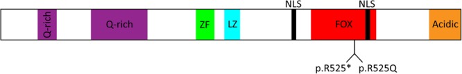
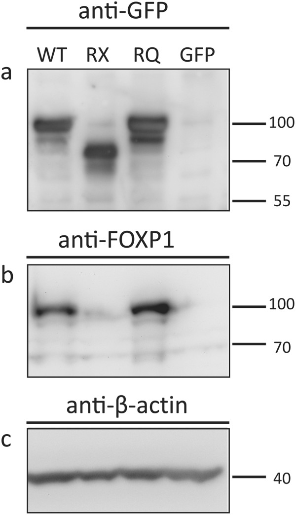
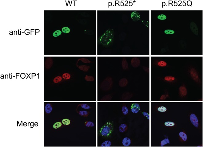
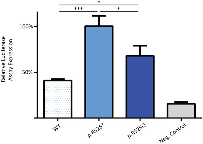
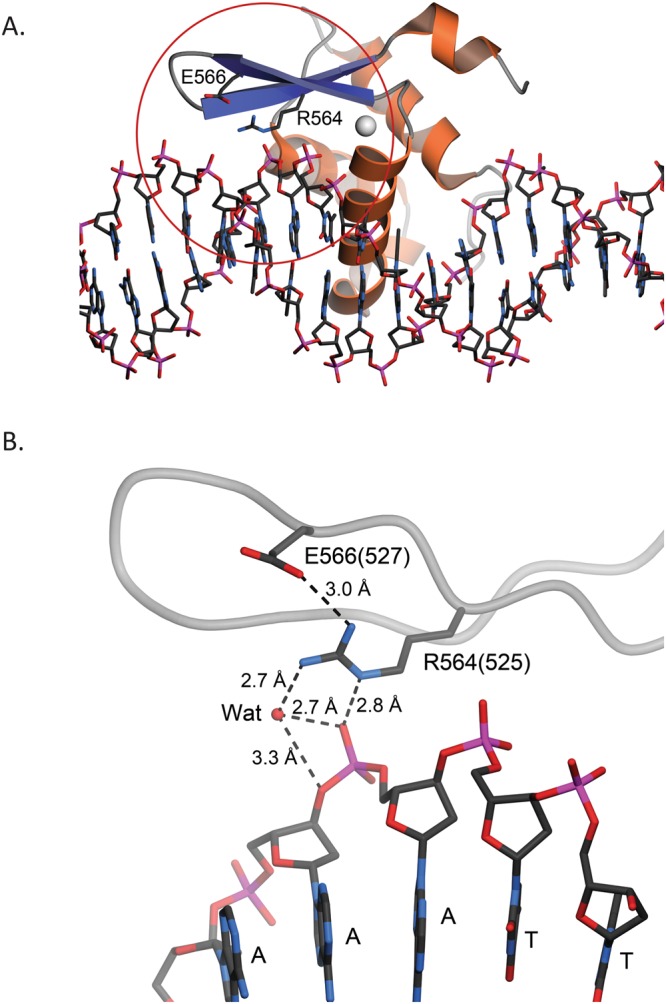
Haploinsufficiency of Forkhead box protein P1 (FOXP1), a highly conserved transcription factor, leads to developmental delay, intellectual disability, autism spectrum disorder, speech delay, and dysmorphic features. Most of the reported FOXP1 mutations occur on the C-terminus of the protein and cluster around to the forkhead domain. All reported FOXP1 pathogenic variants result in abnormal cellular localization and loss of transcriptional repression activity of the protein product. Here we present three patients with the same FOXP1 mutation, c.1574G>A (p.R525Q), that results in the characteristic loss of transcription repression activity. This mutation, however, represents the first reported FOXP1 mutation that does not result in cytoplasmic or nuclear aggregation of the protein but maintains normal nuclear localization.


Conflict of interest statement

The authors declare no competing interests.

Figures

Figure 1
Figure 1
FOXP1 p.R525 mutations map to the FOX DNA-binding domain. FOXP1 protein structure is composed of common conserved domains: an N-terminal Q-rich (polyglutamine) domain, internal zinc-finger (ZF) and leucine zipper (LZ), and a C-terminal forkhead box DNA-binding domain (FOX). Additionally, two nuclear localization signals (NLS) are present at the C-terminus that target the protein to the nucleus under normal conditions. Arginine 525 is localized to the DNA-binding FOX domain nestled between the two NLS.
Figure 2
Figure 2
Expression of FOXP1 transcripts in HeLa cells. Expression constructs containing wild type (WT) FOXP1, p.R525* truncating mutant (RX), p.R525Q missense mutant (RQ) or GFP-only control were transiently transfected into HeLa cells. (a,b) The WT and RQ mutant produce full-length protein while the truncating mutant results in a loss of 152 amino acids at the C-terminus of the protein and a band near 75 kDa. Equal mass was loaded in each lane and (c) beta-actin was used as loading control. Blots are cropped to show bands of interest. Full-length blots are presented in Supplementary Figure 1.
Figure 3
Figure 3
Localization of the FOXP1 proteins in HeLa cells. Expression constructs containing WT FOXP1, truncating p.R525* mutant, and p.R525Q missense mutant were transiently transfected into HeLa cells. WT and p.R525Q mutant properly localize to the nucleus in identical patterns while the p.R525* truncation results in cytoplasmic aggregation.
Figure 4
Figure 4
Luciferase activity resulting from transcriptional repression of the SV40 promoter. The bars represent the relative transcriptional activity of the SV40 promoter resulting from co-transfection with plasmids containing the FOXP1 nonsense mutants. The results are mean values ± SEM of experiments performed in technical triplicate. RX and RQ variants result in severely diminished transcriptional repression of the SV40 promoter as compared to WT levels.
Figure 5
Figure 5
Residue R525 of human FOXP1 is predicted to interact with both DNA and nearby residues. (A) Chain K of the forkhead domain of human FOXP2 (PDB 2A07) bound to DNA. DNA is depicted in ball-and-stick and FoxP2 is shown as a ribbon diagram with α-helices colored orange and β-strands colored blue. The forkhead domain of FoxP2 shares 87% sequence identity with human FoxP1. Residue R564 in FOXP2 (corresponding to R525 in FOXP1) is located at the protein-DNA interface. The red circle shows the area that is depicted in panel B. (B) A closer view of the environment of R564 (R525 in human FOXP1). Residue numbers in PDB 2A07 are indicated; their corresponding human FOXP1 numbering shown in parentheses. Potential hydrogen bonding interactions are shown in dotted lines with distances indicated in Ångstroms. “Wat” indicates an ordered water molecule mediating H-bonds between protein and DNA. R525 in human FOXP1 is likely to make similar contacts, which the R525Q mutation likely disrupts. Figure made with POVScript+.

Similar articles

Cited by

References

    1. Myers A, et al. FOXP1 haploinsufficiency: Phenotypes beyond behavior and intellectual disability? American journal of medical genetics. Part A. 2017;173:3172–3181. doi: 10.1002/ajmg.a.38462. - DOI - PubMed
    1. Siper PM, et al. Prospective investigation of FOXP1 syndrome. Molecular autism. 2017;8:57. doi: 10.1186/s13229-017-0172-6. - DOI - PMC - PubMed
    1. Hu H, et al. Foxp1 is an essential transcriptional regulator of B cell development. Nature immunology. 2006;7:819–826. doi: 10.1038/ni1358. - DOI - PubMed
    1. Shi C, et al. Integrin engagement regulates monocyte differentiation through the forkhead transcription factor Foxp1. The Journal of clinical investigation. 2004;114:408–418. doi: 10.1172/JCI21100. - DOI - PMC - PubMed
    1. Shu W, et al. Foxp2 and Foxp1 cooperatively regulate lung and esophagus development. Development. 2007;134:1991–2000. doi: 10.1242/dev.02846. - DOI - PubMed

Publication types

MeSH terms