

Spinal Enumeration by Morphologic Analysis of Spinal Variants: Comparison to Counting in a Cranial-To-Caudal Manner
- PMID: 30386145
- PMCID: PMC6201970
- DOI: 10.3348/kjr.2018.19.6.1140
Spinal Enumeration by Morphologic Analysis of Spinal Variants: Comparison to Counting in a Cranial-To-Caudal Manner
Abstract
Objective: To compare the spinal enumeration methods that establish the first lumbar vertebra in patients with spinal variants.
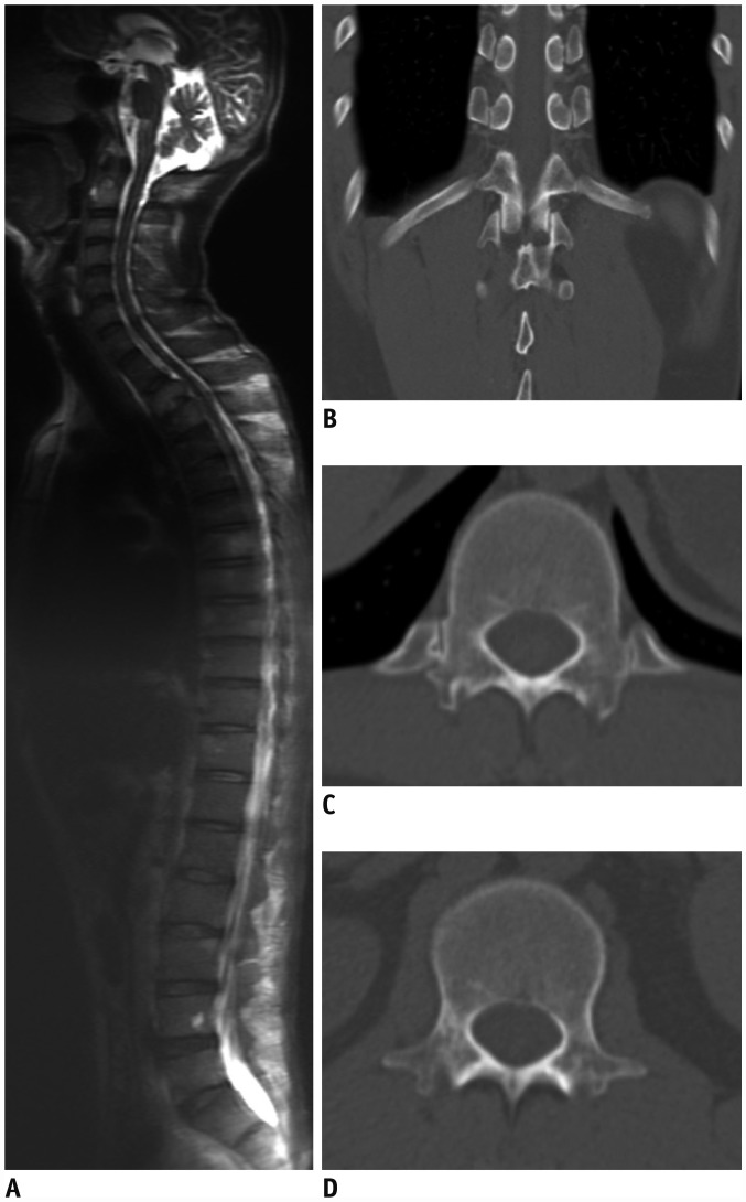
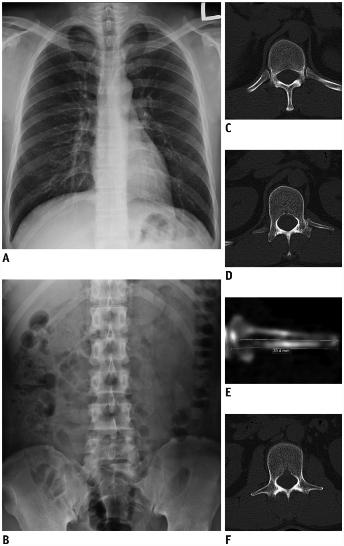
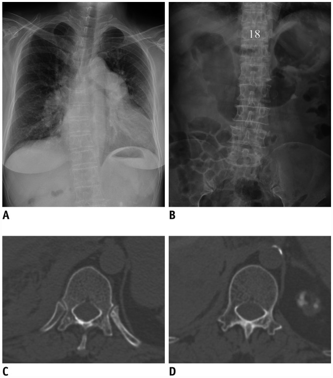
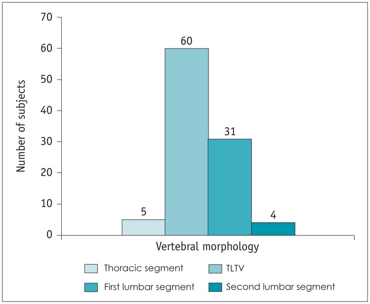
Materials and methods: Of the 1446 consecutive patients who had undergone computed tomography of the spine from March 2012 to July 2016, 100 patients (62 men, 38 women; mean age, 47.9 years; age range, 19-88 years) with spinal variants were included. Two radiologists (readers 1 and 2) established the first lumbar vertebra through morphologic analysis of the thoracolumbar junction, and labeled the vertebra by counting in a cranial-to-caudal manner. Inter-observer agreement was established. Additionally, reader 1 detected the 20th vertebra under the assumption that there are 12 thoracic vertebra, and then classified it as a thoracic vertebra, lumbar vertebra, or thoracolumbar transitional vertebra (TLTV), on the basis of morphologic analysis.
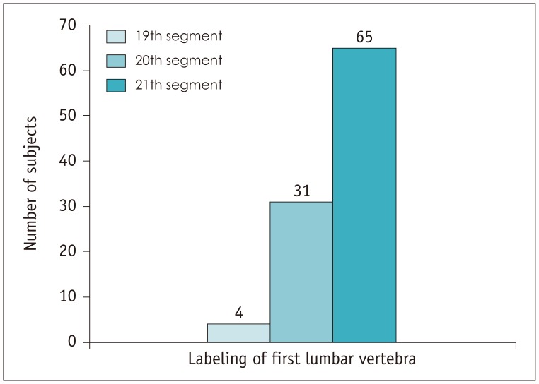
Results: The first lumbar vertebra, as established by morphologic analysis, was labeled by each reader as the 21st segment in 65.0% of the patients, as the 20th segment in 31.0%, and as the 19th segment in 4.0%. Inter-observer agreement between the two readers in determining the first lumbar vertebra, based on morphologic analysis, was nearly perfect (κ value: 1.00). The 20th vertebra was morphologically classified as a TLTV in 60.0% of the patients, as the first lumbar segment in 31.0%, as the second lumbar segment in 4.0%, and as a thoracic segment in 5.0%.
Conclusion: The establishment of the first lumbar vertebra using morphologic characteristics of the thoracolumbar junction in patients with spinal variants was consistent with the morphologic traits of vertebral segmentation.
Keywords: Anatomic variation; Computed tomography; Enumeration; Lumbar vertebra; Spine; Thoracolumbar transitional vertebra.
Figures
Comment in
-
RE: Spinal Enumeration by Morphologic Analysis of Spinal Variants: Comparison to Counting in a Cranial-To-Caudal Manner.Korean J Radiol. 2019 Apr;20(4):693-694. doi: 10.3348/kjr.2018.0834. Korean J Radiol. 2019. PMID: 30887751 Free PMC article. No abstract available.
Similar articles
-
Thoracolumbar junction: morphologic characteristics, various variants and significance.Br J Radiol. 2016 Aug;89(1064):20150784. doi: 10.1259/bjr.20150784. Epub 2016 Jan 19. Br J Radiol. 2016. PMID: 26670155 Free PMC article.
-
Frequency of Coexistent Spinal Segment Variants: Retrospective Analysis in Asymptomatic Young Adults.AJNR Am J Neuroradiol. 2023 Dec 29;45(1):119-126. doi: 10.3174/ajnr.A8071. AJNR Am J Neuroradiol. 2023. PMID: 38123916 Free PMC article.
-
Transitional vertebrae and numerical variants of the spine : prevalence and relationship to low back pain or degenerative spondylolisthesis.Bone Joint J. 2021 Jul;103-B(7):1301-1308. doi: 10.1302/0301-620X.103B7.BJJ-2020-1760.R1. Bone Joint J. 2021. PMID: 34192932
-
Spine segmentation and enumeration and normal variants.Radiol Clin North Am. 2012 Jul;50(4):587-98. doi: 10.1016/j.rcl.2012.04.003. Epub 2012 May 3. Radiol Clin North Am. 2012. PMID: 22643386 Review.
-
Anatomical variability, morphofunctional characteristics, and clinical relevance of accessory ossicles of the back: implications for spinal pathophysiology and differential diagnosis.J Orthop Surg Res. 2025 Mar 6;20(1):240. doi: 10.1186/s13018-024-05407-2. J Orthop Surg Res. 2025. PMID: 40050963 Free PMC article. Review.
Cited by
-
The morphological consequences of segmentation anomalies in the human sacrum.Am J Biol Anthropol. 2022 Apr;177(4):690-707. doi: 10.1002/ajpa.24466. Epub 2021 Dec 29. Am J Biol Anthropol. 2022. PMID: 36787761 Free PMC article.
-
RE: Spinal Enumeration by Morphologic Analysis of Spinal Variants: Comparison to Counting in a Cranial-To-Caudal Manner.Korean J Radiol. 2019 Apr;20(4):693-694. doi: 10.3348/kjr.2018.0834. Korean J Radiol. 2019. PMID: 30887751 Free PMC article. No abstract available.
References
-
- Narita Y, Kuratani S. Evolution of the vertebral formulae in mammals: a perspective on developmental constraints. J Exp Zool B Mol Dev Evol. 2005;304:91–106. - PubMed
-
- Hanson EH, Mishra RK, Chang DS, Perkins TG, Bonifield DR, Tandy RD, et al. Sagittal whole-spine magnetic resonance imaging in 750 consecutive outpatients: accurate determination of the number of lumbar vertebral bodies. J Neurosurg Spine. 2010;12:47–55. - PubMed
-
- Galis F. Why do almost all mammals have seven cervical vertebrae? Developmental constraints, hox genes, and cancer? J Exp Zool. 1999;285:19–26. - PubMed
-
- Akbar JJ, Weiss KL, Saafir MA, Weiss JL. Rapid MRI detection of vertebral numeric variation. AJR Am J Roentgenol. 2010;195:465–466. - PubMed
-
- Thawait GK, Chhabra A, Carrino JA. Spine segmentation and enumeration and normal variants. Radiol Clin North Am. 2012;50:587–598. - PubMed
