

Case Reports
doi: 10.1016/j.radcr.2018.10.009.
eCollection 2019 Jan.
Renal cell carcinoma presenting as epistaxis from a nasal cavity metastasis
Affiliations
- PMID: 30386450
- PMCID: PMC6205054
- DOI: 10.1016/j.radcr.2018.10.009
Item in Clipboard
Case Reports
Renal cell carcinoma presenting as epistaxis from a nasal cavity metastasis
Radiol Case Rep.
.
Abstract
We present a 58-year-old gentleman who initially presented to the otolaryngology clinic with new onset epistaxis revealing a palpable facial mass that was subsequently biopsied revealing metastatic renal cell carcinoma. We hope to present an interesting case highlighting the rarity of this disease and unusual presentation in which the presence of the primary renal cell carcinoma was recognized only after biopsy.
Keywords: Metastatic renal cell carcinoma; Paranasal sinuses.
Figures
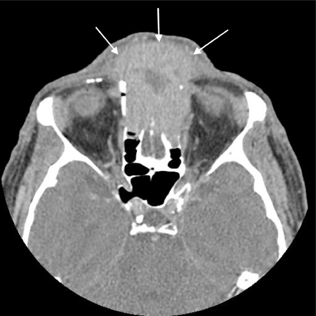
Axial contrast-enhanced computed tomography through the orbits shows an expansile enhancing soft tissue mass occupying the ethmoid sinuses and soft tissues of the anterior face. This image also demonstrates erosion of the anterior ethmoid air cells and nasal bones.
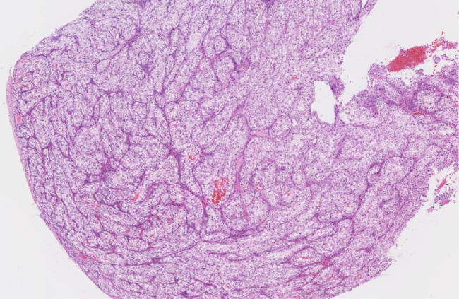
Low-power hematoxylin and eosin image of the lesion demonstrates heterogeneous grouped clear and/or eosinophilic cytoplasm cells with networks of small, thin-walled, ``chicken wire'' vasculature.
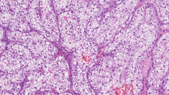
High-power hematoxylin and eosin of the lesion demonstrates heterogeneous group clear and/or eosinophilic cytoplasm cells with network of small, thin-walled, ``chicken wire'' vasculature. Clusters of variably cohesive large epithelioid cells with large, eccentric, and irregular nuclei, and prominent nucleoli.
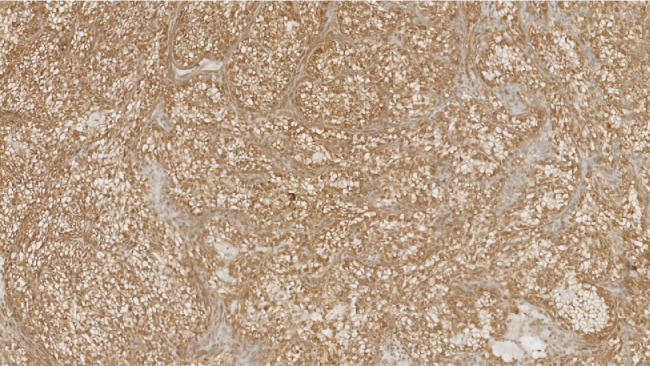
Positive renal cell carcinoma immunohistochemical stain supportive of metastatic renal cell carcinoma.
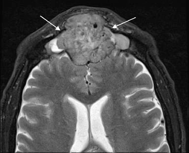
Axial T2-weighted image shows a heterogeneous mass in the frontal sinus with extension through the posterior wall and involving the extra-axial space of the anterior cranial fossa. Note the presence of several flow voids indicating vascularity.
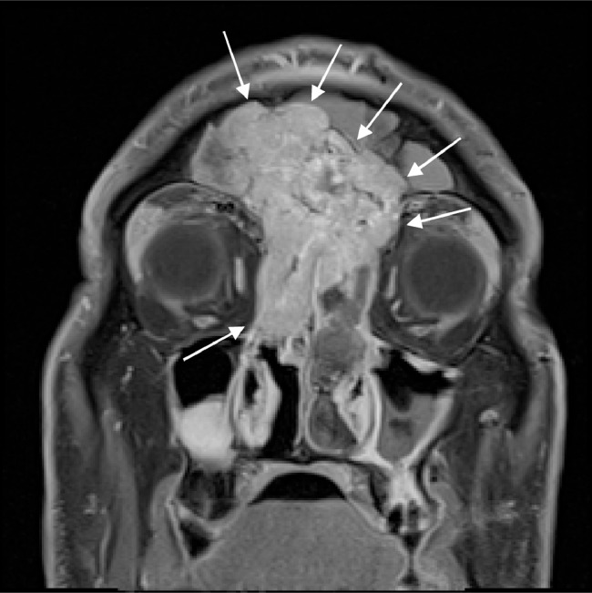
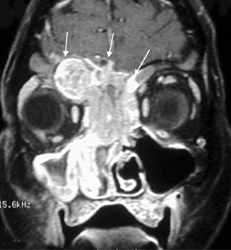
Coronal postcontrast T1W image shows a large infiltrating enhancing mass centered in the sinonasal cavity and eroding through the cribriform plate and ethmoid sinus. Enhancing soft tissue is also extending into the left extraconal orbital fat.
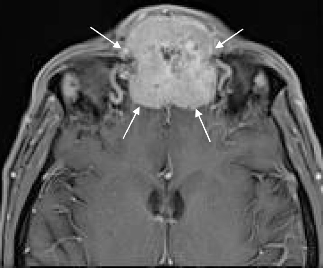
Axial postcontrast T1WI again shows an avidly enhancing mass extending through the frontal sinus and touching the dura of the frontal lobes.
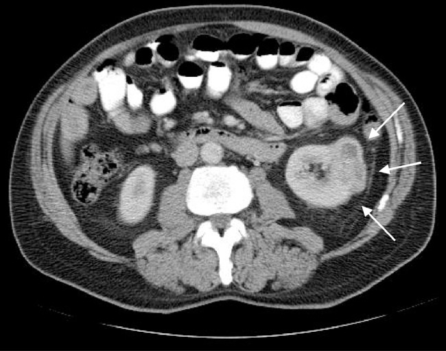
Axial contrast-enhanced CT of the abdomen shows an exophytic enhancing mass at the interpole of the kidney indicating the site of primary malignancy.
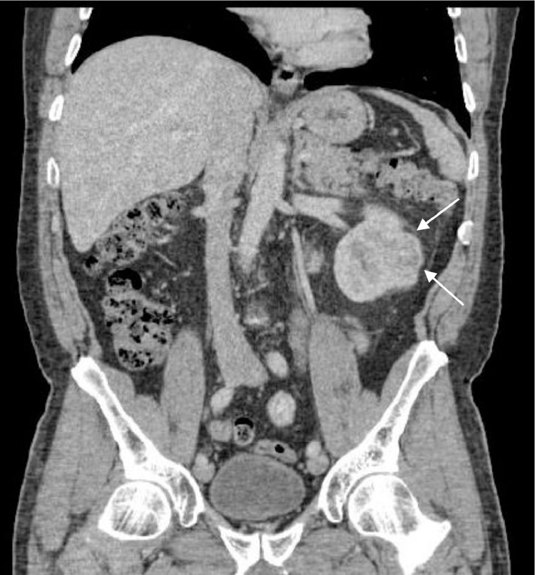
Coronal contrast-enhanced CT of the abdomen shows the solid enhancing mass at the interpole of the left kidney indicating the site of primary malignancy.
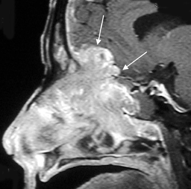
Sagittal contrast-enhanced T1W image of the nasal cavity shows an enhancing mass invading through the cribriform plate and anterior cranial fossa with intracranial extension.
Coronal contrast-enhanced T1W image of the nasal cavity shows an enhancing mass centered in the paranasal sinuses and extending superiorly into the anterior cranial fossa.
References
-
- Bechara G.R., Resende Junior J.A.D., Gouveia H.A., de Souza T.A. Metastasis to paranasal sinuses as the first presenting sign of renal cell carcinoma. Open J Urol. 2012;2:28–31.
-
- Bechara G.R., Resende Junior J.A.D., Gouveia H.A., de Souza T.A. Metastasis to paranasal sinuses as the first presenting sign of renal cell carcinoma. Open J Urol. 2012;2:28–31.
Publication types
LinkOut - more resources
Full Text Sources
