Editorial
. 2018 Dec;285(24):4575-4589.
doi: 10.1111/febs.14688. Epub 2018 Nov 19.

The structure of PghL hydrolase bound to its substrate poly-γ-glutamate

Affiliations
Editorial

The structure of PghL hydrolase bound to its substrate poly-γ-glutamate

Sneha Ramaswamy et al. FEBS J. 2018 Dec.

Abstract

The identification of new strategies to fight bacterial infections in view of the spread of multiple resistance to antibiotics has become mandatory. It has been demonstrated that several bacteria develop poly-γ-glutamic acid (γ-PGA) capsules as a protection from external insults and/or host defence systems. Among the pathogens that shield themselves in these capsules are Bacillus anthracis, Francisella tularensis and several Staphylococcus strains. These are important pathogens with a profound influence on human health. The recently characterised γ-PGA hydrolases, which can dismantle the γ-PGA-capsules, are an attractive new direction that can offer real hope for the development of alternatives to antibiotics, particularly in cases of multidrug resistant bacteria. We have characterised in detail the cleaving mechanism and stereospecificity of the enzyme PghL (previously named YndL) from Bacillus subtilis encoded by a gene of phagic origin and dramatically efficient in degrading the long polymeric chains of γ-PGA. We used X-ray crystallography to solve the three-dimensional structures of the enzyme in its zinc-free, zinc-bound and complexed forms. The protein crystallised with a γ-PGA hexapeptide substrate and thus reveals details of the interaction which could explain the stereospecificity observed and give hints on the catalytic mechanism of this class of hydrolytic enzymes.

Keywords: PGA-hydrolase; antimicrobial drug; biofilm inhibitor; poly-γ-glutamate; virulence.


Conflict of interest statement

The authors declare no conflict of interest.

Figures

Figure 1
Figure 1
PghL is a folded and relatively stable protein. (A) Size exclusion chromatography profile of WT‐PghL. The concentration of the protein used was 0.05 mg·mL −1. (B) Comparison of the far‐UV spectrum of apo (grey) and native PghL (black) at 30 °C. (C) Thermal scan of PghL monitored by CD. Black curve: PghL in 20 mm sodium phosphate, at pH 6.0 using a protein concentration of 5 μm. The calculated melting point was 55 °C. Grey curve: PghL in 5 mm Hepes and 150 mm NaF at pH 6.0 using a protein concentration of 5 μm. The calculated melting point was 53 °C.
Figure 2
Figure 2
PghL‐catalysed hydrolysis of γ‐PGA. Representative chromatograms showing the appearance of reaction products over time. HPLC chromatograms for: (A) Time point 0: only the Sanger reagent is present. (B) After 1 h: small peaks with the retention times of dimers (γ‐GluGlu) and trimers (γ‐Glu‐γ‐GluGlu) appear. (C) After 3 h: peaks attributable to dimers and trimers are well defined; other peaks attributable to higher molecular weight oligomers emerge from a broad peak caused by the progressively degraded polymeric material. (D) After 6 h: peaks attributable to up to hexamers are clearly visible. Signals of putative hepta‐ and octapeptide are distinguishable as shoulder peaks. (E) After 24 h: only peaks assigned to the di‐, tri‐, tetra‐ and pentapeptide are visible. (F) chromatogram of a mixture of authentic di‐ (4), tri‐(3), tetra‐(2) and penta‐peptides (1) used as reference compounds is resolved. Peak 5 is due to the excess Sanger's reagent used for pre‐column derivatisation. Peak 6 accompanies peak 5 as an unidentified impurity.
Figure 3
Figure 3
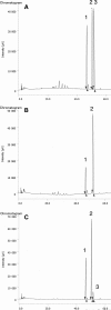
Stereochemical outcome of PghL‐catalysed hydrolysis of γ‐PGA. Chromatograms were obtained after acidic hydrolysis of the samples and pre‐column derivatisation of the resulting glutamic acid with Nα‐(2,4‐dinitro‐5‐fluorophenyl)‐L‐valinamide. (A) Starting γ‐PGA after acidic hydrolysis; 1 = L‐Glu, 2 =  D‐Glu, 3: Nα‐(2,4‐dinitro‐5‐fluorophenyl)‐L‐valinamide. (B) γ‐GluGlu from enzymatic reaction after acidic hydrolysis; 1 = L‐Glu, 2 = Nα‐(2,4‐dinitro‐5‐fluorophenyl)‐L‐valinamide. (C) High‐molecular weight fraction from PghL‐catalysed reaction after acidic hydrolysis; 1 = L‐Glu, 2 =  D‐Glu, 3: Nα‐(2,4‐dinitro‐5‐fluorophenyl)‐L‐valinamide.
Figure 4
Figure 4
PghL crystal structures. Top panel: Ribbon diagrams for apoPghL (in yellow, left), native PghL (in deep teal, middle) and PghL‐γ‐PGA complex structure (in light pink, right; γ‐PGA shown in blue). Residues involved in zinc coordination are shown as sticks. Bottom panel: 2Fo‐Fc maps for apoPghL with the protein in yellow (left),native PghL with the protein in deep teal and sodium citrate (FLC) in light orange (middle)and PghL‐γ‐PGA in light pink and γ‐PGA in blue (right). The maps were contoured at 2σ.
Figure 5
Figure 5
Zinc coordination site and comparison of PghL with carboxypeptidases. (A) Residues involved are shown as sticks. Apo PghL (left, in yellow), native PghL (middle, in deep teal; FLC: sodium citrate) and PghL‐γ‐PGA complex structure (right, in light pink; γ‐PGA shown in blue). Distances are in Å. (B) Superposition of PghL (in pink) and PghP (PDB code: 3a9l, in white). (C) Superposition of PghL (in pink) and carboxypeptidase A (PDB code: 3cpa, in grey) as superposed by the Dali server (http://ekhidna.biocenter.helsinki.fi/dali_server/). Zinc atoms are shown as grey or red spheres.
Figure 6
Figure 6
The structure of a PghL‐γ‐PGA complex. (A) Omit map contoured at 1σ showing the electron density for the hexapeptide; PghL is shown in ribbon representation (light pink). (B) PghL‐γ‐PGA complex structure with the enzyme in light pink and γ‐PGA shown in blue. Residues involved in hexapeptide coordination are shown as sticks. The zinc ion is shown in red. (C) Close‐up and network of interactions. The view is approximately rotated by 120° as compared to (B). (D) Carboxypeptidase A (3cpa; shown in grey) interacting with a tyrosine residue that mimics the substrate represented in the same view as PghL. The two structures were first structurally superposed. The zinc ion is shown in red. (E) Formula of the hexapeptide indicating the interactions with the enzyme and the zinc ion. Interactions that involve the protein side chains, and are thus sequence‐specific, are boxed. The alpha carbons of PGA are labelled sequentially.
Figure 7
Figure 7
Sequence alignment of the γ‐PGA cleaving proteins from B. subtilis. The numbering of the PghL construct used in this study is indicated in the last row, whereas the numbering according to the UNIPROT entries are given in the row above. The residues involved in zinc binding and γ‐PGA sequence‐specific interactions are indicated with red stars and plus symbols respectively.
Figure 8
Figure 8
The key role of Arg171Ser in γ‐PGA anchoring. (A) Close‐up of the interaction between Arg171 and Glu3 of γ‐PGA. (B) Degradation by wild‐type and Arg171Ser mutant PghL. γ‐PGA from B. subtilis was treated with 100 ng of either wild‐type or Arg171Ser mutant hydrolase for 1 h at 37 °C and separated on a 2% agarose gel in TAE buffer. Identity of the enzyme is indicated above.

References

    1. Candela T & Fouet A (2006) Poly‐gamma‐glutamate in bacteria. Mol Microbiol 60, 1091–1098. - PubMed
    1. Mamberti S, Prati P, Cremaschi P, Seppi C, Morelli CF, Galizzi A, Fabbi M & Calvio C (2015) γ‐PGA hydrolases of phage origin in Bacillus subtilis and other microbial genomes. PLoSONE. 10, e0130810. - PMC - PubMed
    1. Green BD, Battisti L, Koehler TM, Thorne CB & Ivins BE (1985) Demonstration of a capsule plasmid in Bacillus anthracis . Infect Immun 49, 291–297. - PMC - PubMed
    1. Fouet A & Mock M (1996) Differential influence of the two Bacillus anthracis plasmids on regulation of virulence gene expression. Infect Immun 64, 4928–4932. - PMC - PubMed
    1. Jang J, Cho M, Lee H, Cha K, Chun J, Hong K, Park J & Rhie G (2013) Monoclonal antibody against the poly‐gamma‐D‐glutamic acid capsule of Bacillus anthracis protects mice from enhanced lethal toxin activity due to capsule and anthrax spore challenge. Biochim Biophys Acta 1830, 2804–2812. - PubMed

Publication types