Review
. 2019 Jun 20:88:811-837.
doi: 10.1146/annurev-biochem-013118-111654. Epub 2018 Nov 2.

Botulinum and Tetanus Neurotoxins

Affiliations
Review

Botulinum and Tetanus Neurotoxins

Min Dong et al. Annu Rev Biochem. .

Abstract

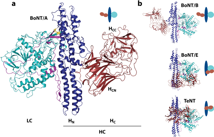
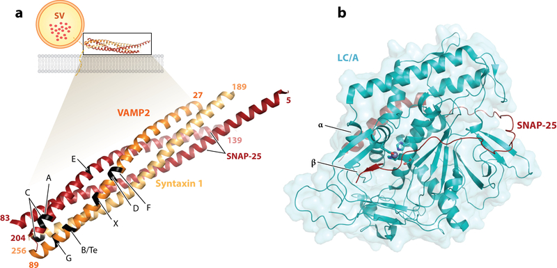
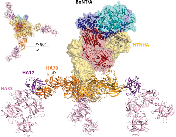
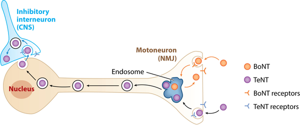
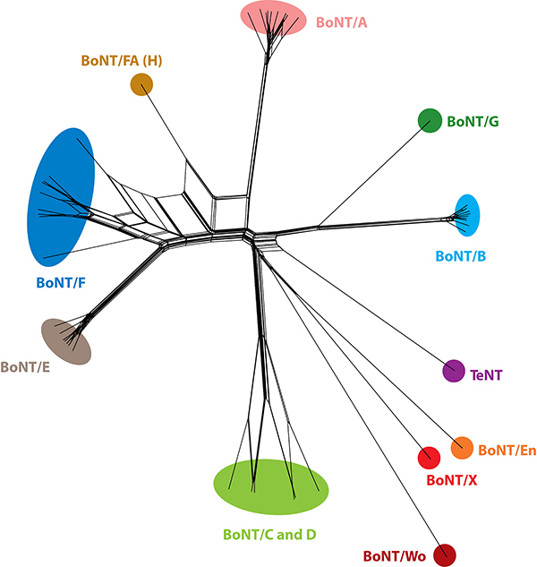
Botulinum neurotoxins (BoNTs) and tetanus neurotoxin (TeNT) are the most potent toxins known and cause botulism and tetanus, respectively. BoNTs are also widely utilized as therapeutic toxins. They contain three functional domains responsible for receptor-binding, membrane translocation, and proteolytic cleavage of host proteins required for synaptic vesicle exocytosis. These toxins also have distinct features: BoNTs exist within a progenitor toxin complex (PTC), which protects the toxin and facilitates its absorption in the gastrointestinal tract, whereas TeNT is uniquely transported retrogradely within motor neurons. Our increasing knowledge of these toxins has allowed the development of engineered toxins for medical uses. The discovery of new BoNTs and BoNT-like proteins provides additional tools to understand the evolution of the toxins and to engineer toxin-based therapeutics. This review summarizes the progress on our understanding of BoNTs and TeNT, focusing on the PTC, receptor recognition, new BoNT-like toxins, and therapeutic toxin engineering.

Keywords: bacterial toxin; botulinum neurotoxin; clostridium; protein engineering; tetanus neurotoxin; toxin.


Figures

Figure 1
Figure 1
(a) Structure of the botulinum neurotoxin A (BoNT/A) [Protein Data Bank (PDB) 3BTA]. The light chain (LC, catalytic domain) is shown in cyan. The heavy chain (HC) is composed of the translocation domain (HN) shown in blue with the belt in purple, and of the binding domain (HC) in red. The two subdomains of HC are indicated with the C-terminal (HCC) and N-terminal (HCN) regions. The disulfide bond linking light and heavy chains is shown as yellow sticks. The zinc ion is shown as a gray sphere. (b) The crystal structures of BoNT/B (PDB 1EPW), BoNT/E (PDB 3FFZ), and tetanus neurotoxin (TeNT) (PDB 5N0B). Domains are colored as in panel a, with a schematic representation highlighting the domain arrangements in the clostridial neurotoxins.
Figure 2
Figure 2
(a) Schematic of the SNARE complex membrane fusion event and cleavage sites for BoNTs/TeNT. VAMP2 (orange), SNAP-25 (red), and syntaxin 1 (yellow) (PDB 1N7S) form a complex that mediates fusion of the vesicular membrane with the presynaptic membrane (lipid bilayer in gray) and allows neurotransmitter release (represented as red dots in the SV). The structures of the SNARE complex (PDB 1N7S) with VAMP2, SNAP-25, and syntaxin 1 are enlarged, with the cleavage site for each toxin indicated. It should be noted that the toxins cleave their substrate only when in their free form, with the complex being resistant to proteolysis. (b) Structure of the LC/A (cyan) in complex with SNAP-25 (red) (PDB 1XTG). The conserved active site residues (HExxH…E) are shown as sticks, with the zinc ion as a gray sphere. The exosites (α and β), important for substrate binding, are indicated. Abbreviations: BoNTs, botulinum neurotoxins; LC/A, light chain of botulinum neurotoxin A; PDB, Protein Data Bank; SNARE, soluble N-ethylmaleimide-sensitive factor attachment protein receptor; SV, synaptic vesicle; TeNT, tetanus neurotoxin.
Figure 3
Figure 3
Structure of the L-PTC/A complex, as described by Lee et al. (23), with the M-PTC complex (PDB 3V0A) made of BoNT/A (LC in cyan, HN in blue, and HC in red), the NTNHA (yellow surface), and the HA complex comprised of HA70 (orange), HA17 (purple), and HA33 (pink) (assembled from PDB 4LO4 and 4LO7). The top left corner inset shows the sample complex from below. Abbreviations: BoNT/A, botulinum neurotoxin A; HA, hemagglutinin component; HC, the receptor-binding domain; HN, translocation domain; LC, light chain; L-PTC, large progenitor toxin complex; M-PTC, minimal progenitor toxin complex; NTNHA, nontoxic nonhemagglutinin protein; PDB, Protein Data Bank.
Figure 4
Figure 4
Dual receptor binding. (a) Schematic depicting activity-facilitated binding and entry of BoNTs into neurons: dual receptor recognition of the presynaptic motoneuron, receptor-mediated endocytosis followed by pH-induced conformational change that allows translocation of LC in the cytosol, and cleavage of one of the SNARE proteins by LC. (b) BoNT/B (red) binding to Syt (orange) and ganglioside (dots) from PDB 4KBB. Conserved GBS residues are highlighted in cyan; important hydrophobic residues F47 and F54 of Syt are shown as sticks. (c) BoNT/DC (red) binding to Syt (orange) and the Sial. (dots) from the superposition of PDB 4ISR and PDB 5LR0, respectively. Residues of the hydrophobic loop involved in lipid interaction are shown as sticks (Y1251-W1252-F1253). (d) BoNT/A (red) binding to gSV2 (yellow) and ganglioside (yellow dots) from the superposition of PDB 5JLV and 2VU9, respectively. Conserved GBS residues and F953 are highlighted in cyan. Glycan linked to N559 of SV2C are shown as orange dots. The lipid bilayer is represented in gray. Dotted lines indicate the continuation of the protein receptors toward their transmembrane domain. Abbreviations: BoNT, botulinum neurotoxin; GBS, ganglioside binding site; gSV2, glycosylated synaptic vesicle glycoprotein 2; LC, light chain; PDB, Protein Data Bank; Sial., sialyl-T antigen; SNARE, soluble N-ethylmaleimide-sensitive factor attachment protein receptor; SV2, synaptic vesicle glycoprotein 2; Syt, synaptotagmin.
Figure 5
Figure 5
Retrograde transport. Schematic representation of the intracellular pathway followed by the clostridial neurotoxins. BoNTs mainly act at the NMJ (orange arrows), whereas TeNT undergoes retrograde transport along the axon (black arrows) and transcytosis to reach the inhibitory interneurons of the CNS (blue arrows). Abbreviations: BoNTs, botulinum neurotoxins; CNS, central nervous system; NMJ, neuromuscular junction; TeNT, tetanus neurotoxin.
Figure 6
Figure 6
A phylogenetic split network covering the clostridial neurotoxins and selected homologs. The diagram illustrates their potential evolutionary relationships, as well as conflicts arising from chimerisms, based on their protein sequences (133). The sequences were clustered with UCLUST to 98% identity. Abbreviations: BoNT, botulinum neurotoxin; TeNT, tetanus neurotoxin.

References

    1. Alouf JE. 2006. A 116-year story of bacterial protein toxins (1888–2004): from “diphtheritic poison” to molecular toxinology In The Comprehensive Sourcebook of Bacterial Protein Toxins, ed. Alouf J, Popoff M, pp. 3–21. Burlington, MA: Academic; 3rd ed.
    1. Gill DM. 1982. Bacterial toxins: a table of lethal amounts. Microbiol. Rev. 46: 86–94 - PMC - PubMed
    1. Schantz EJ, Johnson EA. 1992. Properties and use of botulinum toxin and other microbial neurotoxins in medicine. Microbiol. Rev. 56: 80–99 - PMC - PubMed
    1. Johnson EA. 1999. Clostridial toxins as therapeutic agents: benefits of nature’s most toxic proteins. Annu. Rev. Microbiol. 53: 551–75 - PubMed
    1. Arnon SS, Schechter R, Inglesby TV, Henderson DA, Bartlett JG, et al. 2001. Botulinum toxin as a biological weapon: medical and public health management. JAMA 285: 1059–70 - PubMed

Publication types

LinkOut - more resources