. 2018 Nov 2;13(11):e0206568.
doi: 10.1371/journal.pone.0206568. eCollection 2018.

Validation of near infrared fluorescence (NIRF) probes in vivo with dual laser NIRF endoscope

Affiliations

Validation of near infrared fluorescence (NIRF) probes in vivo with dual laser NIRF endoscope

Manisha Shrivastav et al. PLoS One. .

Erratum in

Abstract

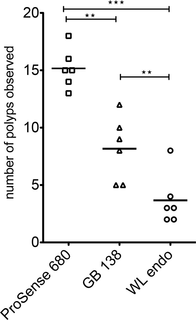
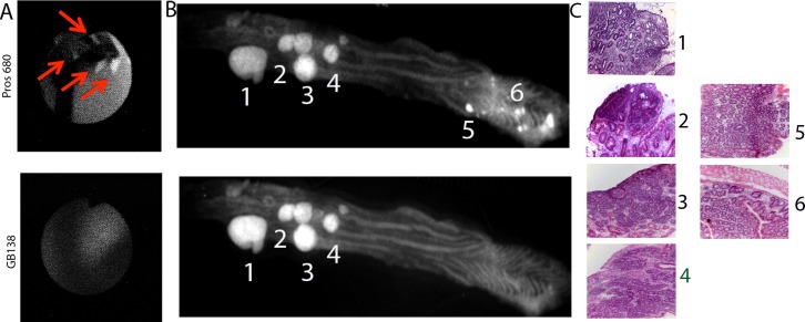
Purpose: The development of NIRF cathepsin activity probes offered the ability to visualize tumor associated tumor reaction and act as a surrogate marker to delineate the dysplastic lesions. One major type is a NIRF substrate of cathepsins (SBP), which is involved in catalytic way to produce high levels of fluorescence emission. The other major type (ABP) reacts with active cathepsins in stoichiometric manner since they bind covalently with their active center. Little is known about the sensitivity and the specificity of the NIRF probes to detect autochthonous developed dysplastic lesions. Dual laser NIRF endoscope provides a good tool to determine the efficiency of various NIRF probes in vivo in the same lesions.

Experimental design: In the current study, we validated both types of NIRF probes by using the dual laser NIRF endoscope to detect lesions colon cancer mouse model (TS4Cre/cAPC +/lox).

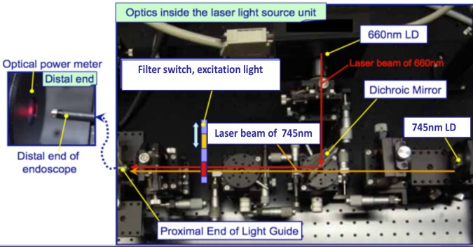
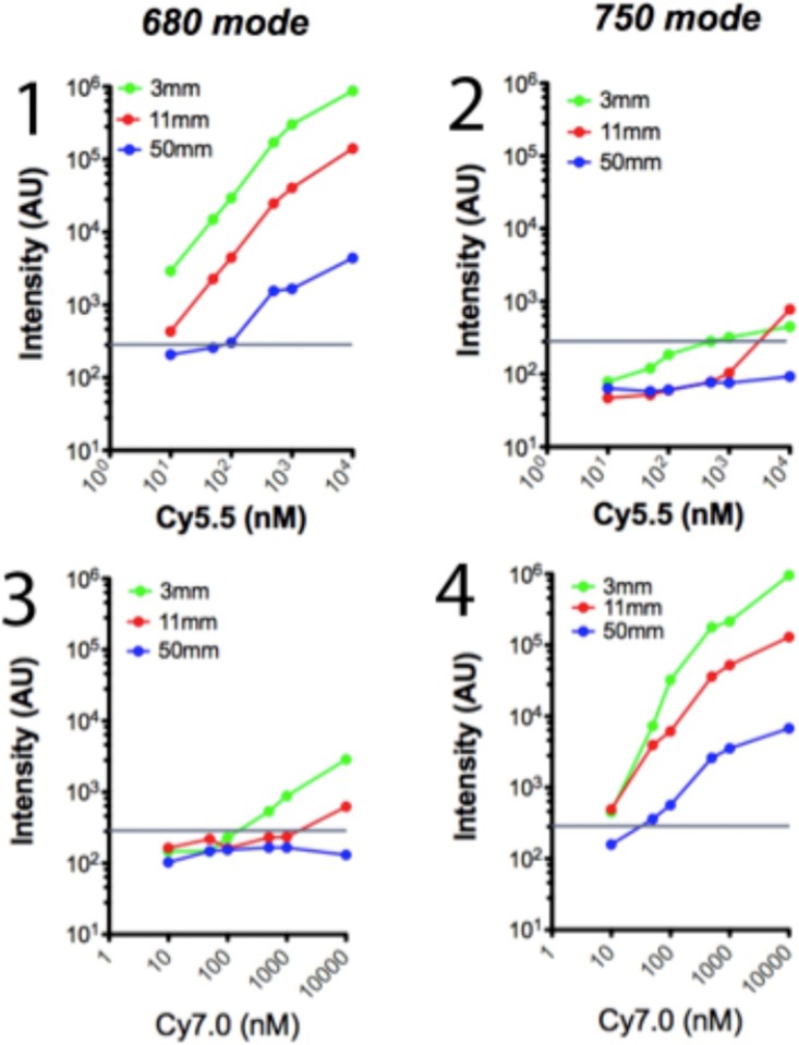
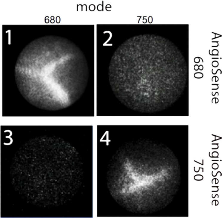
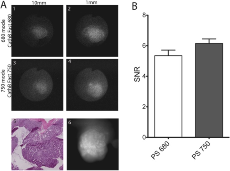
Results: The dual laser NIRF endoscope is emitting equal power with both lasers. It can detect with the same efficiency in 680 mode, as well as, 750 mode when NIFR probes of the same scaffold in vivo. When SBP and ABP were used, our results showed both probes are efficient enough to detect large polyps but small dysplastic lesions could not efficiently imaged with the ABP.

Conclusions: The dual laser NIRF endoscope is a powerful tool to validate probes. The probes that react catalytically with the active center of cathepsins are more efficient than the ones that react stoichiometrically in detecting small lesions.


Conflict of interest statement

The authors have declared that no competing interests exist.

Figures

Fig 1
Fig 1. Optics inside the laser excitation light source unit.
Yellow pointers are indicating the two lasers, the dichroic filter and the filter switch. The light path of 660nm is illustrated with red arrows and the 745nm path is illustrated with brown arrows.
Fig 2
Fig 2. Analysis of cross talk of the two lasers In vitro.
Phantoms have been created containing 101, 5x101, 102, 5x10², 103 and104 nM of fluorophore Cy 5.5 (n = 6) and Cy 7.0 (n = 6). All phantoms were examined with the fiberscope tip being at 3mm (green dots and lines), 11mm (red dots and lines) and 50mm (blue dots and lines) The Cy 5.5 phantoms were examined with the 680 mode (Fig 2, 1) and 750 mode (Fig 2, 2), and the Cy 7.0 were examined with 680 mode (Fig 2, 3) and 750 mode (Fig 2, 4). The images of the phantoms were recorded and their image intensity were calculated with image J and recorded as intensity arbitrary units (AU). The horizontal line represents the electronic noise of the endoscopy system.
Fig 3
Fig 3. Representative images of vessels injected with AngioSense 680 (1&2) or AngioSense 750 (3&4) All mice were either imaged with 680 mode (1&3) and 750 mode (2&4).
Fig 4
Fig 4. Representative images of colon polyp incubated with both Cath B fast 680 and Cath B fast 750 and imaged with 680 mode (1&2) and 750 mode (3&4) at 10mm (1&3) or 1mm (3&4).
The same polyp was imaged for reflectance fluorescence at 680nm (6), and the tissue was stained with H&E (5). The cumulative results of SNR of 6 mice imaged. There is no significant difference of the SNR of the two modes of examination.
Fig 5
Fig 5. Number of polyps detected in 6 mice injected with both ProSense 680 and GB138 according to materials and methods.
The mice were endoscopied with white light (WL endo), 750 mode (GB138) and 680 mode (ProSense 680).
Fig 6
Fig 6. Representative images of mouse colon.
A) endoscopic images of the small polyps visible with 680 mode and not with 750 mode. B) flayed open colon imaged with reflectance fluorescence under 660nm and 750 nm light. C) H&E of the areas with high emissions.

References

    1. Rutter MD. Surveillance programmes for neoplasia in colitis. J Gastroenterol. 2011;46 Suppl 1: 1–5. 10.1007/s00535-010-0309-2 - DOI - PubMed
    1. Mayinger B, Neumann F, Kastner C, Degitz K, Hahn EG, Schwab D. Early detection of premalignant conditions in the colon by fluorescence endoscopy using local sensitization with hexaminolevulinate. Endoscopy. 2008;40: 106–109. 10.1055/s-2007-967019 - DOI - PubMed
    1. Yoshii S, Nojima M, Nosho K, Omori S, Kusumi T, Okuda H, et al. Factors associated with risk for colorectal cancer recurrence after endoscopic resection of t1 tumors. Clin Gastroenterol Hepatol. 2014;12: 292–302.e3. 10.1016/j.cgh.2013.08.008 - DOI - PubMed
    1. Choi PM, Nugent FW, Schoetz DJ, Silverman ML, Haggitt RC. Colonoscopic surveillance reduces mortality from colorectal cancer in ulcerative colitis. YGAST. 1993;105: 418–424. - PubMed
    1. Cairns SR, Scholefield JH, Steele RJ, Dunlop MG, Thomas HJW, Evans GD, et al. Guidelines for colorectal cancer screening and surveillance in moderate and high risk groups (update from 2002). Gut. 2010;59: 666–689. 10.1136/gut.2009.179804 - DOI - PubMed

Publication types