Review
. 2019 Aug 1;40(29):2444-2454.
doi: 10.1093/eurheartj/ehy670.

Non-invasive imaging of the coronary arteries

Affiliations
Review

Non-invasive imaging of the coronary arteries

Philip D Adamson et al. Eur Heart J. .

Abstract

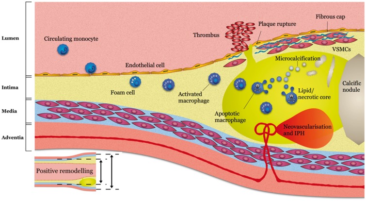
Non-invasive imaging of the coronary arteries is an enterprise in rapid development. From the research perspective, there is great demand for in vivo techniques that can reliably identify features of high-risk plaque that may offer insight into pathophysiological processes and act as surrogate indicators of response to therapeutic intervention. Meanwhile, there is clear clinical need for greater accuracy in diagnosis and prognostic stratification. Fortunately, ongoing technological improvements and emerging data from randomized clinical trials are helping make these elusive goals a reality. This review provides an update on the current status of non-invasive coronary imaging with computed tomography, magnetic resonance, and positron emission tomography with a focus on current clinical applications and future research directions.

Keywords: Computed tomography; Coronary heart disease; Magnetic resonance imaging; Positron emission tomography.


Figures

Figure 1
Figure 1
Imaging targets of high-risk plaque. Reused from Adamson et al. Circulating monocytes migrate into early intimal thickening where they phagocytose lipid becoming foam cells and activated macrophages detectable on 68Ga-DOTATATE positron emission tomography. Vascular remodelling can be detected on computed tomography coronary angiography prior to luminal stenosis developing. As the lipid core develops this can be detected as low-density signal on computed tomography coronary angiography. The resulting hypoxic environment prompts neovascularization with friable vessels prone to intraplaque haemorrhage, both of which can be detected on magnetic resonance coronary angiography. A necrotic core develops with microvesicles arising from apoptotic macrophages and vascular smooth muscle cells giving rise to microcalcifications detectable on 18F-fluoride positron emission tomography before coalescing into more stable calcific nodules detectable on computed tomography calcium scans. Rupture of the fibrous cap may result in intraluminal thrombosis detectable on magnetic resonance coronary angiography.
Figure 2
Figure 2
Complementary roles of non-invasive coronary imaging. The use of non-invasive coronary imaging can be considered in three contexts, each with specific objectives that may inform the choice of imaging modality. (A) Computed tomography angiography provides accurate assessment of coronary stenosis that can guide management of patients with suspected stable angina and rule out Type 1 myocardial infarction in patients with potential acute coronary syndrome. (B) Coronary artery calcium scanning is able to reliably quantify overall atherosclerotic burden and improve risk stratification in asymptomatic individuals. (C) T1-weighted magnetic resonance coronary angiography can identify features of atherosclerotic instability including intracoronary thrombus and intraplaque haemorrhage and may be of value in suspected acute coronary syndrome or asymptomatic risk stratification. (D) Positron emission tomography can employ specific tracers designed to target markers of plaque vulnerability that may improve prognostic assessment or act as a surrogate of therapeutic response in asymptomatic patients.
Figure 3
Figure 3
Adverse coronary plaque characteristics identified on computed tomography coronary angiography. Coronary atherosclerotic plaque features detected using computed tomography coronary angiography including (A) positive remodelling—defined as an outer vessel diameter (large yellow line) 10% greater than the mean diameter of the segments immediately proximal (small yellow line) and distal to the plaque; (B) low attenuation plaque—defined as a focal central area of plaque with an attenuation density of <30 Hounsfield Units (yellow arrow); (C) spotty calcification—defined as focal calcification within the coronary artery wall <3 mm in maximum diameter (yellow arrow); and (D) the ‘napkin ring’ sign—defined as a central area of low attenuation plaque with a peripheral rim of high attenuation (yellow arrow).
Figure 4
Figure 4
Coronary atherosclerosis T1-weighted characterization with integrated anatomical reference (CATCH). T1-weighted magnetic resonance coronary angiogram of a patient who presented with an inferior myocardial infarction shows evidence of a focal high intensity lesion (arrows) in the right coronary artery on magnetic resonance imaging (A and B). Subsequent coronary angiogram demonstrated occlusion of the mid-right coronary artery (C) with restoration of flow following thrombus aspiration (D).
Figure 5
Figure 5
Focal 18F-fluoride and 18F-fluorodeoxyglucose uptake in patients with myocardial infarction and stable angina. (Top row, AC) Patient with acute ST-segment elevation myocardial infarction with (A) proximal occlusion (red arrow) of the left anterior descending artery on invasive coronary angiography and (B) intense focal 18F-fluoride uptake (yellow-red) at the site of the culprit plaque (red arrow) on the combined positron emission and computed tomography coronary angiography (PET-CTCA). Corresponding 18F-fluorodeoxyglucose PET-CT image (C) showing no uptake at the site of the culprit plaque. Note the significant myocardial uptake overlapping with the coronary artery (yellow arrow) and uptake within the oesophagus (blue arrow). (Bottom row) Patient with anterior non-ST-segment elevation myocardial infarction with (D) culprit (red arrow; left anterior descending artery) and bystander non-culprit (white arrow; circumflex artery) lesions on invasive coronary angiography that were both stented during the index admission. Only the culprit lesion had increased 18F-NaF uptake on PET-CT (E) after percutaneous coronary intervention. Corresponding 18F-fluorodeoxyglucose PET-CT (F) showing no uptake either at the culprit or the bystander stented lesion. Note intense uptake within the ascending aorta. Adapted from Joshi et al.

References

    1. Townsend N, Wilson L, Bhatnagar P, Wickramasinghe K, Rayner M, Nichols M.. Cardiovascular disease in Europe: epidemiological update 2016. Eur Heart J 2016;37:3232–3245. - PubMed
    1. Adamson PD, Dweck MR, Newby DE.. The vulnerable atherosclerotic plaque: in vivo identification and potential therapeutic avenues. Heart 2015;101:1755–1766. - PubMed
    1. Pyke D, Symons C.. Calcification of the aortic valve and of the coronary arteries. Br Heart J 1951;13:355–363. - PMC - PubMed
    1. Rifkin RD, Parisi AF, Folland E.. Coronary calcification in the diagnosis of coronary artery disease. Am J Cardiol 1979;44:141–147. - PubMed
    1. Gerber TC, Carr JJ, Arai AE, Dixon RL, Ferrari VA, Gomes AS, Heller GV, McCollough CH, McNitt-Gray MF, Mettler FA, Mieres JH, Morin RL, Yester MV.. Ionizing radiation in cardiac imaging: a science advisory. Circulation 2009;119:1056–1065. - PubMed

Publication types

MeSH terms