. 2018 Nov 28;140(47):16140-16151.
doi: 10.1021/jacs.8b08711. Epub 2018 Nov 14.

A Dual-Function Antibiotic-Transporter Conjugate Exhibits Superior Activity in Sterilizing MRSA Biofilms and Killing Persister Cells

Affiliations

A Dual-Function Antibiotic-Transporter Conjugate Exhibits Superior Activity in Sterilizing MRSA Biofilms and Killing Persister Cells

Alexandra Antonoplis et al. J Am Chem Soc. .

Abstract

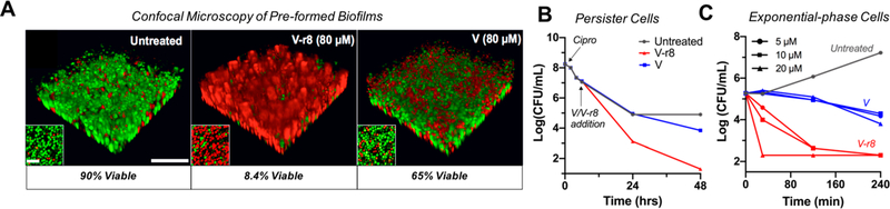
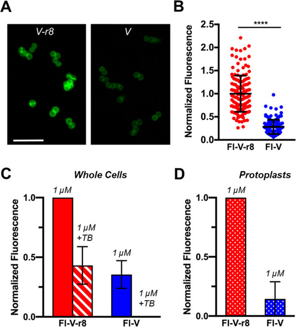
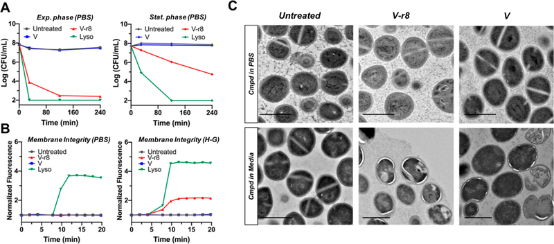
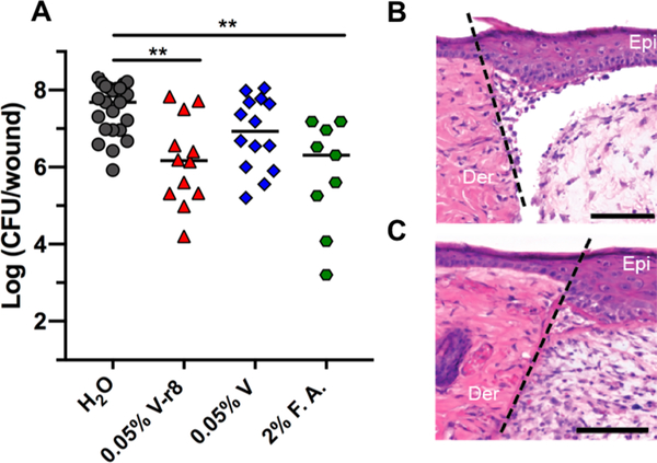
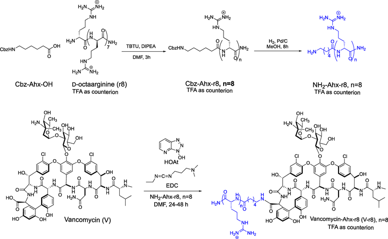
New strategies are urgently needed to target MRSA, a major global health problem and the leading cause of mortality from antibiotic-resistant infections in many countries. Here, we report a general approach to this problem exemplified by the design and synthesis of a vancomycin-d-octaarginine conjugate (V-r8) and investigation of its efficacy in addressing antibiotic-insensitive bacterial populations. V-r8 eradicated MRSA biofilm and persister cells in vitro, outperforming vancomycin by orders of magnitude. It also eliminated 97% of biofilm-associated MRSA in a murine wound infection model and displayed no acute dermal toxicity. This new dual-function conjugate displays enhanced cellular accumulation and membrane perturbation as compared to vancomycin. Based on its rapid and potent activity against biofilm and persister cells, V-r8 is a promising agent against clinical MRSA infections.


Conflict of interest statement

The authors declare no competing financial interest.

Figures

Figure 1.
Figure 1.
V–r8 kills biofilm bacteria, persister cell, and exponential-phase bacteria more effectively than vancomycin (V). (A) 3D-reconstructed confocal images of untreated, V–r8-treated and V-treated MRSA USA400 MW2 biofilms in TSB for 5 h. Insets show bacterial cells from the bottom plane of each image. Biofilm bacteria were stained with SYTO 9 (green: live) and PI (red: dead). Quantification of fluorescence showed that significantly more bacteria were killed by V–r8 than by V as compared to the untreated biofilm. Scale bars represent 20 μm in the main panels and 5 μm in the insets. (B) MRSA USA 300 LAC persister cell time-kill curves for compounds with final concentrations of 10 μM introduced 6 h postciprofloxacin treatment (40 μM). (C) Time-kill kinetic analysis of V or V–r8-treated exponential-phase MRSA USA400 MW2 at treatment concentrations of 5, 10, or 20 μM. Data presented in (A)–(C) are from representative experiments.
Figure 2.
Figure 2.
MRSA USA400 MW2 bacteria treated with Fl-V–r8 exhibits greater cell-associated and protoplast-associated fluorescence than Fl-V. (A) Confocal microscopy of bacteria treated with 5 μM FlV–r8 for 5 min and (B) analysis of fluorescence intensities of individual bacteria imaged as in (A). Intensity values were normalized to the mean intensity of Fl-V–r8-treated cells. Error bars represent standard deviations. P < 0.0001 determined using a Mann–Whitney test. (C) FACS analysis of MRSA whole cells treated with Fl-V–r8 or Fl-V. (D) FACS analysis of protoplasts prepared from MRSA whole cells treated with Fl-V–r8 and Fl-V as in (C). In (C) and (D), each bar represents median normalized fluorescence values from two experiments, with data normalized to the highest fluorescence value in each experiment. Error bars represent the range of normalized fluorescence values obtained in two experiments.
Figure 3.
Figure 3.
Evaluation of V–r8 activity as a function of bacterial growth phase and treatment condition and its influence on membrane and cellular integrity. (A) Comparative time-kill kinetics of MRSA USA400 MW2 harvested from exponential-phase or stationary phase and resuspended in PBS for treatment followed by CFU enumeration on agar. (B) Evaluation of PI uptake and fluorescence as a reporter for the perturbation of membrane barrier function upon treatment with V–r8, V, or lysostaphin. Experiments were performed with stationary-phase MRSA USA400 MW2 resuspended in either PBS or HEPES–glucose (H–G) buffer. In (A) and (B), V and V–r8 treatments were performed at 10 μM, lysostaphin treatments were performed at 125 μg/mL (all ∼10X MIC), and data are from representative experiments. (C) TEM images of untreated, V–r8, or V-treated stationary-phase MRSA USA400 MW2 in PBS or exponential-phase bacteria in tryptic soy broth. Treatments were performed for 90 min at final concentrations of 8 μM in the PBS experiment (top) or 4 μM in the tryptic soy broth experiment (bottom). Scale bars = 1 μm.
Figure 4.
Figure 4.
In vivo evaluation of V–r8 in a skin wound biofilm model. (A) Each data point represents Log(CFU/wound) from one mouse, with the median values indicated by bars. Data were compiled from two to three independent experiments containing four to five animals per treatment group. Statistical analysis was performed using the nonparametric Kruskal–Wallis test with Dunn’s post ad hoc test for intergroup comparisons. **P < 0.01. To examine in vivo cytotoxicity, sterile wounds were generated and inoculated with water (B) or 0.05% V–r8 (C) in the absence of bacteria. The epidermis and dermis layers from mice treated with V–r8 appeared similar to those from mice treated with water and showed no signs of necrosis or apoptosis by hematoxylin and eosin staining. Wound healing was comparable between groups indicated by dermal granulation tissue formation and epidermal healing. A small increase in neutrophil infiltration was observed in V–r8-treated mice as compared to untreated mice. Dotted line represents the site of wounding. Epi: skin epidermis; Der: skin dermis. Scale bars = 100 μm. Images are representative of three independent samples.
Scheme 1.
Scheme 1.
Synthesis of Vancomycin Conjugate V–r8

References

    1. Klein EY; Mojica N; Jiang W; Cosgrove SE; Septimus E; Morgan DJ; Laxminarayan R Trends in Methicillin-Resistant Staphylococcus Aureus Hospitalizations in the United States, 2010–2014. Clin. Infect. Dis 2017, 65, 1921–1923. - PubMed
    1. Stefani S; Chung DR; Lindsay JA; Friedrich AW; Kearns AM; Westh H; MacKenzie FM Meticillin-Resistant Staphylococcus Aureus (MRSA): Global Epidemiology and Harmonisation of Typing Methods. Int. J. Antimicrob. Agents 2012, 39, 273–282. - PubMed
    1. Hassoun A; Linden PK; Friedman B Incidence, Prevalence, and Management of MRSA Bacteremia across Patient Populations-a Review of Recent Developments in MRSA Management and Treatment. Crit. Care London Engl 2017, 21, 211. - PMC - PubMed
    1. Tattevin P; Schwartz BS; Graber CJ; Volinski J; Bhukhen A; Bhukhen A; Mai TT; Vo NH; Dang DN; Phan TH; Basuino L; Perdreau-Remington F; Chambers HF; Diep BA Concurrent Epidemics of Skin and Soft Tissue Infection and Bloodstream Infection Due to Community-Associated Methicillin-Resistant Staphylococcus Aureus. Clin. Infect. Dis 2012, 55, 781–788. - PMC - PubMed
    1. Fisher RA; Gollan B; Helaine S Persistent Bacterial Infections and Persister Cells. Nat. Rev. Microbiol 2017, 15, 453–464. - PubMed

Publication types

MeSH terms