Review
. 2018 Nov 1;175(4):908-920.
doi: 10.1016/j.cell.2018.08.071.

Two to Tango: Dialog between Immunity and Stem Cells in Health and Disease

Affiliations
Review

Two to Tango: Dialog between Immunity and Stem Cells in Health and Disease

Shruti Naik et al. Cell. .

Abstract

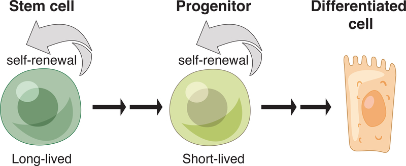
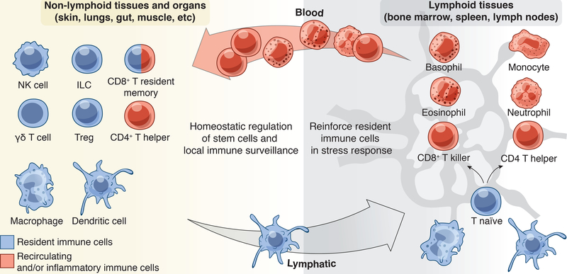
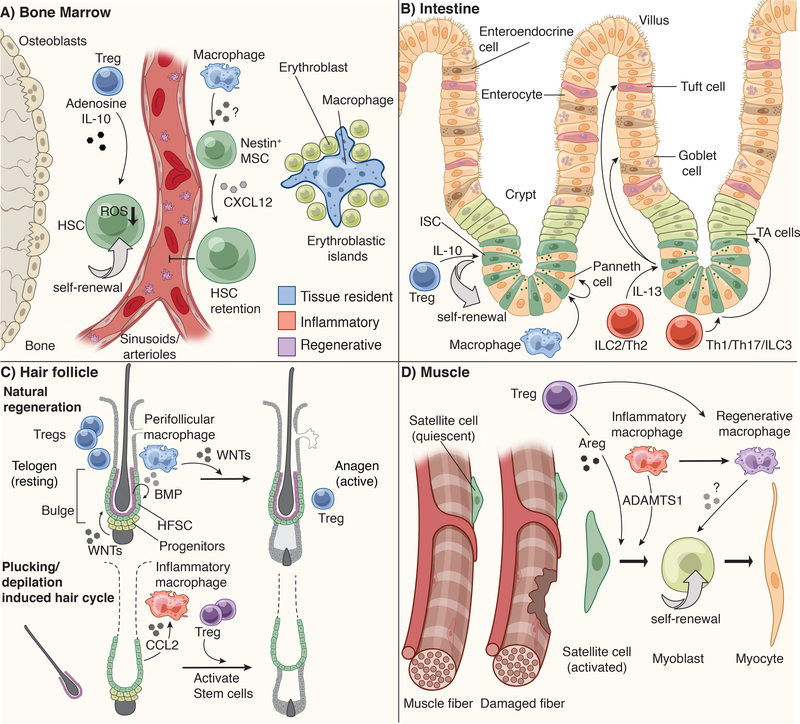
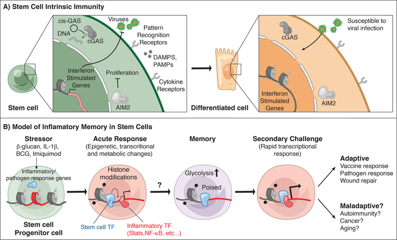
Stem cells regenerate tissues in homeostasis and under stress. By taking cues from their microenvironment or "niche," they smoothly transition between these states. Immune cells have surfaced as prominent members of stem cell niches across the body. Here, we draw parallels between different stem cell niches to explore the context-specific interactions that stem cells have with tissue-resident and recruited immune cells. We also highlight stem cells' innate ability to sense and respond to stress and the enduring memory that forms from such encounters. This fascinating crosstalk holds great promise for novel therapies in inflammatory diseases and regenerative medicine.


Figures

Figure 1:
Figure 1:. Tissue stem cell hierarchy
Long-lived stem cells have the ability to self-renew and give rise to short-term progenitors. These short-term progenitors can also replicate themselves and generate differentiated progeny. They are largely responsible for coordinating tissue homeostasis.
Figure 2:
Figure 2:. Tissue resident, recirculating, and inflammatory immune cells
A constellation of immune cells inhabits tissues, the composition of which varies by tissue site and the inflammatory status of the tissue. Extra-lymphoid tissues and in particular epithelial barrier tissues such as the skin, lungs and gut, house the greatest number of resident immune sentinels. This includes dendritic cells, macrophages, innate lymphoid cell (ILC) subsets, γδ T cells and regulatory T cells (Tregs) that seed tissues early in life. With age and exposure to commensals and pathogens, tissues also acquire CD8+ T resident memory cells (TRM) and recirculating CD4+ T helper subsets. During an acute stress response, inflammatory macrophages/monocytes, neutrophils, basophils, and eosinophils are recruited to the damage to reinforce the function of resident cells. Lymphoid organs such as the lymph node and spleen are epicenters for naïve or unprimed T cells. These cells are primed by dendritic cells to differentiate into effectors and migrate into tissues via blood where they enact their effector functions.
Figure 3:
Figure 3:. Niche specific immune-stem cell interactions
A) Hematopoietic stem cells (HSCs) reside in the bone marrow (BM) in close apposition of sinusoids and arterioles. They are retained in this niche by Nestin+ mesenchymal stem cell (MSC)-derived CXCL12. Bone marrow macrophages are critical for maintaining the production of CXCL12 and retaining HSCs in this niche. Bone marrow Tregs are also localized close to HSCs and produce IL-10 and Adenosine to control HSC quiescence and pool size. In the absence of Tregs, HSCs produce more reactive oxygen species (ROS) and consequently increase their numbers. Erythroblast progenitors in the BM aggregate around a central macrophage, which provides cues to direct their maturation into erythrocytes. B) Intestinal stem cells (ISCs) reside in crypt structures at the base of intestinal villi and are flanked by Paneth cells and transit amplifying (TA) cells. ISCs give rise to all intestinal lineages, including goblet cells, enteroendocrine cells, tuft cells, enterocytes and Paneth cells. However, when ISCs at the base of the crypt are ablated, these progenitors can replenish niche vacancies. Intestinal macrophages promote the differentiation of ISCs to Paneth cells, while resident Tregs promote ISC self-renewal via IL-10. CD4+ T helper (Th)1 and Th17 cells and innate lymphoid cells (ILC) type 3 promote the production of transit amplifying progeny, while Th2 and ILC2 promote differentiation of tuft and goblet cells via IL-13. C) The hair follicle is one of the few systems in mammals where tissue regeneration happens in natural bursts (the hair cycle) and in the absence of injury. The bulge region of the hair follicle is home to hair follicle stem cells (HFSCs). At the bulge base (hair germ), ‘primed stem cells’ give rise to the short-term progenitors that undergo natural cyclical bouts of active tissue regeneration (anagen). Growth inhibitory signals such as bone morphogenic proteins (BMPs), supplied by both the dermis and the inner bulge niche layer, keep stem cells in quiescence throughout most of the hair cycle. In the resting phase of the hair cycle (telogen), WNT signals and BMP inhibitor levels in the hair germ accumulate until stem cells become activated to launch a new tissue regenerative cycle. Perifollicular macrophages are a crucial source of WNTs and tip the balance in favor of regeneration. By contrast, Tregs are highest in telogen when stem cells are at the height of quiescence; Tregs reach a low point at the height of the hair growth phase. Hair plucking removes the inner bulge and its quiescence signals, thereby precociously inducing anagen. During this injury response, inflammatory macrophages are also found near the bulge, where they appear to promote hair cycling and disseminate distress signals to adjacent unplucked follicles. Regenerative Tregs also appear to facilitate the progression to anagen following hair depilation. D) Muscle stem cells (satellite cells) are quiescent at steady state and only activate following injury. Regenerative Tregs and inflammatory macrophages (M1) support satellite cell self-renewal and activation via amphiregulin (Areg) and ADAMTS1, respectively. As repair progresses, Tregs facilitate the switch from inflammatory M1 to regenerative M2 macrophages, which promote myoblast self-renewal and differentiation.
Figure 4:
Figure 4:. Stem cell intrinsic immunity and memory
A) Stem cells express a variety of cytokine and pattern recognition receptors that can sense damage associated and pathogen associated molecular patterns (DAMPs, PAMPs) and cytokine signals from immune cells. These include both surface associated receptors and intracellular sensors such as cGAS and absent in melanoma 2 (AIM2) which bind double stranded DNA. AIM2 limits intestinal stem cell proliferation by modulating AKT. Hematopoietic stem cells express a circular RNA “cis-GAS” to regulate cGAS activation and limit exhaustion. SCs also express high levels of interferon-stimulated genes (ISGs) in contrast to differentiated progeny, and these baseline ISGs protect SCs from viral infections. B) Stem cells are trained by a variety of acute inflammatory encounters: directly by β-glucan, IL-1β, Bacillus Calmette–Guérin (BCG) vaccine for Mycobacterium tuberculosis, and indirectly by Toll like receptor 7 (TLR7) agonist (Imiquimod) induced inflammation. These stimuli activate inflammatory transcription factors (STATs, NF-κB), which likely facilitate the remodeling of chromatin and acquisition of histone modifications at inflammatory stress response genes resulting in dramatic transcriptional and metabolic changes to the SCs and altered activation and cellular output. Upon resolution, stem cells retain changes to a subset of chromatin loci including altered accessibility and histone modifications. These changes may be maintained by homeostatic transcription factors in the absence of overt inflammation. Inflammation-experienced stem cells also exhibit heightened glycolytic activity and in the case of HSCs exhibit bias towards the myeloid lineage. During a secondary challenge, stem cells and their progeny exhibit heighted responses at genes corresponding with memory chromatin domains. This adaptive behavior promotes vaccine and pathogen responses and in the case of epithelial stem cells augmented wound healing. On the other hand, a negative consequence of such memory may be increased predisposition to autoimmunity, cancer or aging.

References

    1. Agudo J, Park ES, Rose SA, Alibo E, Sweeney R, Dhainaut M, Kobayashi KS, Sachidanandam R, Baccarini A, Merad M, et al. (2018). Quiescent Tissue Stem Cells Evade Immune Surveillance. Immunity 48, 271–285.e275. - PMC - PubMed
    1. Ali N, Zirak B, Rodriguez RS, Pauli ML, Truong H-A, Lai K, Ahn R, Corbin K, Lowe MM, Scharschmidt TC, et al. (2017). Regulatory T Cells in Skin Facilitate Epithelial Stem Cell Differentiation. Cell 169, 1119–1129.e11. - PMC - PubMed
    1. Alvarado AG, and Lathia JD (2016). Taking a Toll on Self-Renewal: TLR-Mediated Innate Immune Signaling in Stem Cells. Trends in Neurosciences 39, 463–471. - PubMed
    1. Aparicio-Domingo P, Romera-Hernandez M, Karrich JJ, Cornelissen F, Papazian N, Lindenbergh-Kortleve DJ, Butler JA, Boon L, Coles MC, Samsom JN, et al. (2015). Type 3 innate lymphoid cells maintain intestinal epithelial stem cells after tissue damage. The Journal of Experimental Medicine 212, 1783–1791. - PMC - PubMed
    1. Arnold L, Henry A, Poron F, Baba-Amer Y, van Rooijen N, Plonquet A, Gherardi RK, and Chazaud B (2007). Inflammatory monocytes recruited after skeletal muscle injury switch into antiinflammatory macrophages to support myogenesis. The Journal of Experimental Medicine 204, 1057–1069. - PMC - PubMed

Publication types