. 2019 Jan:20:296-306.
doi: 10.1016/j.redox.2018.10.019. Epub 2018 Oct 24.

Irisin alleviates liver ischemia-reperfusion injury by inhibiting excessive mitochondrial fission, promoting mitochondrial biogenesis and decreasing oxidative stress

Affiliations

Irisin alleviates liver ischemia-reperfusion injury by inhibiting excessive mitochondrial fission, promoting mitochondrial biogenesis and decreasing oxidative stress

Jianbin Bi et al. Redox Biol. 2019 Jan.

Erratum in

Abstract

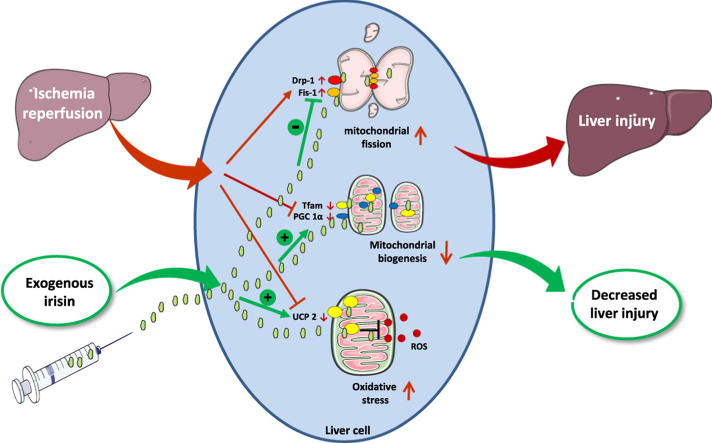
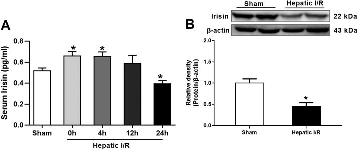
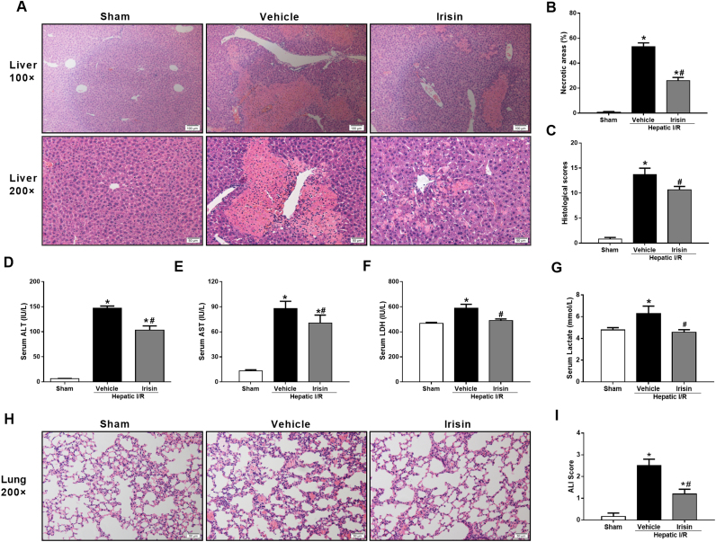
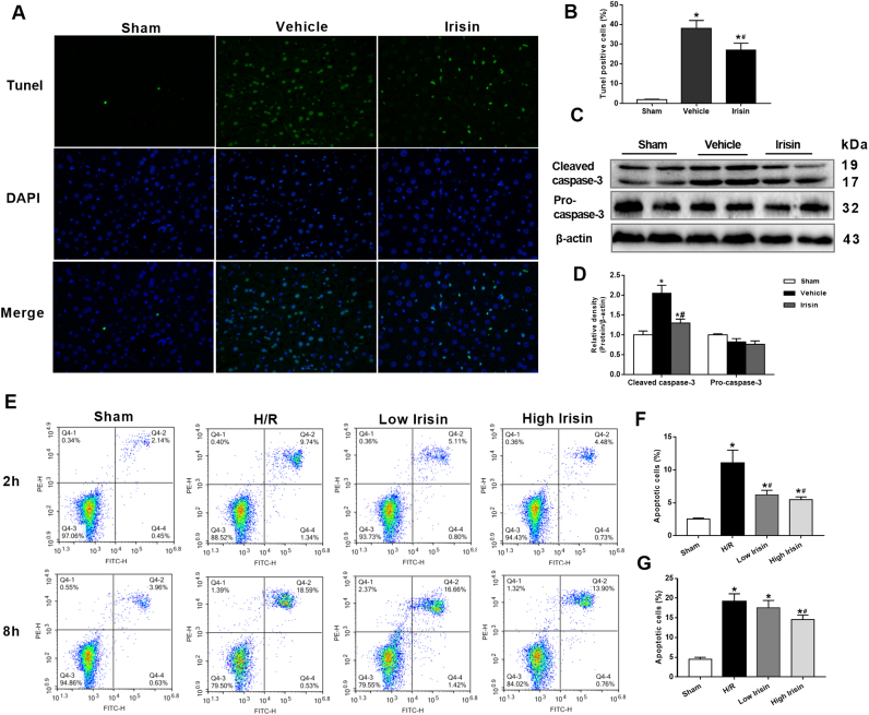
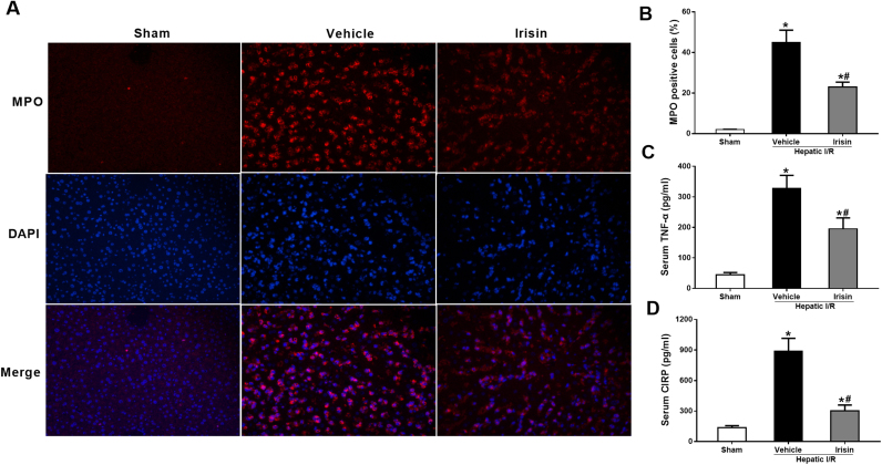
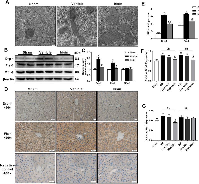
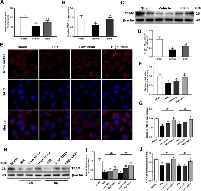
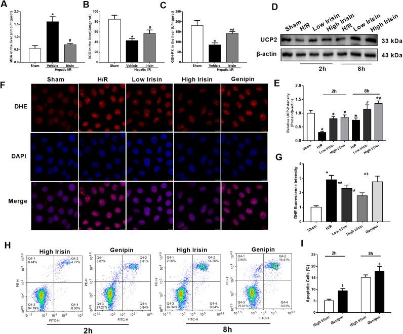
Current management of liver ischemia-reperfusion (I/R) injury is mainly based on supportive care and no specific treatment is available. Irisin, a recently identified hormone, plays pivotal roles in energy expenditure and oxidative metabolism; however, it remains unknown whether irisin has any protective effects on hepatic I/R injury. In this study, we found that serum and liver irisin levels were markedly decreased at 24 h after hepatic I/R. Treatment with exogenous irisin improved liver function, reduced liver necrosis and cell apoptosis, and relieved inflammatory response after hepatic I/R. Meanwhile, exogenous irisin markedly inhibited mitochondrial fission related protein dynamin related protein 1 (drp-1) and fission 1 (Fis-1) expression in hepatic I/R. Additionally, treatment with exogenous irisin increased mitochondrial content and increased mitochondrial biogenesis related peroxisome proliferative activated receptor-γ (PPARγ) co-activator 1α (PGC-1α) and mitochondrial transcription factor (TFAM) expression. Furthermore, irisin decreased oxidative stress by upregulating uncoupling proteins (UCP) 2 expression in hepatic I/R. The results reveal that treatment with exogenous irisin alleviated hepatic I/R injury by restraining mitochondrial fission, promoting mitochondrial biogenesis and relieving oxidative stress. Irisin treatment appears to be a novel and promising therapeutic approach for hepatic I/R injury.

Keywords: Hepatic I/R; Irisin; Mitochondrial homeostasis; Oxidative stress.


Figures

fx1
Graphical abstract
Fig. 1
Fig. 1
Alterations in irisin levels after hepatic ischemia-reperfusion (I/R). Partial (70%) liver arterial/portal venous blood was interrupted for 60 min. Blood samples were harvested at 0 h, 4 h, 12 h and 24 h after reperfusion. A, Serum irisin levels; B, Western blot analysis of liver irisin expressions. n = 6, mean ± SEM, *P < 0.05 versus sham group.
Fig. 2
Fig. 2
Treatment with exogenous irisin attenuates organ injury after hepatic ischemia-reperfusion (I/R). Irisin treatment in mice was conducted by intravenous administration (250 μg/kg, a single dose) at the beginning of reperfusion. The liver, lung and blood samples were harvested at 24 h after reperfusion. A, Hematoxylin and eosin staining (H&E) of representative liver sections (magnification 100× and 200×); B, Percentage of necrotic areas; C, Liver histological scores; D-G, The serum levels of alanine aminotransferase (ALT), aspartate aminotransferase (AST), lactic dehydrogenase (LDH) and lactate in each group, respectively;H, Hematoxylin and eosin staining (H&E) of representative lung sections (magnification 200×); I, Lung injury scores. n = 6, mean ± SEM, *P < 0.05 versus sham group, #P < 0.05 versus vehicle group.
Fig. 3
Fig. 3
Treatment with exogenous irisin decreases apoptosis after hepatic ischemia-reperfusion (I/R). Irisin treatment in mice was conducted by intravenous administration (250 μg/kg, a single dose) at the beginning of reperfusion. The liver tissues were harvested at 24 h after reperfusion. A, transferase-mediated deoxyuridine triphosphate-biotin nick end labeling (TUNEL) fluorescence staining (green), the corresponding nuclear counterstaining (blue) and both channels merged of representative liver sections (magnification 400×); B, percentage of TUNEL positive cells; C and D, Western blot analysis of liver cleaved caspase 3 and pro-caspase 3 expression; n = 6, mean ± SEM, *P < 0.05 versus sham group, #P < 0.05 versus vehicle group. E-G, Flow cytometry analysis of HL-7702 cell apoptotic percentage at 2 h and 8 h after reoxygenation. n = 3, mean ± SEM, *P < 0.05 versus sham group, #P < 0.05 versus H/R group.
Fig. 4
Fig. 4
Treatment with exogenous irisin downregulates inflammatory responses after ischemia-reperfusion (I/R). Irisin treatment in mice was conducted by intravenous administration (250 μg/kg, a single dose) at the beginning of reperfusion. The liver tissues and blood samples were harvested at 24 h after reperfusion. A, Myeloperoxidase (MPO) immunofluorescence staining (green), the corresponding nuclear counterstaining (blue) and both channels merged of representative liver sections (magnification 400×); B, analysis of MPO fluorescence intensity; C, serum tumor necrosis factor α (TNF-α) levels; D, Serum cold-inducible RNA binding protein (CIRP) levels in each group. n = 6, mean ± SEM, *P < 0.05 versus sham group, #P < 0.05 versus vehicle group.
Fig. 5
Fig. 5
Treatment with exogenous irisin inhibits excessive mitochondrial fission after hepatic ischemia-reperfusion (I/R). Irisin treatment in mice was conducted by intravenous administration (250 μg/kg, a single dose) at the beginning of reperfusion The liver tissues were harvested at 24 h after reperfusion. A, Transmission electron microscope (TEM) of liver tissues (magnification 50,000×); B and C, Western blot analysis of liver dynamin related protein 1 (drp1), fission 1 (Fis-1) and mitofusin 2 (Mfn-2) expression. D and E,Immunohistochemistry and its semiquantitative analysis for evaluating the liver dynamin related protein 1 (drp1) and fission 1 (Fis-1) expression of representative liver sections (magnification 400×). n = 6, mean ± SEM, *P < 0.05 versus sham group, #P < 0.05 versus vehicle group. F and G, qPCR analysis of drp-1 and Fis-1 expression in HL-7702 cells at 2 h and 8 h after reoxygenation. n = 3, mean ± SEM, *P < 0.05 versus sham group, #P < 0.05 versus H/R group.
Fig. 6
Fig. 6
Treatment with exogenous irisin increases mitochondrial content by promoting mitochondrial biogenesis after hepatic ischemia-reperfusion (I/R). Irisin treatment in mice was conducted by intravenous administration (250 μg/kg, a single dose) at the beginning of reperfusion. The liver tissues were harvested at 24 h after reperfusion. A, Liver mtDNA copy numbers assessed by quantitative polymerase chain reaction (qPCR). B, qPCR analysis of liver peroxisome proliferative activated receptor-γ (PPARγ) co-activator 1α (PGC-1α) levels. C and D, Western blot analysis of liver transcription factor (TFAM) expression. n = 6, mea ± SEM, *P < 0.05 versus sham group, #P < 0.05 versus vehicle group. E and F, MitoTracker Red CMXRos fluorescence staining and its fluorescence intensity of HL-7702 cells at 2 h after reoxygenation; G, qPCR analysis of PGC-1α levels in HL-7702 cells at 2 h and 8 h after reoxygenation. H and I, Western blot analysis of TFAM expression in HL-7702 cells at 2 h and 8 h after reoxygenation; J, ATP content of HL-7702 cells at 2 h and 8 h after reoxygenation. n = 3, mean ± SEM, *P < 0.05 versus sham group, #P < 0.05 versus H/R group.
Fig. 7
Fig. 7
Treatment with exogenous irisin reduces oxidative stress after hepatic ischemia-reperfusion (I/R). Irisin treatment in mice was conducted by intravenous administration (250 μg/kg, a single dose) at the beginning of reperfusion. The liver tissues were harvested at 24 h after reperfusion. A-C, the malonaldehyde (MDA), superoxide dismutase (SOD) and glutathione (GSH) levels in the liver in each group, respectively. n = 6, mean ± SEM, *P < 0.05 versus sham group, #P < 0.05 versus vehicle group. D and E, Western blot analysis of UCP 2 expression in HL-7702 cells at 2 h and 8 h after reoxygenation; F and G, DHE fluorescence staining and its fluorescence intensity of HL-7702 cells at 2 h after reoxygenation; H and I, Flow cytometry analysis of HL-7702 cell apoptosis percentage at 2 h and 8 h after reoxygenation. n = 3, mean ± SEM, *P < 0.05 versus sham group, #P < 0.05 versus H/R group, $P < 0.05 versus high irisin group.

References

    1. Mendes-Braz M., Elias-Miro M., Jimenez-Castro M.B., Casillas-Ramirez A., Ramalho F.S., Peralta C. The current state of knowledge of hepatic ischemia-reperfusion injury based on its study in experimental models. J. Biomed. Biotechnol. 2012:298657. - PMC - PubMed
    1. Halladin N.L. Oxidative and inflammatory biomarkers of ischemia and reperfusion injuries. Dan. Med. J. 2015;62:B5054. - PubMed
    1. Chen S.D., Yang D.I., Lin T.K., Shaw F.Z., Liou C.W., Chuang Y.C. Roles of oxidative stress, apoptosis, PGC-1alpha and mitochondrial biogenesis in cerebral ischemia. Int. J. Mol. Sci. 2011;12:199–7215. - PMC - PubMed
    1. Archer S.L. Mitochondrial dynamics--mitochondrial fission and fusion in human diseases. N. Engl. J. Med. 2013;369:2236–2251. - PubMed
    1. Toyama E.Q., Herzig S., Courchet J., Lewis T.L., Jr., Loson O.C., Hellberg K., Young N.P., Chen H., Polleux F., Chan D.C. Metabolism. amp-activated protein kinase mediates mitochondrial fission in response to energy stress. Science. 2016;351:275–281. - PMC - PubMed

Publication types

MeSH terms