. 2019 Jan:335:22-29.
doi: 10.1016/j.cellimm.2018.10.008. Epub 2018 Oct 26.

In vivo administration of recombinant BTNL2-Fc fusion protein ameliorates graft-versus-host disease in mice

Affiliations

In vivo administration of recombinant BTNL2-Fc fusion protein ameliorates graft-versus-host disease in mice

Cheng Cui et al. Cell Immunol. 2019 Jan.

Abstract

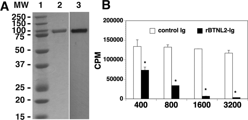
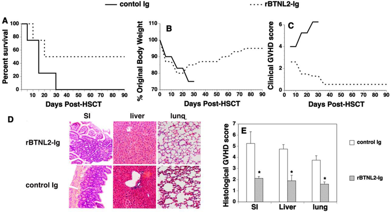
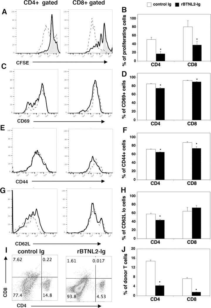
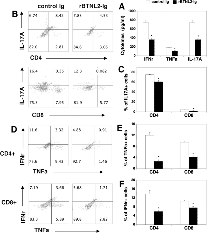
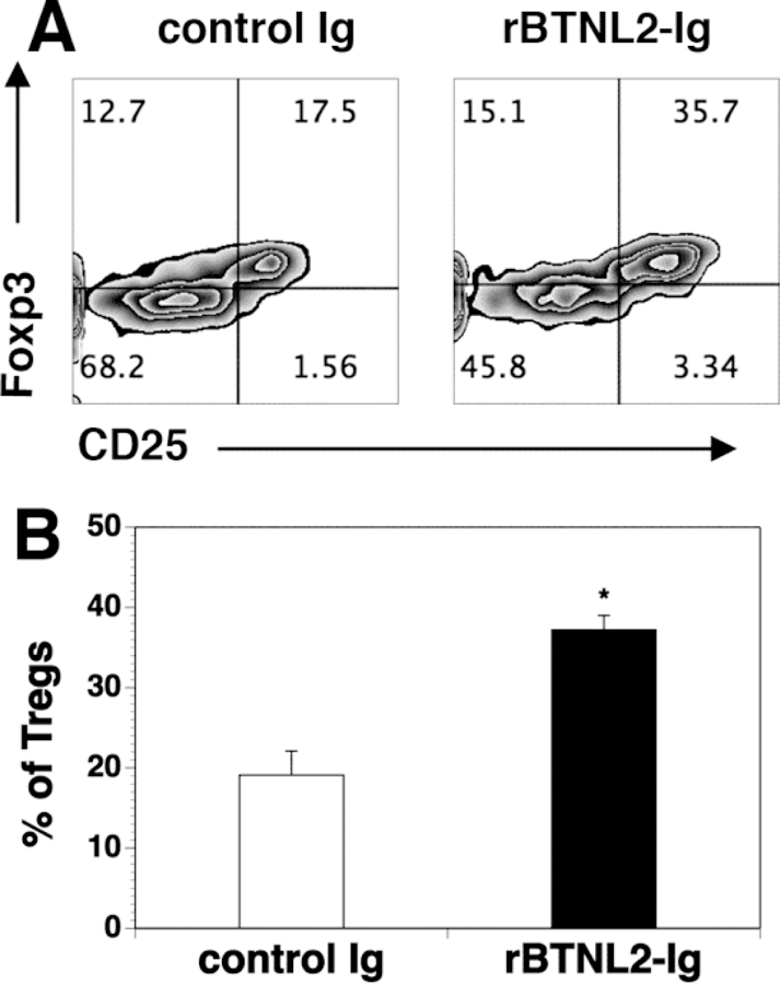
Although hematopoietic stem cell transplantation (HSCT) has been widely used in the treatment of many diseases, graft-versus-host disease (GVHD) remains a major complication after allogeneic HSCT. Butyrophilin-like 2 (BTNL2) protein has been reported to have the ability to inhibit T cell proliferation in vitro; its ability to inhibit T cell responses in vivo has not been determined. We show here that in vivo administration of recombinant BTNL2-IgG2a Fc (rBTNL2-Ig) fusion protein ameliorates GVHD in mice. This is related to the ability of rBTNL2-Ig to inhibit T cell proliferation, activation and Th1/Th17 cytokine production in vivo. Furthermore, rBTNL2-Ig treatment increases the generation of regulatory T cells. Our results suggest that rBTNL2-Ig has the potential to be used in the prevention and treatment of patients with GVHD.

Keywords: Activation; Butyrophilin-like 2; Graft-versus-host disease; Hematopoietic stem cell transplantation; Regulatory T cells; T cell proliferation.


Conflict of interest statement

Declaration of conflicting interests: The Authors declare that there is no conflict of interest.

Figures

Figure 1.
Figure 1.
Characterization of purified rBTNL2-Ig protein. (A) Gel and blot show purified rBTNL2-Ig protein; Lane 1: MW markers; 2: Coomassie blue-stained SDS-PAGE; 3: Western blot with an anti-mouse IgG2a antibody. (B) rBTNL2-Ig protein inhibits T cell proliferation in vitro. T cells were purified from splenocytes of C57BL/6 mice by magnetic separation. The cells were cultured on plates pre-coated with anti-CD3 antibody (1 µg/ml) and indicated doses (ng/ml) of rBTNL2-Ig or control Ig for 3 days. [3H] thymidine (1 µCi/well) was added to the cultures 12 hours before harvest. T cell proliferation was measured by [3H] thymidine incorporation. Results are expressed as counts per minute (CPM). The data are expressed as mean ± SD and representative of 3 independent experiments. * P<0.05 compared with control Ig.
Figure 2.
Figure 2.
rBTNL2-Ig ameliorates GVHD. Lethally irradiated BALB/c recipients were injected with 5X10⁶ BM and 2.5X10⁶ spleen cells from C57BL/6 mice, as well as 50 μg rBTNL2-Ig or control Ig at day 0. The recipients were then injected i.p. with 50 μg rBTNL2-Ig or control Ig at 3-day intervals for 30 days. (A-C) Recipients were monitored for (A) survival (A Kaplan- Meier survival curve is shown), (B) weight change, and (C) clinical GVHD. (D, E) In separate experiments, recipients given 50 μg rBTNL2-Ig or control Ig at 3-day intervals from days 0–12 were euthanized 2 weeks after HSCT. The SI, liver and lung were analyzed for histologic damage. (D) Representative photomicrographs (the magnification was X200), and (E) mean ± SD of histopathology scores. Pooled data from 3 separate experiments are represented; with 4–5 mice per group in each experiment. * P<0.05 compared with control Ig-treated mice.
Figure 3.
Figure 3.
rBTNL2-Ig inhibits T-cell proliferation and activation in response to alloantigens in mice. (A-H) Lethally irradiated BALB/c mice were injected i.v. with 5X10⁶ BM cells and 10X106 splenic cells labelled (A, B) with or (C-H) without CFSE from C57BL/6 mice. The recipients were injected i.v. at day 0 and i.p. at day 2 with 50 μg rBTNL2-Ig or control Ig. At Day 4 post-transplant, the percentage of (A, B) CFSElo (shaded histograms: splenic cells labelled with CFSE before injection into mice), (C, D) CD69+, (E, F) CD44hi, and (G, H) CD62Llo cells in donor CD4 and CD8 T cells (H-2Kb+CD4+, or H-2Kb+CD8+) in the spleen were examined by flow cytometry. Dot lines: control Ig; solid lines: rBTNL2-Ig. (I, J) Lethally irradiated BALB/c mice were injected with BM and splenic cells, and then given 50 μg rBTNL2-Ig or control Ig as in Figure 2D. The percentages of donor CD4+ and CD8+ T cells were analyzed 2 weeks after HSCT. (A, C, E, G, I) Representative flow cytometric profiles and (B, D, F, H, J) mean ± SD from one of three independent experiments with similar results. * P<0.05 compared with control Ig group.
Figure 4.
Figure 4.. rBTNL2-Ig reduces Th1 and Th17 cytokine production in vivo
(A) Serum cytokine profile in control- or BTN-treated recipients. Lethally irradiated BALB/c mice were injected with BM and splenic cells, and then injected with 50 μg rBTNL2-Ig or control Ig as in Figure 2D. Serum was harvested at day 14 after HSCT and indicated cytokines were measured by ELISA. (B-F) Lethally irradiated BALB/c recipients were injected i.v. with 5X10⁶ BM and 2.5X10⁶ spleen cells from C57BL/6 mice. The recipients were then injected i.p. with 50 μg rBTNL2-Ig or control Ig at 3-day intervals for 27 days. At Day 30 after BMT, intracellular cytokine profiles of splenic CD4+ and CD8+ T cells were analyzed. (B, D) Representative flow cytometry profiles, (C, E, F) statistical analysis showing the percentages of IL-17A-, TNFα- and IFNγ-producing CD4+ or CD8+ T cells. * P<0.05 compared with control Ig group.
Figure 5.
Figure 5.. rBTNL2-Ig treatment increases the percentage of Tregs in GVHD recipients.
Lethally irradiated BALB/c recipients were injected i.v. with 5X10⁶ BM and 2.5X10⁶ spleen cells from C57BL/6 mice. The recipients were treated with 50 μg rBTNL2-Ig or control Ig at 3- day intervals from days 0–12 as in Figure 2D. Fourteen days after BMT, the spleens were harvested and analyzed for CD4+CD25+Foxp3+ Tregs. (A) Flow cytometry files showing the expression of CD25 and Foxp3 in gated donor CD4+ cells; (B) Mean ± SD for the percentage of Tregs from one of three independent experiments with similar results. * P<0.05 compared with control Ig group.

Similar articles

Cited by

References

    1. Shlomchik WD, Graft-versus-host disease, Nat. Rev. Immunol, 7 (2007) 340–352. - PubMed
    1. Ferrara JL, Levine JE, Reddy P, Holler E, Graft-versus-host disease, Lancet, 373 (2009) 1550–1561. - PMC - PubMed
    1. Welniak LA, Blazar BR, Murphy WJ, Immunobiology of allogeneic hematopoietic stem cell transplantation, Annu. Rev. Immunol, 25 (2007) 139–170. - PubMed
    1. Coghill JM, Sarantopoulos S, Moran TP, Murphy WJ, Blazar BR, Serody JS, Effector CD4+ T cells, the cytokines they generate, and GVHD: something old and something new, Blood, 117 (2011) 3268–3276. - PMC - PubMed
    1. Warren EH, Deeg HJ, Dissecting graft-versus-leukemia from graft-versus-host-disease using novel strategies, Tissue Antigens, 81 (2013) 183–193. - PMC - PubMed

Publication types

MeSH terms

LinkOut - more resources