Review
. 2018 Dec;30(12):2898-2909.
doi: 10.1105/tpc.18.00523. Epub 2018 Nov 1.

Evolution and Structural Characteristics of Plant Voltage-Gated K+ Channels

Affiliations
Review

Evolution and Structural Characteristics of Plant Voltage-Gated K+ Channels

Timothy Jegla et al. Plant Cell. 2018 Dec.

Abstract

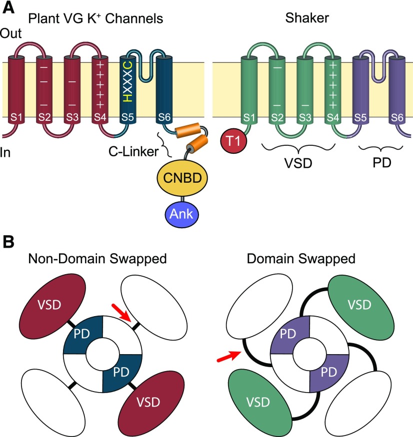
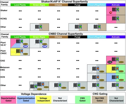
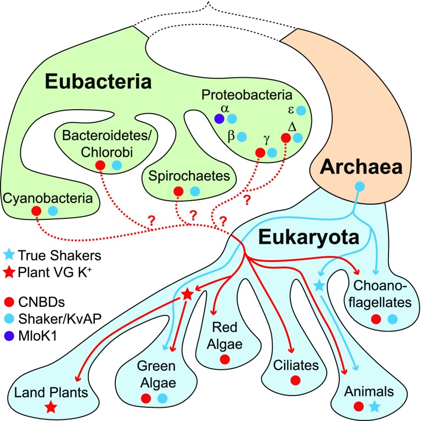
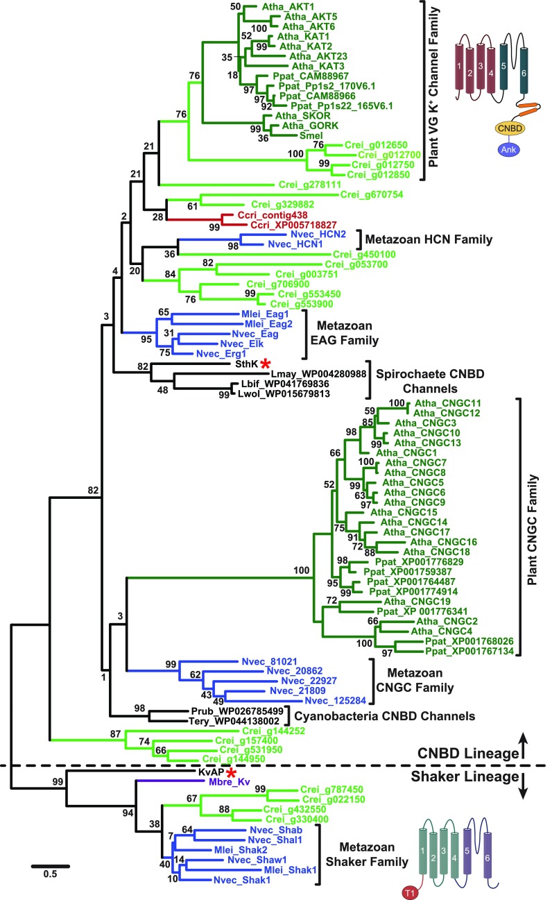
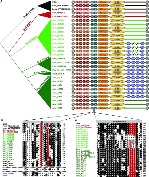
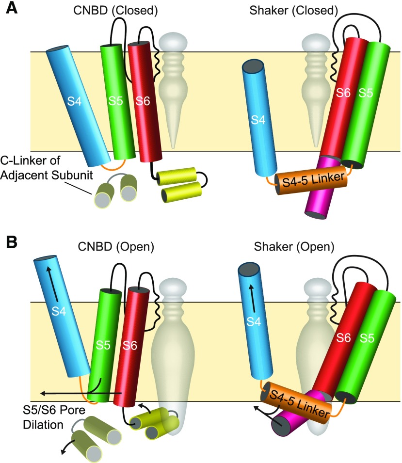
Plant voltage-gated K+ channels have been referred to as "plant Shakers" in reference to animal Shaker channels, the first K+ channels identified. Recent advances in our knowledge of K+ channel evolution and structure have significantly deepened the divide between these plant and animal K+ channels, suggesting that it is time to completely retire the "plant Shaker" designation. Evolutionary genomics reveals that plant voltage-gated K+ channels and metazoan Shakers derive from distinct prokaryotic ancestors. The plant channels belong to a lineage that includes cyclic nucleotide-gated channels and metazoan ether-à-go-go and hyperpolarization-activated, cyclic nucleotide-gated channels. We refer to this lineage as the CNBD channel superfamily, because all these channels share a cytoplasmic gating domain homologous to cyclic nucleotide binding domains. The first structures of CNBD superfamily channels reveal marked differences in coupling between the voltage sensor and ion-conducting pore relative to metazoan Shaker channels. Viewing plant voltage-gated K+ channel function through the lens of CNBD superfamily structures should lead to insights into how these channels are regulated.


Figures

Figure 1.
Figure 1.
Comparison of the Domain Architecture of Plant VG K+ Channels and Metazoan Shaker Channels. (A) Side view schematic drawings of channel subunits. The plasma membrane is represented by the tan horizontal boxes; extracellular and intracellular sides are marked “Out” and “In”; and transmembrane domains within the subunits are depicted with cylinders. In both channels, the S1-S4 transmembrane segments comprise the voltage sensor domain (VSD, marked on Shaker); the S5-S6 transmembrane segments form the channel PD (marked on Shaker); and the extracellular loop between S5 and S6 forms the pore’s selectivity filter. Basic voltage-sensing gating charges reside in S4 and are indicated by + signs. The S5 of plant voltage-gated K+ channels includes an HXXXC amino acid motif that is highly conserved in CNBD superfamily channels but is absent from Shaker channels. Plant VG K+ channels and Shakers also differ in the composition of their cytoplasmic domains. The C terminus of plant VG K+ channels has a CNBD connected to the PD through a conserved helical linker (C-linker). In contrast, Shaker channels have neither a CNBD nor a C-linker and contain a distinctive N-terminal tetramerization domain (T1), which plays a role in subunit assembly. Many plant VG K+ channels also contain a series of Ankyrin repeats (Ank) in the C terminus distal to the CNBD, which are also absent from Shaker channels. (B) Aerial view—from the extracellular membrane face—of channel tetramers showing the relative positions of VSDs within the channel tetramers. Structural analysis of multiple metazoan CNBD superfamily channels shows the VSD is positioned directly adjacent to the PD from the same subunit and connected by a short S4-S5 linker (red arrow); this arrangement is therefore likely to be conserved in plant VG K+ channels. For Shaker lineage channels, the VSD is domain-swapped and sits nearest the neighboring subunit’s PD, although it still gates the PD from the same subunit through an extended S4-S5 linker (red arrow).
Figure 2.
Figure 2.
Major Families of Eukaryotic Shaker/KvAP Superfamily and CNBD Superfamily Channels Listed by Phylogenetic Group. Characteristic cytoplasmic domains are depicted (Shaker tetramerization domain, T1; CNBD; ankyrin repeat domain, ANK; eag domain, EAG). Selectivity is indicated by the superimposed ion (K+-selective, green, K+; non-selective cation channel, orange, +). Protozoan, fungal and algal channels that have not been functionally expressed are listed as K+-selective or non-selective based on the presence or absence of a canonical K+-selectivity filter sequence. The split color background for each icon indicates voltage dependence on the left (red = depolarization gated; blue = hyperpolarization gated; yellow = voltage-insensitive) and CNBD-mediated cyclic nucleotide gating on the right (green = cyclic nucleotide-gated, brown = not cyclic nucleotide gated). Uncertain voltage- and cyclic nucleotide-gating phenotypes due to lack of conclusive data or lack of functional expression are indicated with gray backgrounds. Note that the plant VG K+ channel family includes both hyperpolarization- and depolarization-gated channels and that direct cyclic nucleotide binding for plant CNGCs is considered an open question. CNBD and Shaker/KvAP superfamily channels that are not members of well-characterized gene families are listed as “Other.” Green algae Shaker/KvAP and CNBD “Other” channels are described in this review in Figures 4 and 5. Remaining “Other” channels are described in the following references: 1) Shaker/KvAP superfamily: choanoflagellates (Li et al., 2015c) and fungi (Prole and Taylor, 2012); 2) CNBD superfamily: choanoflagellates and animals (Fechner et al., 2015), fungi (Avelar et al., 2014), and ciliates (Jegla and Salkoff, 1994, 1995).
Figure 3.
Figure 3.
Proposed Paths for Evolutionary Transfer of CNBD Superfamily and Shaker/KvAP Superfamily Channels from Eubacteria into Extant Eukaryotes Based on Phylogenetic Distribution. Only the most relevant prokaryotic and eukaryotic clades are depicted, for simplicity. The presence of CNBD and Shaker/KvAP superfamily channels is indicated with red and blue circles, respectively. The origin and distribution of the plant VG K+ channels (red stars) and metazoan Shakers (blue stars) are marked. Red and blue lines indicate probable paths for evolutionary inheritance of CNBD and Shaker/KvAP channels, respectively. We propose that Shaker/KvAP channels were inherited directly from the Archaean ancestor of eukaryotes, while CNBD superfamily channels, which are absent from Archaea, must have been acquired through lateral gene transfer from the Eubacteria, although the specific group of origin is unclear (dotted red lines). MLoK1-like K+ channels (purple circle), which include a CNBD but not the C-linker of CNBD superfamily channels, are restricted to α-proteobacteria and do not appear to have been transferred to eukaryotes.
Figure 4.
Figure 4.
Maximum Likelihood Phylogeny Supports Separate Prokaryotic Origins for Both Plant VG K+ Channels and Metazoan Shakers. The phylogeny is based on an alignment of the common VSD-PD transmembrane core shared by channels in the CNBD and Shaker/KvAP superfamilies. It contains algal and other plant channels that have a C-terminal C-linker/CNBD (all group in the CNBD lineage) or no identifiable C-terminal domains (all group in the Shaker lineage; algae only). Structural cartoons next to the plant VG K+ channel clade and the metazoan Shaker clade highlight their distinct domain structure outside this core region. The phylogeny is unrooted but is shown with a root between the CNBD and Shaker/KvAP lineages (separated by dotted line) for display only. The scale bar indicates branch length in substitutions/site, and numbers at nodes indicate % support for the clade in 1000 bootstrap replications. Gene family clades supported by bootstrap analysis are indicated with colored terminal branches (dark green, land plants, Atha, Arabidopsis thaliana, Ppat, Physcomitrella patens, Smel, Selaginella moellendorffii; light green, green algae, Crei, Chlamydomonas reinhardtii; blue, metazoans, MLei, Mnemiopsis leidyi, Nvec, Nematostella vectensis; purple, choanoflagellates, Mbre, Monosiga brevicollis; red, red algae, Ccri, Chondrus crispus; black, eubacteria, Lbif, Leptospira biflexa, Lmay, Leptospira mayottensis, Lwol, Leptospira wolbachii, Prub, Planktothrix rubescens, SthK, Spirochaeta thermophila, Tery, Trichodesmium erythraeum). Gene families discussed in this review are labeled with brackets. Red asterisks mark SthK and KvAP, the prototypical prokaryotic CNBD and Shaker/KvAP lineage channels. Note that most connections between channels from distinct phylogenetic groups within the CNBD superfamily lineage are not supported by bootstrap analysis. Sequences used in the phylogeny were aligned using MUSCLE as implemented in MEGA7 (Kumar et al., 2016) and adjusted by hand as necessary; aligned sequences are presented in the Supplemental Data Set with links to original database sources. The phylogeny was constructed in MEGA7 (Kumar et al., 2016) using Maximum Likelihood methods with an LG substitution matrix (Le and Gascuel, 2008) and a discrete Gamma distribution to model evolutionary rate differences among sites (5 categories, +G = 4.4246); positions with less than 75% sequence coverage were eliminated from the analysis. The phylogeny includes 195 sites, and the tree with the highest log likelihood (-26965.57) is shown.
Figure 5.
Figure 5.
Subunit Structure, S5 Transmembrane Domain and Pore Sequence for Plant VG K+ Channels and Select Algal and Prokaryotic CNBD Channels. (A) Subunit structures compared among prokaryotic CNBD superfamily channels (black, SthK, Spirochaeta thermophila, Lmay, Leptospira mayottensis, Prub, Planktothrix rubescens); predicted CNBD channel sequences from red algae (red,Ccri, Chondrus crispus) and green algae (light green, Crei, Chlamydomonas reinhardtii); and land plants (dark green, Atha, Arabidopsis thaliana, Ppat, Physcomitrella patens, Smel, Selaginella moellendorffii). Amino acid sequences for the channels can be found in the Supplemental Data Set. Evolutionary relationships between the organisms are indicated in the schematic phylogeny at the left margin. Four VSD transmembrane domains (S1-S4) are depicted with red boxes, two pore helices (S5, S6) with blue boxes, the C-linker with an orange rectangle, the CNBD with a yellow ellipse and ankyrin repeats (Ank) with purple elipses. VG K+ channels in land plants contain 4 or 0 ankyrin repeats, while numbers vary in the four ankyrin repeat-containing gene predictions from green algae (broken line, 4 to 6 repeats) and final numbers have not been confirmed by cloning. (B) Alignment of the S5 transmembrane domain of the PD from select CNBD family channels from eubacteria, algae, land plants, and metazoans (Hsap_Elk1, NP_653234; Hsap_HCN1, NP_066550; Hsap_CNGA2, NP_005131) illustrates the HXXXC motif characteristic of CNBD channels with MLoK1 (Mesorhizobium loti, WP_010911524), Shaker (Dmel, Drosophila melanogaster, NP_523393), and KvAP provided for comparison. Dotted lines separate MLoK1 and Dmel_Shaker/KvAP to signify they belong to different gene superfamilies. Identical or conservatively substituted residues present in >50% of the sequences are shaded, and the histidine and cysteine of the HXXXC motif are highlighted in red. Note that the cysteine is offset in Arabidopsis CNGC2 and a prokaryotic sequence from Leptospira mayottensis. Sequence positions are listed at the right margin; sequence names (left margin) contain species prefixes and are colored by phylogenetic group as in Figures 4S and 5A. (C) Amino acid alignment of the pore loop between S5 and S6 with the canonical K+ channel selectivity filter residues (G-Y/F-G-D/N) highlighted in red. Residues conserved or conservatively substituted in >50% of the sequences at other positions are shaded black. Note the lack of conservation in the filter sequence in the four ankyrin repeat-containing green algae orthologs of the plant VG K+ channels (cyan box), suggesting they might not be K+-selective. Sequence names (left) are colored by phylogenetic group as in (A) and (B), and amino acid positions are given at the right margin. Sequences used in the alignments of (B) and (C) can be found in the Supplemental Data Set or have the accession number listed in the figure or legend.
Figure 6.
Figure 6.
Differences in Voltage-Gating between CNBD and Shaker Superfamily Channels Illustrated with Structural Cartoons. (A) Diagrams of closed ion channels shown through the plane of the membrane with one of four subunits illustrated and the central pore cavity shown with a gray space fill. Helices depicted as cylinders and subunit features are color-coded as follows: the S4 gating helix (blue), S4-S5 Linker (orange), S5 outer pore helix (green), S6 inner pore helix (red), C-linker (gold, CNBD only), and post-S6 helix (pink, Shaker only). VSD helices S1-S3 have been removed for clarity. The selectivity filter (black line) lines the outer pore cavity, and the pore is closed on the intracellular side. (B) Open configurations of CNBD and Shaker superfamily channels are shown, with hypothesized gating motions that open the intracellular gate, which are depicted with arrows. In the CNBD superfamily channel (left), outward movement of the S4 allows iris-like rotation of the C-linker away from the central axis of the pore and may also allow outward movement of the S5 and S6. Both movements are predicted to dilate the pore at the intracellular gate. The S4-S5 Linker contacts the C-linker of the adjacent subunit, but the S4 contacts the S5 of the same subunit. Note the CNBD superfamily channel cartoons specifically depict a channel opened by depolarization. In the Shaker superfamily channel (right), outward movement of S4 relieves inward pressure on a tight couple formed by the extended S4-S5 linker helix and post-S6 helix. This allows pore opening by outward flex at an intracellular gating hinge within S6.

References

    1. Aggarwal S.K., MacKinnon R. (1996). Contribution of the S4 segment to gating charge in the Shaker K+ channel. Neuron 16: 1169–1177. - PubMed
    1. Ali R., Zielinski R.E Berkowitz G.A. (2006). Expression of plant cyclic nucleotide-gated cation channels in yeast. J. Exp. Bot. 57: 125–138. - PubMed
    1. Anderson J.A., Huprikar S.S., Kochian L.V., Lucas W.J. Gaber R.F. (1992). Functional expression of a probable Arabidopsis thaliana potassium channel in Saccharomyces cerevisiae. Proc. Natl. Acad. Sci. USA 89: 3736–3740. - PMC - PubMed
    1. Avelar G.M., Schumacher R.I., Zaini P.A., Leonard G., Richards T.A. Gomes S.L. (2014). A rhodopsin-guanylyl cyclase gene fusion functions in visual perception in a fungus. Curr. Biol. 24: 1234–1240. - PMC - PubMed
    1. Baker E.C., Layden M.J., van Rossum D.B., Kamel B., Medina M., Simpson E. Jegla T. (2015). Functional characterization of cnidarian HCN channels points to an early evolution of Ih. PLoS One 10: e0142730. - PMC - PubMed

Publication types

MeSH terms

Substances

LinkOut - more resources