. 2019 Jan;179(1):168-183.
doi: 10.1104/pp.18.00910. Epub 2018 Nov 2.

The SUMO Conjugation Complex Self-Assembles into Nuclear Bodies Independent of SIZ1 and COP1

Affiliations

The SUMO Conjugation Complex Self-Assembles into Nuclear Bodies Independent of SIZ1 and COP1

Magdalena J Mazur et al. Plant Physiol. 2019 Jan.

Abstract

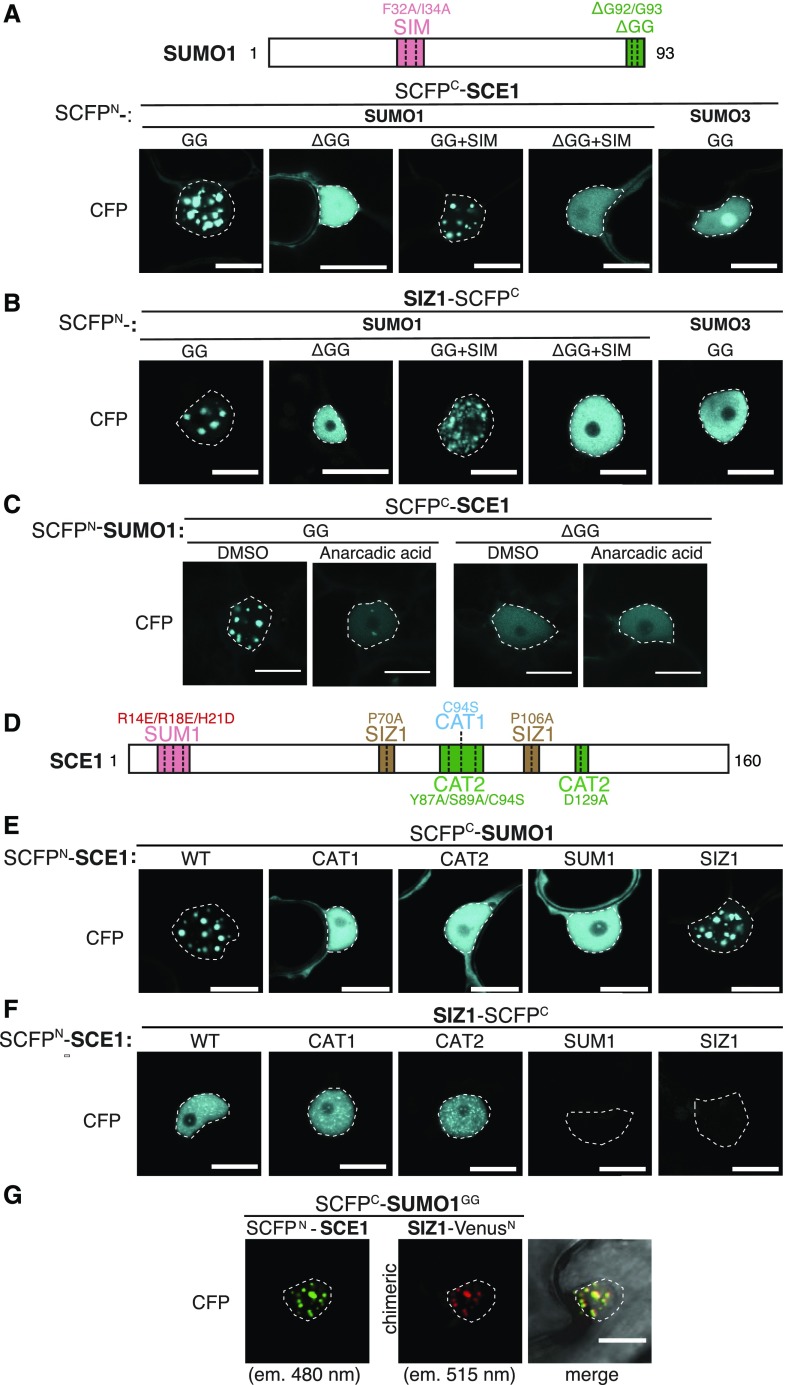
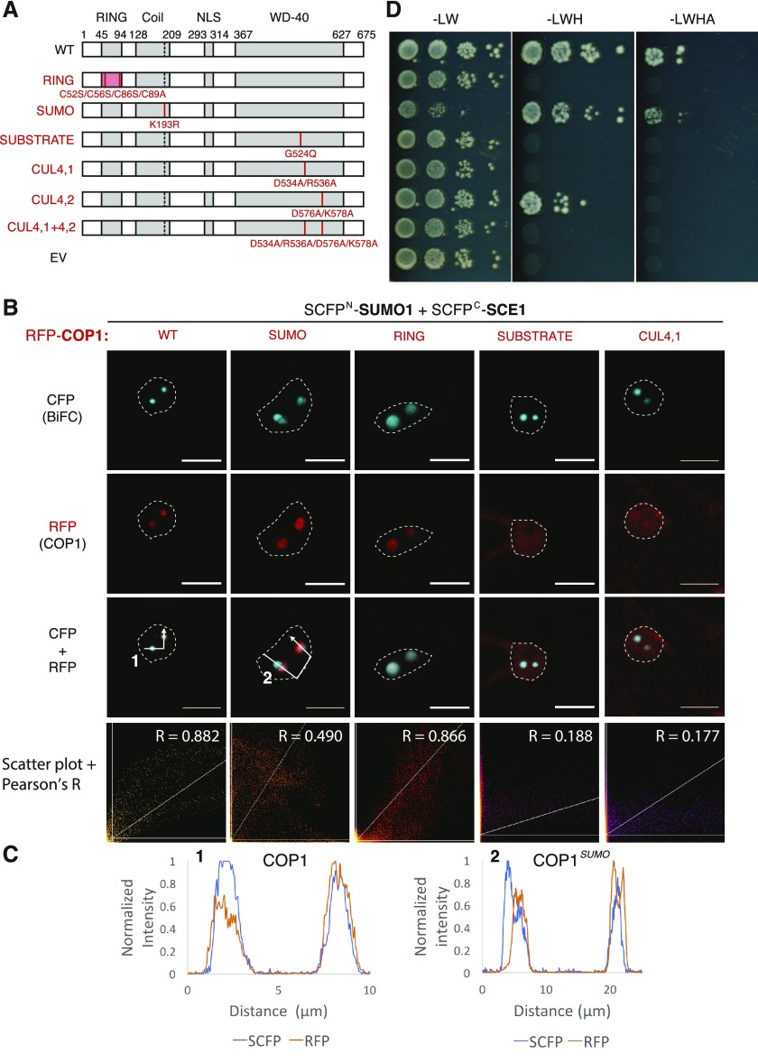
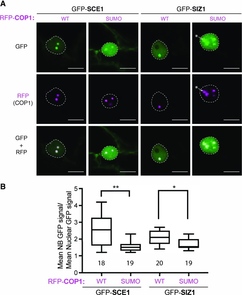
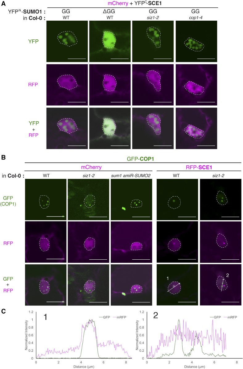
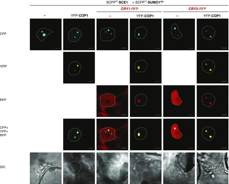
Attachment of the small ubiquitin-like modifier (SUMO) to substrate proteins modulates their turnover, activity, or interaction partners. However, how this SUMO conjugation activity concentrates the proteins involved and the substrates into uncharacterized nuclear bodies (NBs) remains poorly understood. Here, we characterized the requirements for SUMO NB formation and for their subsequent colocalization with the E3 ubiquitin ligase CONSTITUTIVE PHOTOMORPHOGENIC 1 (COP1), a master regulator of plant growth. COP1 activity results in degradation of transcription factors, which primes the transcriptional response that underlies elongation growth induced by darkness and high ambient temperatures (skoto- and thermomorphogenesis, respectively). SUMO conjugation activity alone was sufficient to target the SUMO machinery into NBs. Colocalization of these bodies with COP1 required, in addition to SUMO conjugation activity, a SUMO acceptor site in COP1 and the SUMO E3 ligase SAP and Miz 1 (SIZ1). We found that SIZ1 docks in the substrate-binding pocket of COP1 via two valine-proline peptide motifs, which represent a known interaction motif of COP1 substrates. The data reveal that SIZ1 physically connects COP1 and SUMO conjugation activity in the same NBs that can also contain the blue-light receptors CRYPTOCHROME 1 and CRYPTOCHROME 2. Our findings thus suggest that sumoylation stimulates COP1 activity within NBs. Moreover, the presence of SIZ1 and SUMO in these NBs explains how both the timing and amplitude of the high-temperature growth response is controlled. The strong colocalization of COP1 and SUMO in these NBs might also explain why many COP1 substrates are sumoylated.


Figures

Figure 1.
Figure 1.
SUMO nuclear body formation depends on SCE1 conjugation activity. A, Nuclear localization pattern of SUMO1∙SCE1 BiFC pairs, including mutant variants of SUMO1 and the SUMO3∙SCE1 BiFC pair. Included above is a schematic representation of residues mutated and/or deleted in SUMO1 (GG, mature SUMO; SIM, F32A+I34A; ΔGG, deletion of the C-terminal diGly motif). B, Similar to A; nuclear localization pattern of SUMO1/3-SIZ1 BiFC pairs including mutant variants of SUMO1. C, Nuclear localization pattern of the SUMO1∙SCE1 complex in response to anacardic acid inhibition of SUMO conjugation (100 μm in 1% [v/v] DMSO) 1.5 h postinfiltration. D, Schematic representation of SCE1 mutants and their substitutions. CAT1, catalytic Cys residue mutated; CAT2, binding pocket for the ψKxE SUMO acceptor motif mutated; SUM1, noncovalent association of SUMO disrupted; SIZ1, SIZ1-binding disrupted. E, Nuclear localization pattern of SUMO1∙SCE1 BiFC pairs, including mutant variants of SCE1: SCE1CAT1, SCE1CAT2, SCE1SUM1, and SCE1SIZ1. F, Nuclear localization pattern of SCE1∙SIZ1 BiFC pairs, including mutant variants of SCE1: SCE1CAT1, SCE1CAT2, SCE1SUM1, and SCE1SIZ1. G, Multicolor BiFC of SUMO1GG, SCE1, and SIZ1 showing nuclear localization pattern. The micrographs show the nuclear signal of the reconstituted SCFPN/SCFPC (SCE1∙SUMO1GG) and VenusN/SCFPC (SIZ∙SUMO1GG) fluorophores and their merged signals. The two chimeric BiFC combinations differ in their excitation and emission spectra. The BiFC pairs in A to F were fused to two halves of SCFP (SCFPN + SCFPC) with the orientation of the fusions indicated. Scale bars, 20 µm. All micrographs were taken in N. benthamiana epidermal leaf cells 2 to 3 d post-agro-infiltration with strains expressing the indicated constructs; nuclei are outlined with white lines. Supplemental Figure S3 depicts for A to F an overlay of the DIC and CFP images of the nuclei shown and a zoom-out (A′–F′) depicting the BiFC signal in the entire cell.
Figure 2.
Figure 2.
The SUMO1∙SCE1 BiFC pair colocalizes with COP1 in nuclear bodies when catalytically active. A, Y2H analysis of the interaction between COP1 as a GAL4 BD fusion protein and the SUMO (machinery) proteins fused to the GAL4 AD domain. Yeast growth was scored 3 d after incubation on selective media at 30°C (−Leu [L] and Trp [W], −LW and His [H], −LWH + 1 mm 3-Amino-1,2,4-triazole [3AT], −LWH and Adenine [A]). B and C, Nuclear localization pattern of the SUMO1∙SCE1 BiFC pair in cells overexpressing RFP-COP1. B, ΔGG, conjugation-deficient SUMO variant; C, CAT1, catalytically inactive SCE1. Micrographs show from top-to-bottom the reconstituted BiFC signal, RFP-COP1, and their merged signals. Conditions were identical to those in Figure 1. Scale bars, 10 µm.
Figure 3.
Figure 3.
The COP1-SIZ1 interaction in NBs depends on both the substrate binding pocket and the SUMO acceptor site in COP1. A, Schematic representation of COP1 variants and the protein-protein interaction domain disrupted (left side, red text) with their mutations shown: RING, RING Zn2+-finger binding domain mutant; SUMO, loss of SUMO acceptor site; SUBSTRATE, mutation in the substrate binding groove; CUL4, mutations in the CUL4-binding “WDRX” motifs. WT, Wild type. B, Nuclear localization pattern of the SUMO1∙SCE1 BiFC pair in cells overexpressing functional mutants of RFP-COP1. Micrographs depict from top to bottom the BiFC CFP signal, RFP-COP1, and their combined signals. Bottom row depicts a scatter plot of the RFP/CFP signal intensities per pixel and their Pearson’s correlation coefficient (R). Conditions were identical to those in Figure 1. Scale bars, 10 µm. C, Normalized intensity profiles depict the fluorescence signal intensities for SCFP: (BiFC pair) and RFP-COP1: coexpression of (1) wild-type COP1 or (2) COP1SUMO; the profiles follow the white arrows depicted in B. Note the profiles of RFP-COP1SUMO (A) and SUMO∙SCE1 CFP signals poorly overlap in (2). D, Mapping of the SIZ1 interaction site in COP1 using Y2H analysis of the COP1 variants depicted in B. The COP1 variants were fused to the GAL4 BD, whereas SIZ1 was fused to the GAL4 AD-fusion. Yeast growth was scored 3 d after incubation on selective medium at 30°C.
Figure 4.
Figure 4.
Formation of COP1 + SUMO1∙SIZ1-containing NBs depends on the COP1 substrate-binding pocket that apparently recruits SIZ1 via VP motifs. A, Nuclear localization pattern of the SUMO1∙SIZ1 BiFC pair in cells overexpressing RFP-COP1 variants: COP1SUMO, COP1RING, and COP1SUBSTRATE. See Figure 3A for details on the COP1 variants. Micrographs show from top to bottom the BiFC signal, RFP-COP1, and their merged signals. Conditions were identical to those in Figure 1. Scale bars, 10 µm. B, Schematic representation of SIZ1 variants and the protein-protein interaction domain disrupted (left side) by the mutations introduced: plant homeodomain (PHD) and Pro-Ile-Asn-Ile-Thr (PINIT), both reduced substrate binding; SP-RING, no interaction with SCE1; VP1 and VP2, putative interacting motifs for the COP1 substrate binding groove. C, Mapping of the COP1 interaction site in SIZ1 using Y2H analysis of the SIZ1 variants depicted in B. Similar to that in Figure 3, SIZ1 variants were fused to GAL4 AD-fusion, whereas COP1 was fused to GAL4 BD. Yeast growth was scored after 3 d at 30°C.
Figure 5.
Figure 5.
Colocalization of GFP-tagged SCE1/SIZ1 with RFP-COP1 NBs is compromised following mutation of the COP1 SUMO acceptor site. A, Nuclear localization pattern of GFP-tagged SCE1 and SIZ1 in the presence of RFP-COP1 or the RFP-COP1SUMO SUMO acceptor site mutant (Lys-193Arg). * marks amorphous NBs in the GFP-SIZ1, RFP-COP1SUMO combination. Micrographs from top to bottom: GFP, RFP, and their merged signals. Scale bars, 10 µm; WT, wild type. B, Quantification of the average GFP signal intensity in the NBs per nucleus divided by the average fluorescence signal in the nucleus (with the data of three biological replicates pooled with at least five nuclei per replica). Total number of nuclei analyzed is shown. Significant differences were detected using an unpaired Student’s t test assuming unequal variances; **P < 0.01, *P < 0.05. Conditions were identical to those in Figure 1.
Figure 6.
Figure 6.
SUMO conjugation also triggers NB formation in Arabidopsis, which depends genetically on SIZ1 for recruitment to COP1 NBs. A, Nuclear localization pattern of the SCE1∙SCE1 BiFC pair in Arabidopsis cells (wild type [WT], siz1-2, or cop1-4) transformed using particle bombardment. The BiFC constructs and the Arabidopsis genotypes used are indicated at the top. To detect the transformed cells, tissue was cobombarded with mCherry. The micrographs show from top to bottom the YFP, RFP, and the merged signal; nuclei are outlined with a white line. Four-week-old Arabidopsis rosettes were bombarded. Two days postbombardment, plant material was transferred to a dark box, and the fluorescence signals were examined after a further day. B, Nuclear localization pattern of GFP-COP1 and RFP-SCE1 in Arabidopsis cells (wild type, siz1-2, or sumo1;amiR-SUMO2) transformed using particle bombardment. To detect the transformed cells, tissue was either cobombarded with mCherry or RFP-SCE1. Micrographs with nuclei with GFP-COP1 containing NBs are shown. Scale bars, 10 μm. C, Normalized intensity profiles depict the fluorescence signal intensities of GFP-COP1 and RFP-SCE1 co-expressed in wild type and the siz1-2 mutant, along arrows 1 and 2 in the right two panels of (B).
Figure 7.
Figure 7.
CRY1-mRFP or CRY2-mRFP and the SUMO1∙SCE1 BiFC complex colocalize in COP1 NBs. Micrographs of cells transiently expressing SCFPN-SCE1 and SCFPC-SUMO1GG with or without YFP-COP1 and either CRY1-mRFP or CRY2-mRFP, imaged 3 d post-agro-infiltration. Scale bars, 10 µm.

References

    1. Ballesteros ML, Bolle C, Lois LM, Moore JM, Vielle-Calzada JP, Grossniklaus U, Chua NH (2001) LAF1, a MYB transcription activator for phytochrome A signaling. Genes Dev 15: 2613–2625 - PMC - PubMed
    1. Banani SF, Rice AM, Peeples WB, Lin Y, Jain S, Parker R, Rosen MK (2016) Compositional Control of Phase-Separated Cellular Bodies. Cell 166: 651–663 - PMC - PubMed
    1. Banani SF, Lee HO, Hyman AA, Rosen MK (2017) Biomolecular condensates: Organizers of cellular biochemistry. Nat Rev Mol Cell Biol 18: 285–298 - PMC - PubMed
    1. Bencsath KP, Podgorski MS, Pagala VR, Slaughter CA, Schulman BA (2002) Identification of a multifunctional binding site on Ubc9p required for Smt3p conjugation. J Biol Chem 277: 47938–47945 - PubMed
    1. Bernier-Villamor V, Sampson DA, Matunis MJ, Lima CD (2002) Structural basis for E2-mediated SUMO conjugation revealed by a complex between ubiquitin-conjugating enzyme Ubc9 and RanGAP1. Cell 108: 345–356 - PubMed

Publication types

MeSH terms