. 2018 Dec 28;293(52):20137-20156.
doi: 10.1074/jbc.RA118.004325. Epub 2018 Nov 2.

Binding of a glaucoma-associated myocilin variant to the αB-crystallin chaperone impedes protein clearance in trabecular meshwork cells

Affiliations

Binding of a glaucoma-associated myocilin variant to the αB-crystallin chaperone impedes protein clearance in trabecular meshwork cells

Jeffrey M Lynch et al. J Biol Chem. .

Abstract

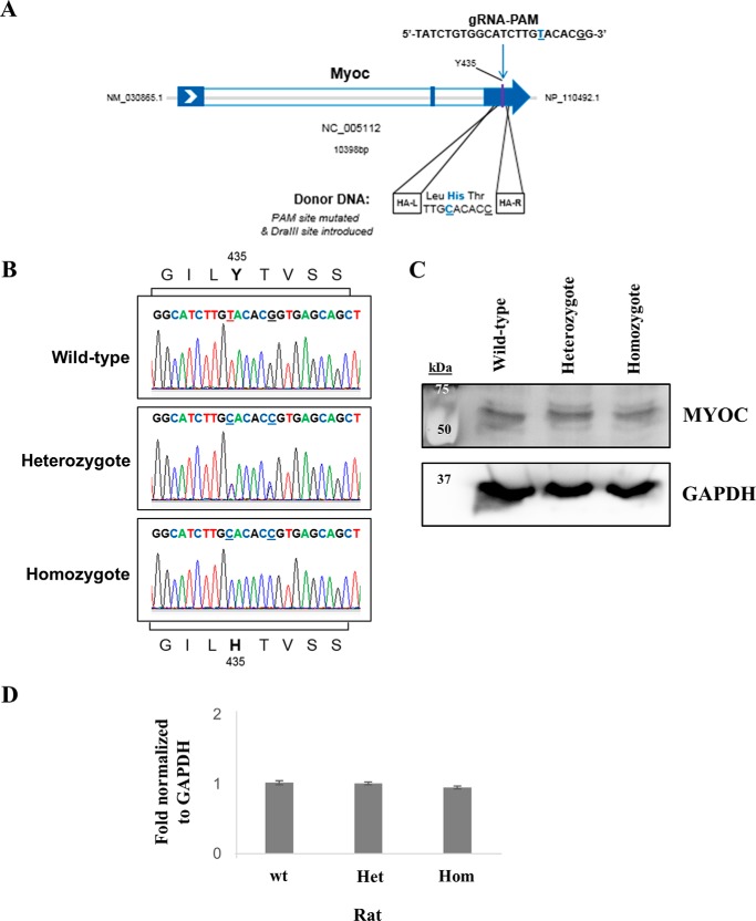
Myocilin (MYOC) was discovered more than 20 years ago and is the gene whose mutations are most commonly observed in individuals with glaucoma. Despite extensive research efforts, the function of WT MYOC has remained elusive, and how mutant MYOC is linked to glaucoma is unclear. Mutant MYOC is believed to be misfolded within the endoplasmic reticulum, and under normal physiological conditions misfolded MYOC should be retro-translocated to the cytoplasm for degradation. To better understand mutant MYOC pathology, we CRISPR-engineered a rat to have a MYOC Y435H substitution that is the equivalent of the pathological human MYOC Y437H mutation. Using this engineered animal model, we discovered that the chaperone αB-crystallin (CRYAB) is a MYOC-binding partner and that co-expression of these two proteins increases protein aggregates. Our results suggest that the misfolded mutant MYOC aggregates with cytoplasmic CRYAB and thereby compromises protein clearance mechanisms in trabecular meshwork cells, and this process represents the primary mode of mutant MYOC pathology. We propose a model by which mutant MYOC causes glaucoma, and we propose that therapeutic treatment of patients having a MYOC mutation may focus on disrupting the MYOC-CRYAB complexes.

Keywords: CRISPR/Cas; aggregation; aging; animal model; cell biology; crystallin; genetic disease; glaucoma; myocilin; secretion.


Conflict of interest statement

Authors are all employees of Novartis Institutes for BioMedical Research (NIBR) and receive salary. As NIBR is a publicly traded pharmaceutical company, the authors may hold stock

Figures

Figure 1.
Figure 1.
MYOC protein is found in human AH, and in the eye tissue, the highest MYOC expression is observed in the trabecular meshwork. A, Western blotting for MYOC protein in human AH collected from different living donor eyes as well as from different deceased donor eyes. All AH samples were from donors of a similar elderly age, and no donors had a MYOC mutation. For the Western blotting, 5 μg of each sample was loaded per well, and anti-MYOC antibody is from R&D Systems. B, human AH sample Western blots were quantified. Error bars are ± S.D., and * indicates t test p < 0.05. C, control immunohistochemistry image of the human TM region and an image stained to show MYOC expression (MYOC indicated by the brown color) in the TM region. High expression of MYOC is evident in the TM. MYOC expression was observed in other regions of the eye (Fig. S1), but at a lower expression than that observed for the TM. Abbreviations used are as follows: AC, anterior chamber; SC, Schlemm's canal.
Figure 2.
Figure 2.
Generation and confirmation of rats with the Myoc Y435H mutation. A, CRISPR/Cas9–based strategy to introduce Y435H point mutation in rat Myoc. B, sequencing traces of Myoc PCR products amplified from rat genomic DNA isolated from tail biopsies. Sequencing results confirm both the Y435H point mutation and the silent (PAM site) mutation in the heterozygote and homozygote animals (sites of mutation are underlined). C, Western blotting of soluble Myoc in rat limbal ring lysates (40 μg of samples) using anti-MYOC antibody from Acris. D, rat limbal ring lysate Western blots were quantified. Error bars are ± S.D., and t tests showed p > 0.1. Abbreviations used are as follows: HA-L, left homology arm; HA-R, right homology arm; Het, heterozygote; Hom, homozygote; wt, WT.
Figure 3.
Figure 3.
Rats with the Myoc Y435H mutation have IOP and TM histology (at 9 months of age) that does not differ from WT rats, and this did not change following prednisolone challenge. A, IOP was monitored in a 4-month-old cohort of 10 WT (wt), 10 heterozygote (Het), and 10 homozygote (Hom) rats before and after implantation of a prednisolone pellet (25 mg for 60 days). Arrow indicates time of implantation. Results are ± S.E. B, top, H&E and trichrome staining of TM of 9-month-old rats that were not treated with prednisolone. Immunohistochemical images for α-SMA, COLIV, and FN1 (red color) indicated that the WT rats did not have tissue expression of these proteins that differed from the rats with a Myoc Y435H mutation. Bottom, H&E and trichrome staining of 9-month-old rat eyes from animals treated with prednisolone. Immunohistochemical images for α-SMA, COLIV, and FN1 for 9-month-old rats that received prednisolone treatment. Abbreviation used is as follows: SC, Schlemm's canal. Scale bars are 100 μm.
Figure 4.
Figure 4.
Mutant MYOC Y435H was not found to adversely impact the rat retina. A, averaged waveforms at each stimulus tested. Arrows indicate where the pSTR and nSTR amplitudes were measured. No significant differences in ganglion cell function as assessed by the pSTR (−6.0 p = 0.110, −5.5 p = 0.231, and −5.0 p = 0.237) and nSTR (−6.0 p = 0.998, −5.5 p = 0.651, and −5.0 p = 0.624) were observed between 18-month-old WT (wt) and mutant Myoc Y435H (Het) animals. B, averaged scotopic waveforms with arrows indicating a- and b-wave measurements. No significant differences in rod/cone photoreceptor function (a-wave, p = 0.696) and on bipolar cell function (b-wave, p = 0.552) were observed between groups. C, representative OCT images; arrows indicate thickness measurement. Thickness measurements were made across the entire retinal image excluding the optic nerve region. Retinal thickness as measured from the NFL to the RPE was not significantly different (p = 0.946) between groups. Statistical analysis was performed using Student's unpaired t test. Abbreviations used are as follows: ERG, electroretinogram; pSTR, ganglion cell response to low-intensity flash stimuli; nSTR, ganglion cell dominated response with contribution from other cell types (e.g. amacrine and/or Muller cells).
Figure 5.
Figure 5.
Soluble CRYAB is reduced in rats with a MYOC Y435H mutation, and MYOC has been found to bind CRYAB. A, representative Western blottings (WB) for CRYAB protein expression in 12-month-old rats indicate that there is more soluble CRYAB detected in WT rat limbal ring lysates compared with MYOC Y435H heterozygote and homozygote rats lysates. 10 μg of each sample was loaded per lane. Representative Western blotting is shown. Quantification of Western blottings indicated approximately 1-fold more soluble CRYAB protein in limbal rings from WT mice compared with rats with the MYOC Y435H mutation. ± S.D. is indicated. B, anti-CRYAB rat limbal ring lysate Western blots were quantified. Error bars are ± S.D., and * indicates t test p < 0.001. C, AH from several different 12-month-old rats were examined for soluble CRYAB protein, and the WT animals had the greatest amount of CRYAB. 10 μg of each sample was loaded per lane. Representative Western blotting is shown. D, anti-CRYAB rat AH Western blottings were quantified. Error bars are ± S.D., and * indicates t test, p < 0.05, and ** indicates t test p = 0.01. E, immunohistochemistry for CRYAB in the trabecular meshwork of 18-month-old WT and heterozygote rats indicates that the WT animals had more detectable CRYAB. F, Western blotting results following immunoprecipitation (IP) indicate that CRYAB binds MYOC. Abbreviations used are as follows: Het, heterozygote; Hom, homozygote; SC, Schlemm's canal; TM, trabecular meshwork; wt, WT.
Figure 6.
Figure 6.
Western blottings for eye lysates from our in vivo models show no differences in endoplasmic reticulum proteins, but aged MYOC Y435H heterozygous rats had less soluble CRYAB detected and more high molecular weight Ub proteins. A, Western blottings using whole-eye lysates isolated from 18-month-old WT and MYOC Y435H heterozygote rats. Arrow indicates expected band location for PDI. B, rat whole-eye lysate Western blottings were quantified. Error bars are ± S.D. t tests for the examined ER proteins showed p > 0.1, and * indicates p < 0.05 and ** p < 0.01. Abbreviations used are as follows: Het, heterozygote; Ub, ubiquitin; wt, WT.
Figure 7.
Figure 7.
In vitro, MYOC protein was found to accumulate in presence of CRYAB. A, NTM5 cells were transiently transfected for 48 h with different MYOC cDNAs ± CRYAB and then MYOC in soluble and insoluble fractions examined by Western blotting. Note that very little GAPDH was detectable in the insoluble fraction. B, bar graph quantifying anti-MYOC Westerns blottings to show percentage of MYOC in soluble and insoluble fractions. C, ThT staining (green) for aggregates in NTM5 cells co-transfected with MYOC and CRYAB suggests the most intense staining is when mutant MYOC is co-expressed with CRYAB. The nucleus is counter-stained with DAPI (blue). D, quantification of number of ThT aggregates relative to cell number ± S.E. and with t test results.
Figure 8.
Figure 8.
In human AH samples, CRYAB appears following stress conditions. A, NTM5 cells transiently transfected for 48 h had cytoplasmic proteins isolated by a hypotonic buffer, and MYOC Y437H was found in the cytoplasmic extract. CALR serves as a control indicating no lysis of the ER. B, Western blotting of human donor aqueous humor (10 μg per sample) indicates that CRYAB expression in AH is elevated during stress conditions. C, IHC suggests that CRYAB protein (red color) is highly expressed in human glaucoma in the trabecular meshwork nearest Schlemm's canal. Nuclei are indicated by hematoxylin staining.
Figure 9.
Figure 9.
Cartoon figure of a cell illustrating simplified mechanism of mutant MYOC-induced glaucoma pathology. Step 1, stresses will result in step 2, activation of MYOC and CRYAB gene expression. Step 3, WT MYOC (dark green) is synthesized in the ER and is a secreted matricellular protein. Step 4, in the case of mutant MYOC (red), it is misfolded and will be retro-translocated from the ER to the cytoplasm for clearance by ERAD, which utilizes the proteasome. In the cytoplasm, mutant MYOC can bind CRYAB. Increased stress as well as time/age will result in diminished efficiency of protein clearance, and this will impact ERAD with feedback signaling to the ER occurring so as to mildly increase expression of ER and ERAD proteins to try and correct the problem. Step 5, with time/age mutant MYOC protein aggregates will accumulate within the cell. These aggregates can compromise cell function and threaten/impact cell viability. Thus, MYOC-induced glaucoma is a protein misfolding disease, and pathology occurs in a manner similar to other aging diseases. Step 6, outside the cell WT MYOC likely acts as a matricellular protein and may have other functions, but additional research is required. Crystallin has been reported (73) to bind and inhibit dexamethasone. Abbreviation used is as follows: DEX, dexamethasone.

References

    1. Quigley H. A., and Broman A. T. (2006) The number of people with glaucoma worldwide in 2010 and 2020. Br. J. Ophthalmol. 90, 262–267 10.1136/bjo.2005.081224 - DOI - PMC - PubMed
    1. Friedman D. S., Wolfs R. C., O'Colmain B. J., Klein B. E., Taylor H. R., West S., Leske M. C., Mitchell P., Congdon N., Kempen J., and Eye Diseases Prevalence Research Group. (2004) Eye diseases prevalence research group. Prevalence of open-angle glaucoma among adults in the United States. Arch. Ophthalmol. 122, 532–538 10.1001/archopht.122.4.532 - DOI - PMC - PubMed
    1. Weinreb R. N., Aung T., and Medeiros F. A. (2014) The pathophysiology and treatment of glaucoma. JAMA 311, 1901–1911 10.1001/jama.2014.3192 - DOI - PMC - PubMed
    1. Meyer A., Béchetoille A., Valtot F., Dupont de Dinechin S., Adam M. F., Belmouden A., Brézin A. P., Gomez L., Bach J. F., and Garchon H. J. (1996) Age-dependent penetrance and mapping of the locus for juvenile and early-onset open-angle glaucoma on chromosome 1q (GLC1A) in a French family. Hum. Genet. 98, 567–571 10.1007/s004390050260 - DOI - PubMed
    1. Sunden S. L., Alward W. L., Nichols B. E., Rokhlina T. R., Nystuen A., Stone E. M., and Sheffield V. C. (1996) Fine mapping of the autosomal dominant juvenile open angle glaucoma (GLC1A) region and evaluation of candidate genes. Genome Res. 6, 862–869 10.1101/gr.6.9.862 - DOI - PubMed

Publication types

MeSH terms