. 2018 Dec;210(4):1287-1299.
doi: 10.1534/genetics.118.301456. Epub 2018 Nov 2.

Transgenerational Epigenetic Inheritance Is Negatively Regulated by the HERI-1 Chromodomain Protein

Affiliations

Transgenerational Epigenetic Inheritance Is Negatively Regulated by the HERI-1 Chromodomain Protein

Roberto Perales et al. Genetics. 2018 Dec.

Abstract

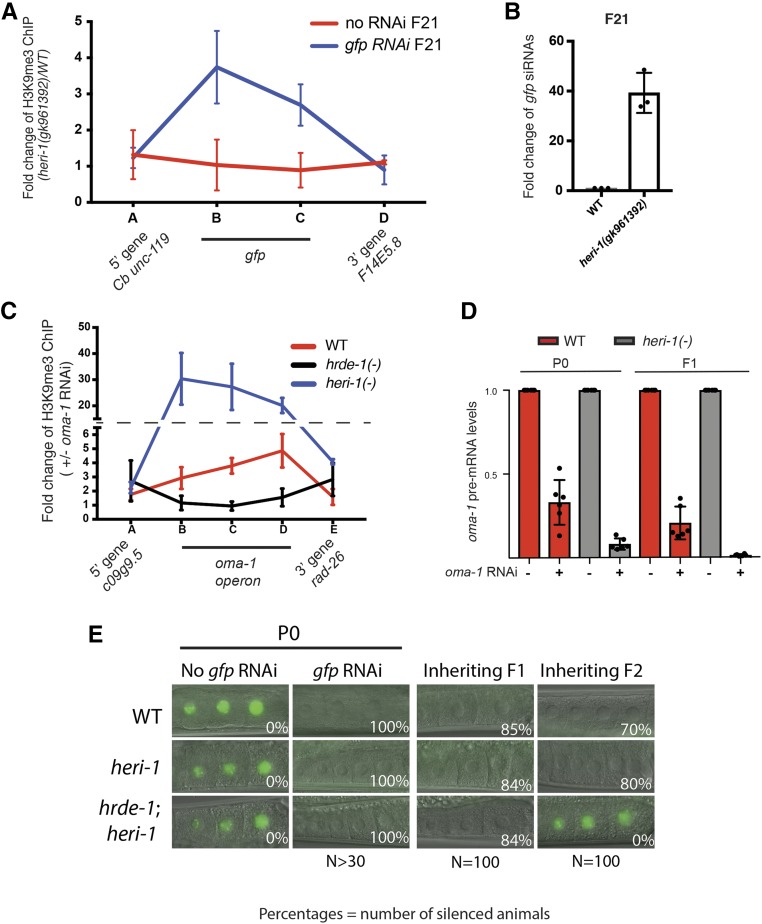
Transgenerational epigenetic inheritance (TEI) is the inheritance of epigenetic information for two or more generations. In most cases, TEI is limited to a small number of generations (two to three). The short-term nature of TEI could be set by innate biochemical limitations to TEI or by genetically encoded systems that actively limit TEI. In Caenorhabditis elegans, double-stranded RNA (dsRNA)-mediated gene silencing [RNAi (RNA interference)] can be inherited (termed RNAi inheritance or RNA-directed TEI). To identify systems that might actively limit RNA-directed TEI, we conducted a forward genetic screen for factors whose mutation enhanced RNAi inheritance. This screen identified the gene heritable enhancer of RNAi (heri-1), whose mutation causes RNAi inheritance to last longer (> 20 generations) than normal. heri-1 encodes a protein with a chromodomain, and a kinase homology domain that is expressed in germ cells and localizes to nuclei. In C. elegans, a nuclear branch of the RNAi pathway [termed the nuclear RNAi or NRDE (nuclear RNA defective) pathway] promotes RNAi inheritance. We find that heri-1(-) animals have defects in spermatogenesis that are suppressible by mutations in the nuclear RNAi Argonaute (Ago) HRDE-1, suggesting that HERI-1 might normally act in sperm progenitor cells to limit nuclear RNAi and/or RNAi inheritance. Consistent with this idea, we find that the NRDE nuclear RNAi pathway is hyperresponsive to experimental RNAi treatments in heri-1 mutant animals. Interestingly, HERI-1 binds to genes targeted by RNAi, suggesting that HERI-1 may have a direct role in limiting nuclear RNAi and, therefore, RNAi inheritance. Finally, the recruitment of HERI-1 to chromatin depends upon the same factors that drive cotranscriptional gene silencing, suggesting that the generational perdurance of RNAi inheritance in C. elegans may be set by competing pro- and antisilencing outputs of the nuclear RNAi machinery.

Keywords: RNAi; chromatin; small RNAs; transgenerational epigenetic inheritance.


Figures

Figure 1
Figure 1
A genetic screen to identify heritable enhancers of RNAi (heri) genes. pie-1::h2b::gfp and oma-1(zu405ts) can be silenced heritably by RNAi (Vastenhouw et al. 2006; Alcazar et al. 2008). We treated pie-1::h2b::gfp;oma-1(zu405ts) animals with the mutagen EMS. Control animals were left untreated. After two generations, animals were fed bacteria expressing double-stranded RNA targeting gfp and oma-1 simultaneously. Embryos from these animals were isolated and shifted to a non-RNAi (OP50) food source and grown at 20°, which is the nonpermissive temperature for oma-1 (zu405ts). Embryos were isolated for an additional five generations until control animals stopped inheriting gfp and oma-1(zu405ts) silencing (gfp ON and embryonic arrest, ∼F7). Mutagenized pools were allowed to grow for two more generations and potential Heri strains (animals were gfp OFF and alive, indicating that pie-1::h2b::gfp and oma-1(zu405ts) were still being silenced) were singled. After five additional generations, those populations still inheriting gene silencing were kept for further study. heri genes were identified by a combination of whole-genome sequencing and bioinformatic analysis (see Materials and Methods and Figure S1). Rare animals within these populations had lost gene silencing at pie-1::h2b::gfp and oma-1(zu405ts). These animals were isolated to establish mutant lines that expressed reporter genes (termed “reset”). EMS, ethyl methanesulfate; RNAi, RNA interference; RT, room temperature.
Figure 2
Figure 2
HERI-1 limits transgenerational epigenetic inheritance. (A) Diagram of HERI-1 domain structure. heri-1 alleles identified in our genetic screen and the heri-1(gk961392) deletion allele used in this study are indicated. Asterisk represents premature stop codon. (B) RNAi inheritance assay (see Materials and Methods) showing percent embryonic viability [oma-1(zu405ts) silencing] over generations after oma-1 RNAi feeding in our starting strain (control, YY565), and heri-1 mutants that were identified in the Heri screen and that had been “reset” for reporter gene expression (see Figure 1). Error bars represent SD of the mean. (C) gfp inheritance assay showing the percentage of animals of the indicated genotypes showing gfp silencing in control and reset heri-1 strains over generations after gfp RNAi exposure. (D) Representative images of oocytes in wild-type and heri-1(gk961392) animals that harbor the pie-1::h2b::gfp transgene during the gfp RNAi inheritance assay. Generations after gfp RNAi are indicated. RNAi, RNA interference.
Figure 3
Figure 3
HERI-1 is expressed in germ cell nuclei. (A) Fluorescent micrographs of the mitotic and transition zones of an adult germline from an animal expressing HERI-1::GFP::3XFLAG and mCHERRY::H2B. Note that HERI-1::GFP::3XFLAG does not colocalize with chromatin in dividing cells (arrows). Distal tip cell is to the left of image. One nuclei (square box) is magnified in panels shown to the right. (B) Micrograph of a larval stage one (L1) and a larval stage two (L2) animal. HERI-1::GFP::3XFLAG is expressed in Z2 and Z3 (arrows) in L1, and in the developing germline of L2 animals (dotted line). In these animals, no HERI-1::GFP signal was observed in the soma. Note that the nongermline fluorescence signal is due to autofluorescence, which is also present in wild-type animals. HERI-1::GFP is expressed throughout the remainder of germline development and no GFP fluorescence could be observed in somatic cells during this time.
Figure 4
Figure 4
HERI-1 inhibits nuclear RNAi. (A) H3K9me3 ChIP-qPCR on heri-1(+) heri-1(−) animals. Data are expressed as a ratio of H3K9me3 ChIP signals detected in heri-1(+) over heri-1(−) mutant animals before gfp RNAi (bottom panel) and 20 generations after gfp RNAi (bottom panel). Data are from three biological replicates and error bars are SD of the mean. (B) Total RNA isolated from animals of the indicated genotypes was used for Custom Taqman assays (Burton et al. 2011) to detect gfp siRNAs 20 generations after gfp RNAi treatment. Signal from WT (starting strain) animals is defined as one. Data are from three biological replicates and error bars are SD. (C) H3K9me3 ChIP was conducted on WT, heri-1 (gk961392), and hrde-1(tm1200) animals in the progeny of animals exposed +/− to oma-1 RNAi. Relative location of primers used for qPCR are indicated. Position along x-axis is not to scale with actual genomic loci (see Figure 5). Data are from three biological replicates and error bars are SD of the mean. Note, a value of one in this assay means that RNAi had no effect on the state of H3K9me3 at this locus. Data show that H3K9me3 is enriched within the oma-1 operon after oma-1 RNAi. oma-1 operon contains three genes; oma-1, spr-2, and c27b76.2. Primer set B and C are in oma-1. Primer set D is in spr-2. (D) qPCR data showing that the oma-1 pre-mRNA is more silenced in heri-1(gk961392) animals than in WT animals after oma-1 RNAi. Error bars are SD of the mean. Note, a value of one would indicate that RNAi had no effect on oma-1 pre-mRNA levels for this experiment. (E) Animals of the indicated genotypes [heri-1(gk961392) and hrde-1(tm1200)] and expressing pie-1::gfp::h2b were exposed to +/− gfp RNAi. Representative micrographs of −1, −2, and −3 oocytes are shown. Percentages of animals inheriting silencing in the indicated generations are shown. ChIP, chromatin immunoprecipitation; qPCR, quantitative PCR; RNAi, RNA interference; siRNA, small interfering RNA; WT, wild-type.
Figure 5
Figure 5
RNAi causes HERI-1 to associate with chromatin of genes undergoing heritable silencing. HERI-1::GFP::3xFlag animals were exposed to +/− oma-1 RNAi and HERI-1 ChIP was conducted on progeny. Fold enrichment of HERI-1 on the oma-1 locus, as well as neighboring loci, is shown and is expressed as a ratio of HERI-1 ChIP signals +/− oma-1 RNAi. Data are from three biological replicates and error bars are SD of the mean. Similar results were obtained when HERI-1::3xFLAG animals were subjected to a similar analysis (Figure S6). A value of one would indicate that RNAi had no effect on HERI-1 ChIP. Note that primer set D does not show RNAi-induced HERI-1 binding, but did show an increase in H3K9me3 in Figure 4C (see Discussion). ChIP, chromatin immunoprecipitation; RNAi, RNA interference; WT, wild-type.
Figure 6
Figure 6
heri-1 mutant animals have defective sperm. (A) Fluorescent micrograph of WT or heri-1(gk961392) oocytes expressing pie-1p::h2b::gfp. Of the heri-1 mutant animals, 20% show a stacked oocytes in either one or both gonad arms. Asterisk indicates vulva. Thick arrows indicate stacked oocytes. Thin arrow indicates an unfertilized oocyte. (B) Quantification of the indicated oocyte defects in wild-type, heri-1(gk961392), hrde-1(tm1200), and double-mutant animals. Error bars are SD of the mean. (C) Number of progeny from heri-1(gk961392) animals that had stacked oocytes in both gonad arms that were selfed (self) or after crossing to WT (N2) males (cross). n = 13 for self and n = 14 for cross. (D) DAPI staining of a heri-1(gk961392);pie-1::h2b::gfp animals that had stacked oocytes in one gonad arm. A magnification of spermatheca from both gonad arms (indicated by box in “merge” panel) is shown below. Arrow indicates gonad arm with stacked oocytes. Asterisk indicates vulva. (E) Images of WT and heri-1(gk961392) animals undergoing spermatogenesis. Spermatogenesis steps are labeled for WT animals. heri-1(gk961392) fail to complete spermatogenesis. “%” refers to the percent of animals displaying spermatogenesis defects. RNAi, RNA interference; WT, wild-type.
Figure 7
Figure 7
Model for the role of HERI-1 in limiting RNA inheritance. The AGO HRDE-1 uses siRNAs as guides to recognize and bind pre-mRNAs with sequence homology to siRNAs. HRDE-1 then recruits downstream silencing factors, such as the NRDEs (unlabeled teal ovals) and chromatin-modifying enzymes, to deposit H3K9me3 as well as another, currently unknown (X), signal on chromatin to mark genes undergoing nuclear RNAi. Note: X could be another chromatin PTM or a chromatin/DNA-binding protein. X and H3K9me3 cooperate to recruit HERI-1 to chromatin. HERI-1 is a pseudokinase that may limit RNAi inheritance by binding and regulating prosilencing proteins (such as HRDE-1 or NRDEs) that are present on genes undergoing nuclear RNAi/RNAi inheritance. The model predicts that nuclear RNAi drives cotranscriptional gene silencing by inhibiting RNA Polymerase II (RNAP II) while, at the same time, limiting this silencing by recruiting negative regulators of the process, such as HERI-1, to sites of nuclear RNAi/RNAi inheritance. NRDE, nuclear RNA defective; RNAi, RNA interference; siRNA, small interfering RNAs; WT, wild-type.

Similar articles

Cited by

References

    1. Alcazar R. M., Lin R., Fire A. Z., 2008. Transmission dynamics of heritable silencing induced by double-stranded RNA in Caenorhabditis elegans. Genetics 180: 1275–1288. 10.1534/genetics.108.089433 - DOI - PMC - PubMed
    1. Alleman M., Sidorenko L., McGinnis K., Seshadri V., Dorweiler J. E., et al. , 2006. An RNA-dependent RNA polymerase is required for paramutation in maize. Nature 442: 295–298. 10.1038/nature04884 - DOI - PubMed
    1. Andersen E. C., Horvitz H. R., 2007. Two C. elegans histone methyltransferases repress lin-3 EGF transcription to inhibit vulval development. Development 134: 2991–2999. 10.1242/dev.009373 - DOI - PubMed
    1. Arribere J. A., Bell R. T., Fu B. X., Artiles K. L., Hartman P. S., et al. , 2014. Efficient marker-free recovery of custom genetic modifications with CRISPR/Cas9 in Caenorhabditis elegans. Genetics 198: 837–846. 10.1534/genetics.114.169730 - DOI - PMC - PubMed
    1. Arteaga-Vazquez M. A., Chandler V. L., 2010. Paramutation in maize: RNA mediated trans-generational gene silencing. Curr. Opin. Genet. Dev. 20: 156–163. 10.1016/j.gde.2010.01.008 - DOI - PMC - PubMed

Publication types