. 2019 Apr;33(4):592-599.
doi: 10.1038/s41433-018-0264-3. Epub 2018 Nov 2.

Sector Retinitis Pigmentosa caused by mutations of the RHO gene

Affiliations

Sector Retinitis Pigmentosa caused by mutations of the RHO gene

Ting Xiao et al. Eye (Lond). 2019 Apr.

Abstract

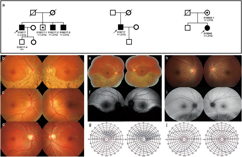
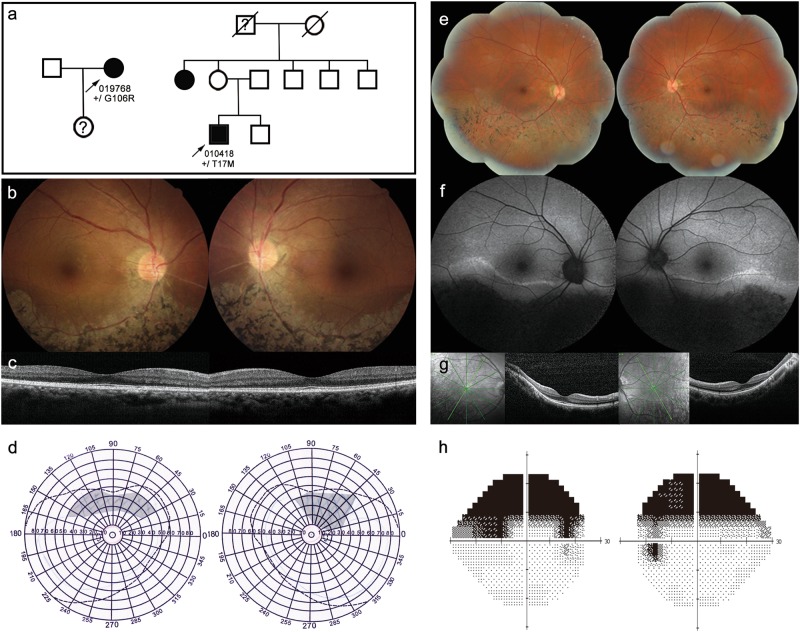
Background: Sector retinitis pigmentosa (RP) is an atypical form of RP in which only one or two quadrants of the retina are involved. The objectives of this study were to report the results of a molecular screening of five unrelated Chinese patients with sector RP and describe the clinical features observed in patients with RHO mutations.

Methods: Five probands that were clinically diagnosed with sector RP were recruited for genetic analysis. They underwent ophthalmic examinations, including best corrected visual acuity, fundus examination, visual field examinations, and electroretinography. A combination of molecular screening methods, including the targeted next-generation sequencing (TES) and sanger-DNA sequencing of RHO, were used to detect mutations. In silico programs were used to analyze the pathogenicity of all the variants.

Results: Three RHO missense mutations (p.T17M, p.L31Q, and p.G106R) were identified in the five unrelated probands. The novel mutation p.L31Q was detected in three unrelated probands. All patients showed bilateral and symmetrical retinal degeneration in the inferior retina and had relatively good visual acuity. Patients with the p.L31Q mutation showed phenotypic variability and variable penetrance.

Conclusion: Our results indicate that RHO mutations are also common in Chinese patients with sector RP. The RHO gene should be given priority during mutation screening analysis for Chinese patients with sector RP.


Conflict of interest statement

The authors declare that they have no conflict of interest.

Figures

Fig. 1
Fig. 1
Pedigrees, fundus appearances, autofluorescence (AF), and visual fields (VF) of three families with sector RP. a Pedigrees and segregation analysis (heterozygous mutation p. L31Q in family 019217, 019717, and 019945). Square denote males; circles denote females; solid symbols indicate affected; open symbols indicate unaffected; open symbols with a spot indicate an asymptomatic carrier; slashed symbols indicate deceased; an arrow below the symbol indicates the proband; + indicates wild-type. b, c, h The color fundus (CF) photos of probands 019217, 019717, and 019945 show symmetrical retinal degenerations in the inferior quadrants in both eyes. b In addition to the retinal degeneration, CF photo of proband 019217 displays a typical glaucoma cup (black arrows) in the left eye. c The CF photo of 019217–1 shows mild RPE changes in the inferior peripheral retina. d The CF photo of 019217–4 displays normal fundus appearances. d, i The AF of proband 019717 and 019945 present symmetrical hypofluorescence areas in the inferior quadrants. g The VF of prband 019717 showed the superior visual field defects corresponded to the affected retina. j The VF of proband 019945 presents normal visual fields
Fig. 2
Fig. 2
Pedigrees, fundus appearances, autofluorescence (AF), macular optic coherence tomography (OCT) images, and visual field (VF) of two sector RP families. a Pedigrees of family 019768 and 010418 the heterozygous mutations p.G106R and p.T17M, respectively. Square denote males; circles denote females; solid symbols indicate affected; open symbols indicate unaffected; open symbols with an interrogation mark indicate suspect symptomatic; slashed symbols indicate deceased; an arrow below the symbol indicates the proband; + indicates wild-type. bd The color fundus photo, OCT, and VF of proband 019768 show symmetrical retinal degeneration and hypofluorescence areas in the inferior quadrant, normal macular appearances, and the superior visual field defects. e, g, h The color fundus photo, OCT, and VF of proband 010418 present similar changes as described above. f The AF of proband 010418 shows symmetrical hypofluorescence areas in the inferior retina
Fig. 3
Fig. 3
ERG recording of a normal control (21 years old) and two patients. a The ERG of the right eye of the normal control. b, c The ERG recordings of patient 019717 and 010418 show different extent of defects of rod and cone function. The upper line of responses denotes the right eye, and the lower line denotes the left eye

Comment in

Similar articles

Cited by

References

    1. Van Woerkom C, Ferrucci S. Sector retinitis pigmentosa. Optometry. 2005;76:309–17. doi: 10.1016/S1529-1839(05)70314-6. - DOI - PubMed
    1. Berson EL, Howard J. Temporal aspects of the electroretinogram in sector retinitis pigmentosa. Arch Ophthalmol. 1971;86:653–65. doi: 10.1001/archopht.1971.01000010655008. - DOI - PubMed
    1. Omphroy CA. Sector retinitis pigmentosa and chronic angle-closure glaucoma: a new association. Ophthalmologica. 1984;189:12–20. doi: 10.1159/000309379. - DOI - PubMed
    1. Fishman GA, Stone EM, Sheffield VC, Gilbert LD, Kimura AE. Ocular findings associated with rhodopsin gene codon 17 and codon 182 transition mutations in dominant retinitis pigmentosa. Arch Ophthalmol. 1992;110:54–62. doi: 10.1001/archopht.1992.01080130056026. - DOI - PubMed
    1. Heckenlively JR, Rodriguez JA, Daiger SP. Autosomal dominant sectoral retinitis pigmentosa. Two families with transversion mutation in codon 23 of rhodopsin. Arch Ophthalmol. 1991;109:84–91. doi: 10.1001/archopht.1991.01080010086038. - DOI - PubMed

Substances