GPCR homology model template selection benchmarking: Global versus local similarity measures
- PMID: 30390544
- PMCID: PMC6449851
- DOI: 10.1016/j.jmgm.2018.10.016
GPCR homology model template selection benchmarking: Global versus local similarity measures
Abstract
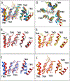
G protein-coupled receptors (GPCR) are integral membrane proteins of considerable interest as targets for drug development. GPCR ligand interaction studies often have a starting point with either crystal structures or comparative models. The majority of GPCR do not have experimentally-characterized 3-dimensional structures, so comparative modeling, also called homology modeling, is a good structure-based starting point. Comparative modeling is a widely used method for generating models of proteins with unknown structures by analogy to crystallized proteins that are expected to exhibit structural conservation. Traditionally, comparative modeling template selection is based on global sequence identity and shared function. However high sequence identity localized to the ligand binding pocket may produce better models to examine protein-ligand interactions. This in silico benchmark study examined the performance of a global versus local similarity measure applied to comparative modeling template selection for 6 previously crystallized, class A GCPR (CXCR4, FFAR1, NOP, P2Y12, OPRK, and M1) with the long-term goal of optimizing GPCR ligand identification efforts. Comparative models were generated from templates selected using both global and local similarity measures. Similarity to reference crystal structures was reflected in RMSD values between atom positions throughout the structure or localized to the ligand binding pocket. Overall, models deviated from the reference crystal structure to a similar degree regardless of whether the template was selected using a global or local similarity measure. Ligand docking simulations were performed to assess relative performance in predicting protein-ligand complex structures and interaction networks. Calculated RMSD values between ligand poses from docking simulations and crystal structures indicate that models based on locally selected templates give docked poses that better mimic crystallographic ligand positions than those based on globally-selected templates in five of the six benchmark cases. However, protein model refinement strategies in advance of ligand docking applications are clearly essential as the average RMSD between crystallographic poses and poses docked into local template models was 9.7 Å and typically less than half of the ligand interaction sites are shared between the docked and crystallographic poses. These data support the utilization of local similarity measures to guide template selection in protocols using comparative models to investigate ligand-receptor interactions.
Keywords: Comparative modeling; Comparative protein modeling; Deorphanization; G protein-coupled receptor; GPCR; Homology modeling; Ligand identification; Template selection.
Copyright © 2018 Elsevier Inc. All rights reserved.
Figures


Similar articles
-
Benchmarking GPCR homology model template selection in combination with de novo loop generation.J Comput Aided Mol Des. 2020 Oct;34(10):1027-1044. doi: 10.1007/s10822-020-00325-x. Epub 2020 Jul 31. J Comput Aided Mol Des. 2020. PMID: 32737667 Free PMC article.
-
Assessing GPCR homology models constructed from templates of various transmembrane sequence identities: Binding mode prediction and docking enrichment.J Mol Graph Model. 2018 Mar;80:38-47. doi: 10.1016/j.jmgm.2017.12.017. Epub 2017 Dec 29. J Mol Graph Model. 2018. PMID: 29306746
-
Reliability of Docking-Based Virtual Screening for GPCR Ligands with Homology Modeled Structures: A Case Study of the Angiotensin II Type I Receptor.ACS Chem Neurosci. 2019 Jan 16;10(1):677-689. doi: 10.1021/acschemneuro.8b00489. Epub 2018 Oct 17. ACS Chem Neurosci. 2019. PMID: 30265513
-
Efficiency of Homology Modeling Assisted Molecular Docking in G-protein Coupled Receptors.Curr Top Med Chem. 2021;21(4):269-294. doi: 10.2174/1568026620666200908165250. Curr Top Med Chem. 2021. PMID: 32901584 Review.
-
Homology modeling of G-protein-coupled receptors with X-ray structures on the rise.Curr Opin Drug Discov Devel. 2010 May;13(3):317-25. Curr Opin Drug Discov Devel. 2010. PMID: 20443165 Review.
Cited by
-
Homology Modeling of Class A G-Protein-Coupled Receptors in the Age of the Structure Boom.Methods Mol Biol. 2021;2315:73-97. doi: 10.1007/978-1-0716-1468-6_5. Methods Mol Biol. 2021. PMID: 34302671
-
A two-stage computational approach to predict novel ligands for a chemosensory receptor.Curr Res Struct Biol. 2020 Oct 9;2:213-221. doi: 10.1016/j.crstbi.2020.10.001. eCollection 2020. Curr Res Struct Biol. 2020. PMID: 34235481 Free PMC article.
-
G Protein-Coupled Receptor-Ligand Pose and Functional Class Prediction.Int J Mol Sci. 2024 Jun 22;25(13):6876. doi: 10.3390/ijms25136876. Int J Mol Sci. 2024. PMID: 38999982 Free PMC article.
-
GPR101: Modeling a constitutively active receptor linked to X-linked acrogigantism.J Mol Graph Model. 2024 Mar;127:108676. doi: 10.1016/j.jmgm.2023.108676. Epub 2023 Nov 21. J Mol Graph Model. 2024. PMID: 38006624 Free PMC article.
-
Benchmarking GPCR homology model template selection in combination with de novo loop generation.J Comput Aided Mol Des. 2020 Oct;34(10):1027-1044. doi: 10.1007/s10822-020-00325-x. Epub 2020 Jul 31. J Comput Aided Mol Des. 2020. PMID: 32737667 Free PMC article.
References
-
- Pierce KL; Premont RT; Lefkowitz RJ Signalling: Seven-transmembrane receptors. Nature Reviews Molecular Cell Biology 2002, 3, 639–650. - PubMed
-
- Shaikh MF Reverse Pharmacology: Fast Track Path of Drug Discovery. Pharmacy & Pharmacology International Journal 2016, 4, 1–2.
Publication types
MeSH terms
Substances
Grants and funding
LinkOut - more resources
Full Text Sources

